


-
With the advantages of non-ionizing and low cost, ultrasound imaging has been widely used in clinical diagnosis and treatment. However, due to the significant velocity changes between cortical bone and soft-tissue, the traditional ultrasound beamforming method under the assumption of constant velocity fails to reconstruct the cortical bone image. The velocity model based beamforming has been used in geophysics and non-destructive testing as an effective way to solve the challenges resulting from the velocity changes in multi-layer structure. Since the cortical bone can be modeled as a three-layer structure consisting of soft tissue, cortical bone and marrow, a multi-layer velocity model based synthetic aperture ultrasound method is introduced for cortical bone imaging. In this study, we first utilize synthetic transmit aperture ultrasound to obtain the full-matrix dataset to increase the signal-to-noise ratio. Second, a three-layer cortical bone velocity model is built with the compressed sensing estimated arriving time delay. The bases of compressed sensing consist of a series of excitation pulses with different delays. The received signals are regarded as a composition of the bases with different weights, thus can be projected into the bases by using compressed sensing. The time-delay of each received element is estimated by compressed sensing. According to the time-delay, the full-matrix dataset is reformed into a zero-offset format. By extracting the bases corresponded with the interface reflected signals, the time-delay between and the thickness values of the interfaces can be estimated. The velocity model can thus be built with the estimated cortical bone thickness. Based on the velocity model and zero-offset data, the phase shift migration method is used to reconstruct the cortical bone image. The finite-difference time-domain (FDTD) method is used to simulate the wave propagation in a 3.4-mm-thick cortical bone. The transmitting pulse is a Gaussian-function enveloped tone-burst signal with 6.25 MHz center frequency and 250 MHz iteration rate. The reconstructed image of simulation shows a clear top interface and bottom interface of cortical bone with correct thickness. Further FDTD simulations are carried out on a 3-mm-to-5-mm-thick cortical bone, and the average relative error of estimated thickness is 4.9% with a 13.5% variance. In vitro experiment is performed on a 3.4-mm-thick bovine bone plate to test the feasibility of the proposed method by using Verasonics platform (128-element linear array). The transmitting pulse is a Gaussian-function enveloped tone-burst signal with 6.25 MHz center frequency and 25 MHz sampling rate. The reconstructed image in experiment reveals a clear top interface and bottom interface of cortical bone with correct thickness. The experiment is repeated several times and the average relative error of estimated thickness is 3.6% with a 5.4% variance. The results of simulation and experiment both indicate that compressed sensing is effective in estimating the delay parameters of the velocity model. Finally, we evaluate the capability of compressed sensing in time-delay estimation, and the result shows that compressed sensing is more accurate than Hilbert transform even in a 20 dB-noise condition. In conclusion, the proposed method can be useful in the thickness estimation and the ultrasound imaging of cortical bone. In vivo experiment and clinical application should be further investigated.
-
Keywords:
- synthetic aperture ultrasound /
- cortical bone imaging /
- velocity model /
- compressed sensing
[1] Bover J, Bailone L, Lopez-Baez V, Benito S, Ciceri P, Galassi A, Cozzolino M 2017 J. Nephrol. 30 677
 Google Scholar
[2] 章轶立, 魏戌, 申浩, 谢雁鸣 2018 中国骨质疏松杂志 24 676
Google Scholar
Zhang Y L, Wei X, Shen H, Xie Y M 2018 Chin. J. Osteoporos. 24 676
Google Scholar
[3] Oo W M, Naganathan V, Bo M T, Hunter D J 2018 Quant. Imaging. Med. Surg. 8 100
Google Scholar
[4] 他得安, 王威琪, 汪源源 2009 应用声学 28 161
Google Scholar
Ta D A, Wang W Q, Wang Y Y 2009 Appl. Acoust. 28 161
Google Scholar
[5] Minonzio J G, Bochud N, Vallet Q, Ramiandrisoa D, Etcheto A, Briot K, Kolta S, Roux C, Laugier P 2019 J. Bone Miner. Res.
Google Scholar
[6] Ta D A, Wang W Q, Wang Y Y, Le L H, Zhou Y Q 2009 Ultrasound. Med. Biol. 35 641
Google Scholar
[7] Xu K L, Minonzio J G, Ta D A, Hu B, Wang W Q, Laugier P 2016 IEEE Trans. Ultrason. Ferroelectr. Freq. Control 63 1514
Google Scholar
[8] Xu K L, Ta D A, He R X, Qin Y X, Wang W Q 2014 Ultrasound. Med. Biol. 40 817
Google Scholar
[9] Bai L, Xu K L, Li D, Ta D A, Le L H, Wang W Q 2018 J. Biomech. 77 83
Google Scholar
[10] Xu K L, Laugier P, Minonzio J G 2018 J. Acoust. Soc. Am. 143 2729
Google Scholar
[11] Chartier L B, Bosco L, Lapointe-Shaw L, Chenkin J 2017 CJEM 19 131
Google Scholar
[12] 刘洋, 郭霞生, 章东, 龚秀芬 2011 声学学报 36 179
Google Scholar
Liu Y, Guo X S, Zhang D, Gong X F 2011 Acta Acustica 36 179
Google Scholar
[13] Li H J, Le L H, Sacchi M D, Lou E H M 2013 Ultrasound. Med. Biol. 39 1482
Google Scholar
[14] Renaud G, Kruizinga P, Cassereau D, Laugier P 2018 Phys. Med. Biol. 63 125010
Google Scholar
[15] Jensen J A, Nikolov S I, Gammelmark K L, Pedersen M H 2006 Ultrasonics 44 e5
Google Scholar
[16] Yu M Y, Li Y, Ma T, Shung K K, Zhou Q F 2017 IEEE Trans. Med. Imaging 36 2171
Google Scholar
[17] Nayak R, Schifitto G, Doyley M M 2017 Med. Phys. 44 4068
Google Scholar
[18] Brandt A H, Hemmsen M C, Hansen P M, Madsen S S, Krohn P S, Lange T, Hansen K L, Jensen J A, Nielsen M B 2015 Ultrasound. Med. Biol. 41 2368
Google Scholar
[19] Taner M T, Koehler F 1969 Geophysics 34 821
[20] Saenger E H, Kocur G K, Jud R, Torrilhon M 2011 Appl. Math. Modell. 35 807
Google Scholar
[21] Gazdag J 1978 Geophysics 43 1342
Google Scholar
[22] Olofsson T 2010 IEEE Trans. Ultrason. Ferroelectr. Freq. Control 57 2522
Google Scholar
[23] 孙宝申, 沈建中 1993 应用声学 3 43
Google Scholar
Sun B S, Shen J Z 1993 Appl. Acoust. 3 43
Google Scholar
[24] Trots I, Nowicki A, Lewandowski M 2010 WASET 4 136
[25] 康荣宗, 田鹏武, 于宏毅 2014 物理学报 63 200701
Google Scholar
Kang R Z, Tian P W, Yu H Y 2014 Acta Phys. Sin. 63 200701
Google Scholar
[26] 刘珍黎, 宋亮华, 白亮, 许凯亮, 他得安 2017 物理学报 66 154303
Google Scholar
Liu Z L, Song L H, Bai L, Xu K L, Ta D A 2017 Acta Phys. Sin. 66 154303
Google Scholar
[27] Liu C C 2014 Ph. D. Dissertation (Shanghai: Fudan University) (in Chinese)
[28] Baron C, Talmant M, Laugier P 2007 J. Acoust. Soc. Am. 122 1810
Google Scholar
[29] Pithioux M, Lasaygues P, Chabrand P 2002 J. Biomech. 35 961
Google Scholar
[30] Qin K H, Yang C, Sun F 2014 IEEE Trans. Ultrason. Ferroelectr. Freq. Control 61 133
Google Scholar
-
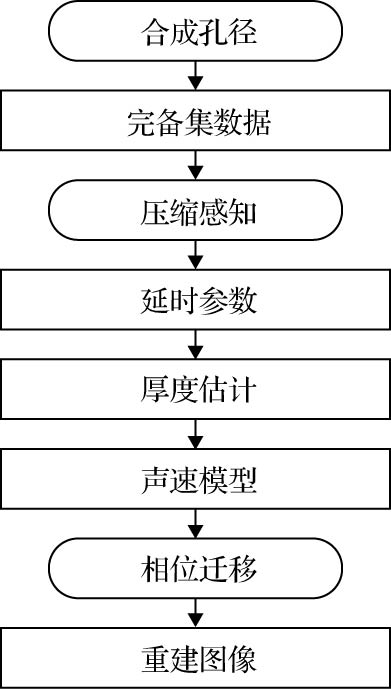
图 1 方法流程图
Figure 1. Flow chart of the proposed method.

图 2 合成孔径超声原理 (a)合成孔径聚焦; (b)发射合成孔径
Figure 2. Principle of synthetic aperture ultrasound: (a) Synthetic aperture focusing technique; (b) synthetic transmit aperture.

图 3 PSM模型 (a)单层介质模型; (b)多层介质模型
Figure 3. PSM model: (a) Single layer model; (b) multi-layer model.

图 4 实验装置示意图
Figure 4. Experiment setup.
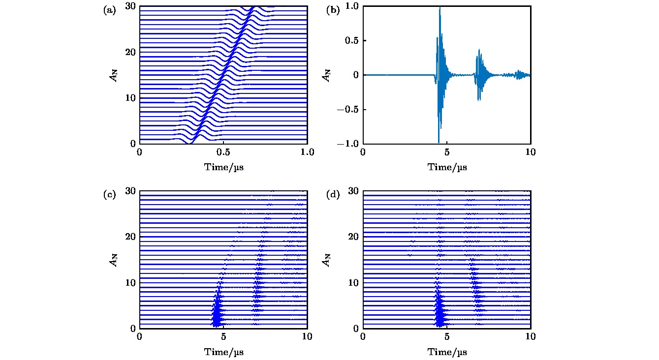
图 5 仿真结果 (a)压缩感知前30个基底; (b)单通道接收信号; (c)单次发射全部通道接收信号; (d)压缩感知调整的单次发射全部通道接收信号
Figure 5. Simulated results: (a) The first 30 bases of compressed sensing; (b) received signal of single element; (c) received signals of all elements; (d) compressed sensing based temporally adjusted received signals of all elements.

图 6 仿真重建结果 (a)未建立声速模型的仿真重建结果; (b)建立声速模型的仿真重建结果
Figure 6. Simulated reconstructed results: (a) Simulated reconstructed result without velocity model; (b) simulated reconstructed result with velocity model.
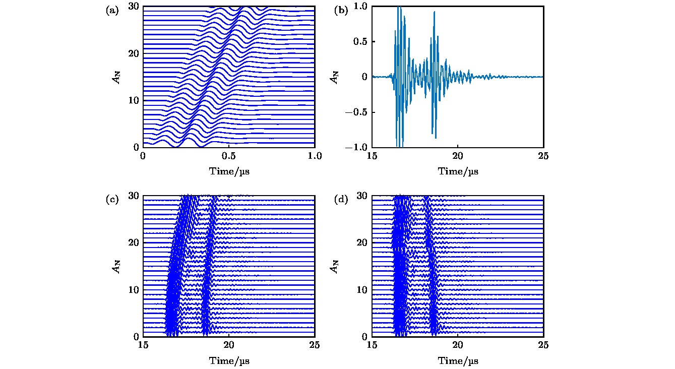
图 7 实验结果 (a)压缩感知前30个基底; (b)单通道接收信号; (c)单次发射全部通道接收信号; (d)压缩感知调整的单次发射全部通道接收信号
Figure 7. Experiment results: (a) The first 30 bases of compressed sensing; (b) received signal of single element; (c) received signals of all elements; (d) compressed sensing based temporally adjusted received signals of all elements.
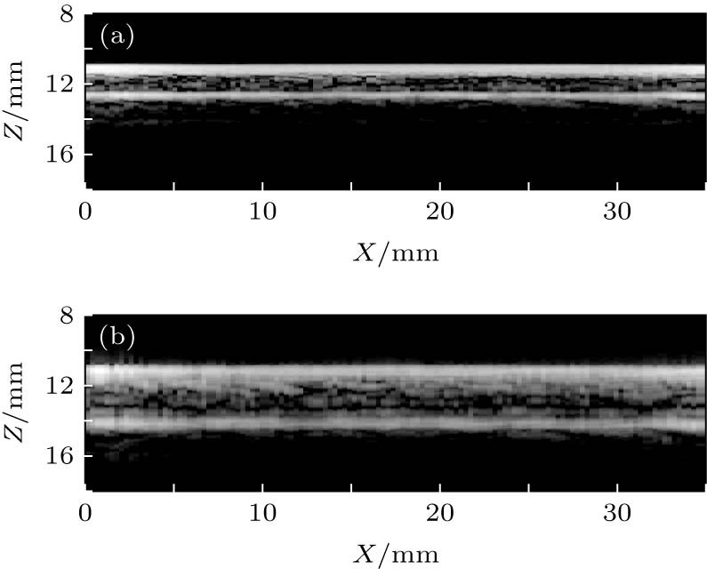
图 8 实验重建结果 (a)未建立声速模型的实验重建结果; (b)建立声速模型的实验重建结果
Figure 8. Experiment reconstructed results: (a) Experiment reconstructed result without velocity model; (b) experiment reconstructed result with velocity model.

图 9 压缩感知与Hilbert变换比较 (a)原始信号及带噪信号; (b)针对原始信号和带噪信号的压缩感知结果; (c)针对原始信号和带噪信号的Hilbert变换结果
Figure 9. Comparison of compressed sensing and Hilbert transform: (a) Origin signal and noisy signal; (b) result of compressed sensing; (c) result of Hilbert transform.
表 1 皮质骨厚度估计及误差
Table 1. Estimation and relative error of cortical bone thickness.
参量 数值 真实厚度/mm 3.0 3.1 3.2 3.3 3.4 3.5 3.6 3.7 3.8 3.9 4.0 4.1 4.2 4.3 4.4 4.5 4.6 4.7 4.8 4.9 估计厚度/mm 2.7 3.2 3.3 3.0 3.4 3.9 3.9 3.3 3.5 4.0 4.1 4.2 4.2 4.0 4.5 4.5 4.3 4.6 4.6 4.7 相对误差/% 10 3.2 3.1 9.1 0 11 8.3 11 7.9 2.6 2.5 2.4 0 6.9 2.3 0 6.5 2.1 4.2 4.1  DownLoad: CSV
-
[1] Bover J, Bailone L, Lopez-Baez V, Benito S, Ciceri P, Galassi A, Cozzolino M 2017 J. Nephrol. 30 677
Google Scholar
[2] 章轶立, 魏戌, 申浩, 谢雁鸣 2018 中国骨质疏松杂志 24 676
Google Scholar
Zhang Y L, Wei X, Shen H, Xie Y M 2018 Chin. J. Osteoporos. 24 676
Google Scholar
[3] Oo W M, Naganathan V, Bo M T, Hunter D J 2018 Quant. Imaging. Med. Surg. 8 100
Google Scholar
[4] 他得安, 王威琪, 汪源源 2009 应用声学 28 161
Google Scholar
Ta D A, Wang W Q, Wang Y Y 2009 Appl. Acoust. 28 161
Google Scholar
[5] Minonzio J G, Bochud N, Vallet Q, Ramiandrisoa D, Etcheto A, Briot K, Kolta S, Roux C, Laugier P 2019 J. Bone Miner. Res.
Google Scholar
[6] Ta D A, Wang W Q, Wang Y Y, Le L H, Zhou Y Q 2009 Ultrasound. Med. Biol. 35 641
Google Scholar
[7] Xu K L, Minonzio J G, Ta D A, Hu B, Wang W Q, Laugier P 2016 IEEE Trans. Ultrason. Ferroelectr. Freq. Control 63 1514
Google Scholar
[8] Xu K L, Ta D A, He R X, Qin Y X, Wang W Q 2014 Ultrasound. Med. Biol. 40 817
Google Scholar
[9] Bai L, Xu K L, Li D, Ta D A, Le L H, Wang W Q 2018 J. Biomech. 77 83
Google Scholar
[10] Xu K L, Laugier P, Minonzio J G 2018 J. Acoust. Soc. Am. 143 2729
Google Scholar
[11] Chartier L B, Bosco L, Lapointe-Shaw L, Chenkin J 2017 CJEM 19 131
Google Scholar
[12] 刘洋, 郭霞生, 章东, 龚秀芬 2011 声学学报 36 179
Google Scholar
Liu Y, Guo X S, Zhang D, Gong X F 2011 Acta Acustica 36 179
Google Scholar
[13] Li H J, Le L H, Sacchi M D, Lou E H M 2013 Ultrasound. Med. Biol. 39 1482
Google Scholar
[14] Renaud G, Kruizinga P, Cassereau D, Laugier P 2018 Phys. Med. Biol. 63 125010
Google Scholar
[15] Jensen J A, Nikolov S I, Gammelmark K L, Pedersen M H 2006 Ultrasonics 44 e5
Google Scholar
[16] Yu M Y, Li Y, Ma T, Shung K K, Zhou Q F 2017 IEEE Trans. Med. Imaging 36 2171
Google Scholar
[17] Nayak R, Schifitto G, Doyley M M 2017 Med. Phys. 44 4068
Google Scholar
[18] Brandt A H, Hemmsen M C, Hansen P M, Madsen S S, Krohn P S, Lange T, Hansen K L, Jensen J A, Nielsen M B 2015 Ultrasound. Med. Biol. 41 2368
Google Scholar
[19] Taner M T, Koehler F 1969 Geophysics 34 821
[20] Saenger E H, Kocur G K, Jud R, Torrilhon M 2011 Appl. Math. Modell. 35 807
Google Scholar
[21] Gazdag J 1978 Geophysics 43 1342
Google Scholar
[22] Olofsson T 2010 IEEE Trans. Ultrason. Ferroelectr. Freq. Control 57 2522
Google Scholar
[23] 孙宝申, 沈建中 1993 应用声学 3 43
Google Scholar
Sun B S, Shen J Z 1993 Appl. Acoust. 3 43
Google Scholar
[24] Trots I, Nowicki A, Lewandowski M 2010 WASET 4 136
[25] 康荣宗, 田鹏武, 于宏毅 2014 物理学报 63 200701
Google Scholar
Kang R Z, Tian P W, Yu H Y 2014 Acta Phys. Sin. 63 200701
Google Scholar
[26] 刘珍黎, 宋亮华, 白亮, 许凯亮, 他得安 2017 物理学报 66 154303
Google Scholar
Liu Z L, Song L H, Bai L, Xu K L, Ta D A 2017 Acta Phys. Sin. 66 154303
Google Scholar
[27] Liu C C 2014 Ph. D. Dissertation (Shanghai: Fudan University) (in Chinese)
[28] Baron C, Talmant M, Laugier P 2007 J. Acoust. Soc. Am. 122 1810
Google Scholar
[29] Pithioux M, Lasaygues P, Chabrand P 2002 J. Biomech. 35 961
Google Scholar
[30] Qin K H, Yang C, Sun F 2014 IEEE Trans. Ultrason. Ferroelectr. Freq. Control 61 133
Google Scholar

 DownLoad:
Catalog
Metrics
- Abstract views: 7806
- PDF Downloads: 118
- Cited By: 0
