


-
The current source reconstruction and magnetic imaging is a new technique to non-invasively obtain spatial information regarding cardiac electrical activity using magnetocardiogram (MCG) signals measured by the superconducting quantum interference device (SQUID) on the human thorax surface. Using MCG signals to reconstruct distributed current sources needs to solve the inverse problem of magnetic field. The beamforming is a type of spatial filter method that has been used for distributed source reconstruction and source imaging in electroencephalogram (EEG) and magnetoencephalogram (MEG). In this paper, the dipole moment of distributed current source is estimated with corresponding each spatial filter based on the cardiac source field model. The purpose is to enhance the intensity contrast of the dipole moment of distributed current sources in distributed source spatial spectrum estimation with beamforming, so that the reconstructed-pseudo sources beyond the heart can be removed for imaging cardiac electric activity well. A new beamforming method of improving intensity contrast (IIC) of distributed source spatial spectrum estimation is developed for imaging cardiac electric activity in P-wave, due to cardiac magnetic signals in P-wave lower than that of the peak value of R-wave, which has a relatively low signal-to-noise ratio (SNR). For enhancing the accuracy of current source reconstruction in P-wave, the IIC divided into two steps: firstly, to introduce the lead-field matrix, which represents the measurement sensor-array sensitivity to magnetic field current sources, into a weight matrix of the spatial filter for making the output estimation of the filter more sensitive to the current sourcedistribution, so as to improve the intensity contrast of the reconstructed distributed sources.Secondly, by setting a threshold of source intensity from experience, to extract the reconstructed source with locally-maximal dipole strength at each time for eliminating the relatively weak pseudo sources in other locations, so as to enhance the accuracy of current source reconstruction during P-wave. In this paper, the IIC and three other methods, including minimum variance beamforming (MVB), suppressing spatial filter output noise-power gain (SONG) and trust region reflective (TRR), are compared by using the theoretical analysis and simulation experiments of MCG current source reconstruction during P-wave. The results show that the IIC has higher intensity contrast of the single source spatial spectrum estimation, and possesses better accuracy of the current source reconstruction. The 61-channel MCG signals of two healthy subjects and their imaging of cardiac electrical activity during P-wave also are analyzed. The result shows that the IIC is better than the other three methods. It is indicated that two healthy subjects have stronger electrical activity in the atrium than that in the ventricle at Ppeak time, also that the electrical activity has the direction feature when the right-atrium is depolarized during P-wave. In summary, the IIC is useful for imaging the cardiac electrical activity. However, it is needed to carry out a further research on patients with local myocardial ischemia and left or right coronary artery stenosis, and to establish the evaluation index for imaging of cardiac electrical activity in such patients. -
Keywords:
- cardiac electrical activity /
- magnetic imaging /
- inverse problem /
- spatial filter /
- current source reconstruction
[1] Cohen D, Edelsack E A, Zimmerman J E 1970 Appl. Phys. Lett. 16 278
 Google Scholar
[2] Van Leeuwen P, Hailer B, Lange S, Klein A, Geue D, Seybold K, Poplutz C, Grönemeyer D 2008 Phys. Med. Biol. 53 2291
Google Scholar
[3] Zhang S L, Wang Y L, Wang H W, Jiang S Q, Xie X M 2009 Phys. Med. Biol. 54 4793
Google Scholar
[4] Chen T, Zhao C, Jiang S Q, van Leeuwen P, Gronemeyer D 2014 Sci. Bull. 59 1123
Google Scholar
[5] 李明, 张朝祥, 张树林, 陈威, 鲁丽, 王毅, 孔祥燕 2017 低温物理学报 39 4
Li M, Zhang C Y, Zhang S L, Chen W, Lu L, Wang Y, Kong X Y 2017 Chin. J. Low Temp. Phys. 39 4
[6] Zhao C, Jiang S Q, Wu Y H, Zhu J J, Zhou D F, Hailer B, Gronemeyer D, van Leeuwen P 2017 IEEE J. Biomed. Health 22 495
[7] Tao R, Zhang S L, Huang X, Tao M F, Ma J, Ma S X, Zhang C X, Zhang T X, Tang F K, Lu J P, Shen C X, Xie X M 2018 IEEE Trans. Biomed. Eng. 66 1658
[8] Zhang S L, Zhang G F, Wang Y L, Zeng J, Qiu Y, Liu M, Kong X Y, Xie X M 2013 Sci. Bull. 58 2917
Google Scholar
[9] Tripp J H 1983 Biomagnetism: An Interdisciplinary Approach (New York: Springer) p101
[10] Sarvas J 1987 Phys. Med. Biol. 32 11
Google Scholar
[11] Sekihara K, Nagarajan S S, Poeppel D, Marantz A 2002 IEEE Trans. Biomed. Eng. 49 1534
Google Scholar
[12] Sekihara K, Sahani M, Nagarajan S S 2005 NeuroImage 25 1056
Google Scholar
[13] Sekihara K, Nagarajan S S 2005 Modeling and Imaging of Bioelectrical Activity: Principles and Applications (New York: Kluwer Academic/Plenum Publishers) p213
[14] Van Veen B, Van Drongelen W, Yuchtman M, Suzuki A 1997 IEEE Trans. Biomed. Eng. 44 867
Google Scholar
[15] Gramfort A, Strohmeier D, Haueison J, Hämäläinen M S, Kowalski M 2013 NeuroImage 70 410
Google Scholar
[16] 王伟远, 蒋式勤, 周大方, 朱嘉辰, 闫玉蕊, 权薇薇 2014 物理学报 63 248701
Google Scholar
Wang W Y, Jiang S Q, Zhou D F, Zhu J C, Yan Y R, Quan W W 2014 Acta Phys. Sin. 63 248701
Google Scholar
[17] Ha T, Kim K, Lim S, Yu K K, Kwon H 2015 IEEE Trans. Biomed. Eng. 62 60
Google Scholar
[18] Nenonen J, Pesola K, Hänninen H, Lauerma K, Takala P, Mäkelä T, Mäkijärvi M, Knuuti J, Toivonen L, Katila T 2001 J. Electrocardiol. 34 37
Google Scholar
[19] Kobayashi K, Uchikawa Y, Nakai K, Yoshizawa M 2004 IEEE Trans. Magn. 40 2970
Google Scholar
[20] Kim K, Lee Y, Kwon H, Kim J, Bae J 2006 Comput. Biol. Med. 36 253
Google Scholar
[21] Zhu J J, Jiang S Q, Wang W Y, Zhao C, Wu Y H, Luo M, Quan W W 2014 Chin. Phys. B 23 048702
Google Scholar
[22] Chen M, Jiang S, Bing L, Zhao C, Hailer B, Grönemeyer D, Van Leeuwen P 2015 Sci. Bull. 60 1235
Google Scholar
[23] Brookes M J, Vrba J, Robinson S E, Stevenson C M, Peters A M, Barnes G R, Hillebrand A, Morris P G 2008 NeuroImage 39 1788
Google Scholar
[24] Kumihashi I, Sekihara K 2010 IEEE Trans. Biomed. Eng. 57 1358
Google Scholar
[25] Nakai K, Kawazoe K, Izumoto H, Tsuboi J, Oshima Y, Oka T, Yoshioka K, Shozushima M, Suwabe A, Itoh M, Kobayashi K, Shimizu T, Yoshizawa M 2005 Int. J. Cardiovasc Imaging 21 555
Google Scholar
[26] Nakai K, Izumoto H, Kawazoe K, Tsuboi J, Fukuhiro Y, Oka T, Yoshioka K, Shozushima M, Itoh M, Suwabe A, Yoshizawa M 2006 Int. J. Cardiovasc Imaging 22 573
Google Scholar
[27] 周大方, 张树林, 蒋式勤 2018 物理学报 67 158702
Google Scholar
Zhou D F, Zhang S L, Jiang S Q 2018 Acta Phys. Sin. 67 158702
Google Scholar
[28] Gross J, Ioannides A A 1999 Phys. Med. Biol. 44 2081
Google Scholar
[29] Horn R A, Johnson C R 2013 Matrix Analysis (New York: Cambridge University Press) p49, p139, p231
[30] Coleman T F, Li Y 1993 SIAM J. Optim. 6 418
[31] Coleman T F, Li Y 1994 Math. Program. 67 189
Google Scholar
[32] 赵晨, 蒋式勤, 石明伟, 朱俊杰 2014 物理学报 63 078702
Google Scholar
Zhao C, Jiang S Q, Shi M W, Zhu J J 2014 Acta Phys. Sin. 63 078702
Google Scholar
[33] Malmivuo J, Plonsey R 1995 Bioelectromagnetism: Principles and Applications of Bioelectric and Biomagnetic Fields (New York: Oxford University Press) p165
[34] Durrer D, Van Dam R T, Freud G E, Janse M J, Meijler F L, Arzbaecher R C 1970 Circulation 41 899
Google Scholar
-
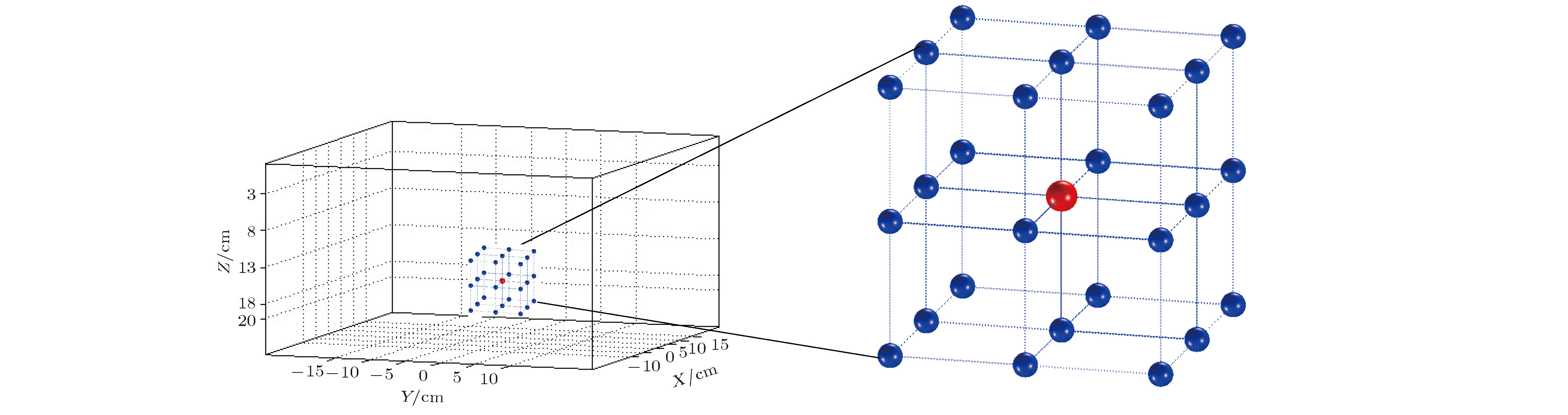
图 1 分布源位置及指定的26个邻域位置, 分别用红色和蓝色表示
Figure 1. The position of the source and 26 specified adjacent positions are shown in red and blue colors, respectively
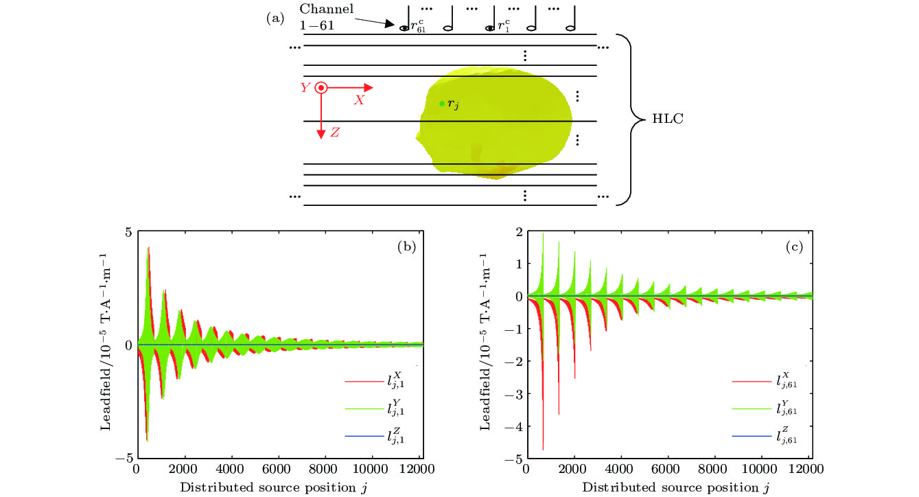
图 2 (a) 水平分层导体. 其中黑色直线表示分层躯干的边界, 黄色椭圆体表示心脏; (b), (c) 测量通道1和61的磁场导联
$l_{j,k}^\zeta , \left( {\zeta = X, Y, Z} \right)$ 曲线. 其中红、绿和蓝色分别表示X, Y和Z方向导联的曲线Figure 2. (a) The horizontally layered conductor (HLC), where the black line indicates the boundary of HLC, and the yellow spheroid the heart; (b),(c) The X, Y and Z lead-field based plots of channels 1 and 61 are expressed in red, green and blue, respectively
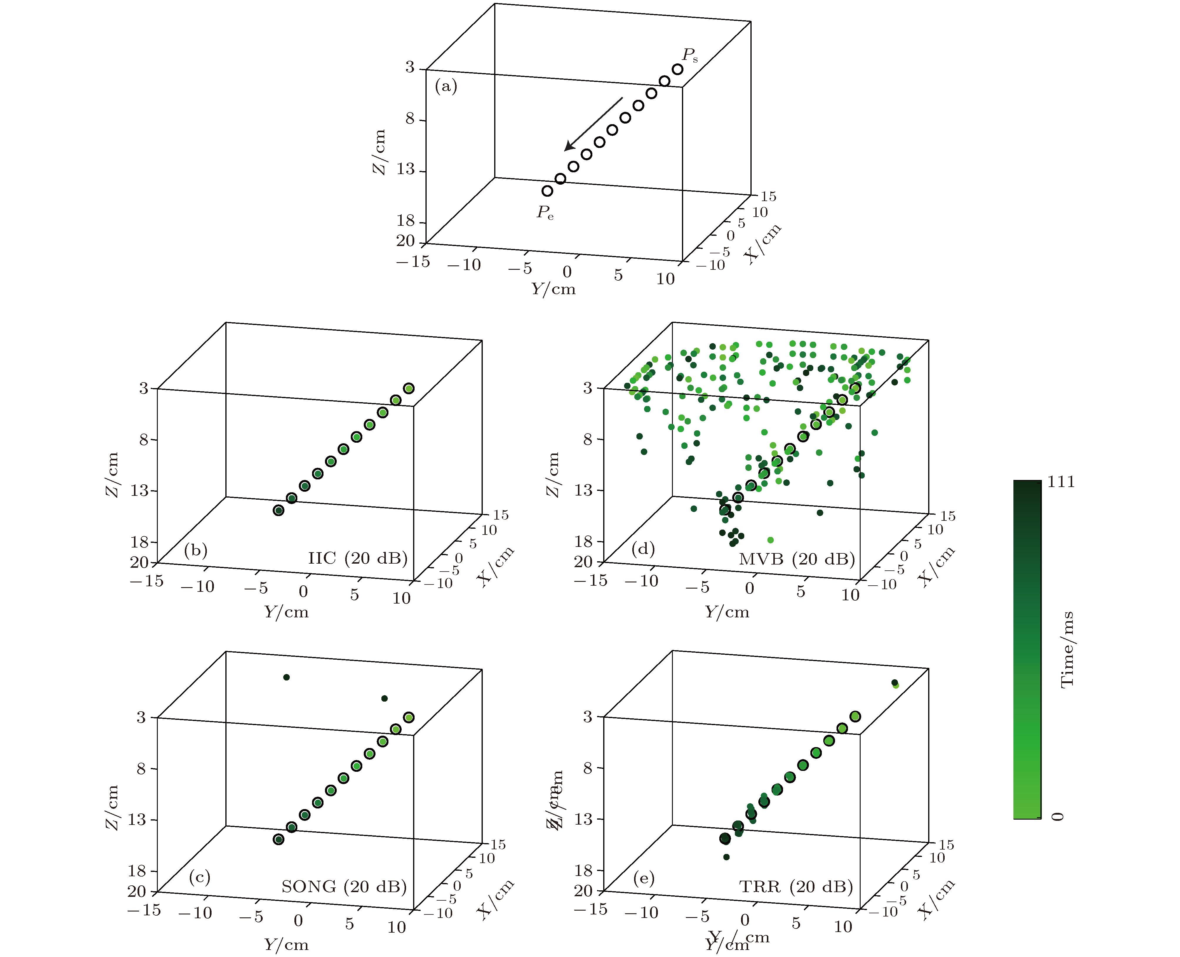
图 3 (a) 黑色圆圈○和箭头表示沿直线方向的参考路径1; (b)−(e) 是SNR = 20 dB时, IIC, SONG, MVB和TRR方法的源重建结果. 黑圈中的浅绿色到深绿色表示重建电流源位置随时间的变化
Figure 3. (a) The black circles ○ and arrow indicate the reference path 1 along the straight line; (b)−(e) The results of the current source reconstruction using IIC, SONG, MVB and TRR methods when SNR is 20 dB. The green points at different color levels denote the reconstructed source locations at different times
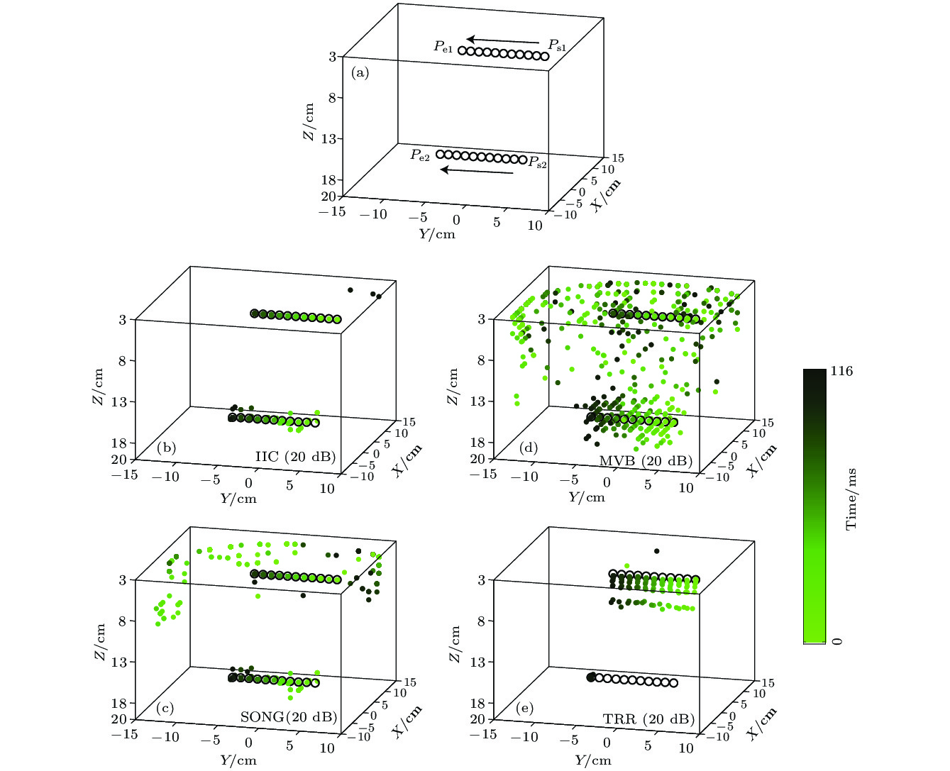
图 4 (a) 黑色圆圈○和箭头表示沿直线方向的参考路径2; (b)−(e) SNR = 20 dB时, IIC,SONG,MVB和TRR方法的源重建结果. 黑圈中的浅绿色到深绿色表示重建电流源位置随时间的变化
Figure 4. (a) The black circles ○ and arrow indicate the reference path 2 along the straight line; (b)−(e) The results of the current source reconstruction using IIC, SONG, MVB and TRR methods when SNR is 20 dB. The green points at different color levels denote the reconstructed source locations at different times
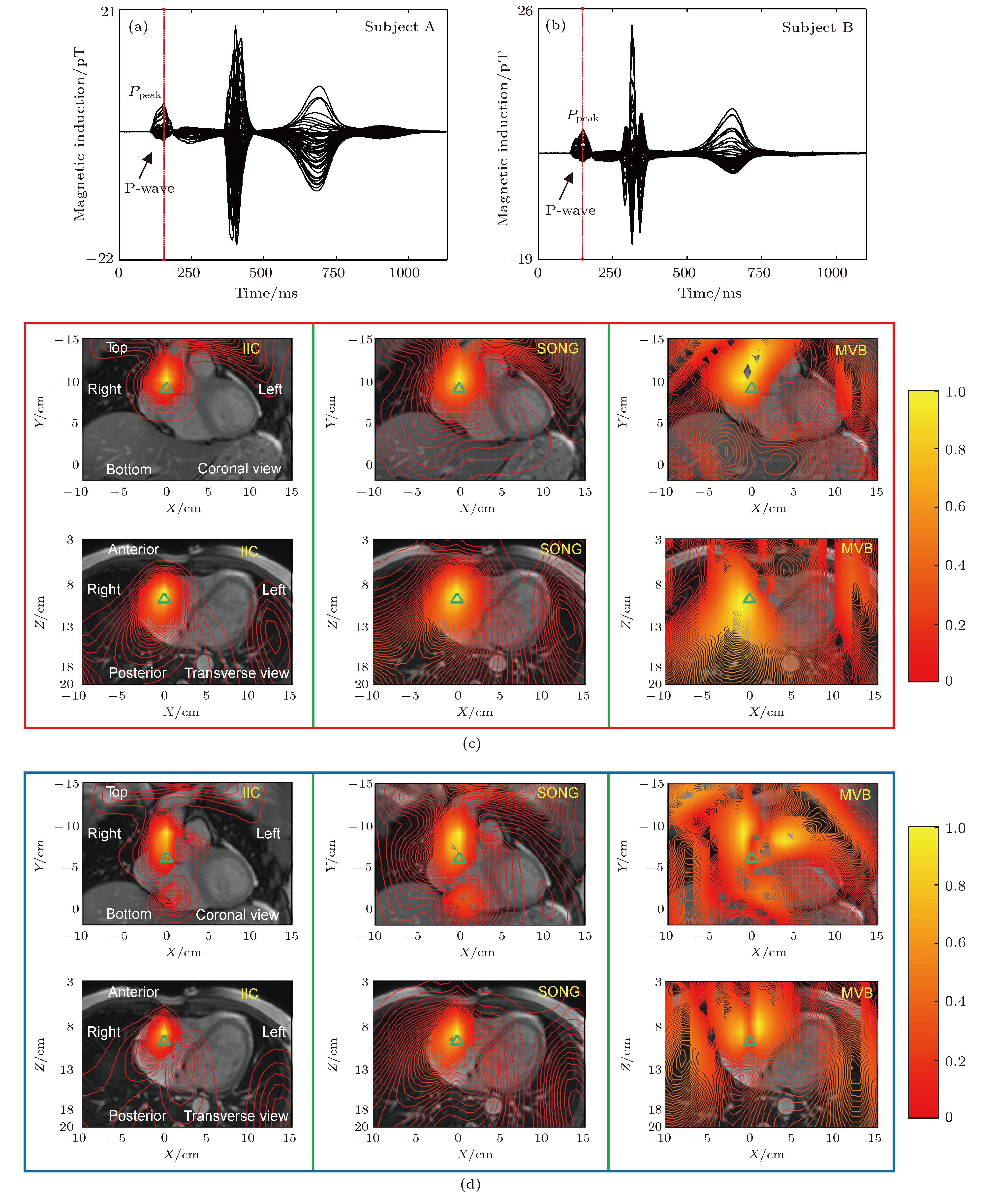
图 5 (a), (b) 健康人A与B的单周期61通道心脏磁场信号及其Ppeak时刻; (c), (d) 用IIC,SONG与MVB方法得到的2个健康人Ppeak时刻源空间谱强度的XY与XZ面等高线图
Figure 5. (a),(b) The 61-channel MCG signals and Ppeak time of single-cycle from healthy subjects A and B; (c),(d) The XY and XZ based spatial spectrum intensity contours from subjects A and B at Ppeak using IIC, SONG or MVB

图 6 (a), (b) 健康人A与B的单周期61通道心脏磁场信号及其Ponset-Ppeak段; (c), (d) 用IIC, SONG, MVB和TRR得到的2个健康人的Ponset-Ppeak段的心脏电流源重建结果. 圆点中的浅红色到深红色表示重建电流源随时间的位置变化. 绿色箭头表示电活动的方向
Figure 6. (a),(b) 61-channel MCG signal and Ponset-Ppeak segment of single-cycle from healthy subjects A and B; (c), (d) The cardiac current sources reconstruction of subjects A and B in Ponset-Ppeak segment using IIC, SONG, MVB or TRR. The reconstructed current sources change the color from white to red with time. The green arrow shows the direction of electrical activity
表 1 参考路径1对应的单源估计误差
Table 1. The error of single source estimation correspond to the reference path 1
参数设置 方法 单源估计误差/cm MV/m/s SNR/dB E EX EY EZ $\sqrt 3 $ 无噪声 IIC 1.29 0.75 0.75 0.75 SONG 1.27 0.74 0.74 0.74 MVB 1.39 0.80 0.80 0.80 TRR 0.72 0.52 0.24 0.43 30 IIC 1.37 0.79 0.79 0.79 SONG 1.73 1.13 0.81 1.04 MVB 5.76 3.08 3.73 3.12 TRR 1.50 0.83 0.17 1.24 20 IIC 1.19 0.68 0.68 0.68 SONG 1.67 1.17 0.69 0.98 MVB 6.73 4.04 3.88 3.74 TRR 2.13 1.17 1.26 1.26  DownLoad: CSV
表 2 参考路径2对应的双电流源估计误差
Table 2. The error of two sources estimation correspond to the reference path 2
参数设置 方法 一条路径的源估计误差/cm 另一路径的源估计误差/cm MV/m/s SNR/dB E EX EY EZ E EX EY EZ 1 无噪声 IIC 1.71 1.07 0.80 1.07 0.82 0 0.82 0 SONG 1.70 1.07 0.79 1.07 1.05 0.47 0.81 0.47 MVB 2.73 0.85 2.45 0.84 2.80 1.53 1.66 1.64 TRR * * * * * * * * 30 IIC 1.73 1.06 0.86 1.06 1.76 1.03 0.80 1.18 SONG 1.98 1.09 1.26 1.07 2.18 1.33 1.01 1.40 MVB 5.00 2.47 4.15 1.30 4.93 1.84 3.27 3.20 TRR * * * * * * * * 20 IIC 1.66 0.96 0.92 1.00 2.44 1.52 1.38 1.32 SONG 6.51 2.26 6.01 1.04 4.13 2.04 3.01 1.96 MVB 5.62 2.58 4.84 1.24 5.20 2.05 3.57 3.17 TRR * * * * * * * *
DownLoad: CSV
-
[1] Cohen D, Edelsack E A, Zimmerman J E 1970 Appl. Phys. Lett. 16 278
Google Scholar
[2] Van Leeuwen P, Hailer B, Lange S, Klein A, Geue D, Seybold K, Poplutz C, Grönemeyer D 2008 Phys. Med. Biol. 53 2291
Google Scholar
[3] Zhang S L, Wang Y L, Wang H W, Jiang S Q, Xie X M 2009 Phys. Med. Biol. 54 4793
Google Scholar
[4] Chen T, Zhao C, Jiang S Q, van Leeuwen P, Gronemeyer D 2014 Sci. Bull. 59 1123
Google Scholar
[5] 李明, 张朝祥, 张树林, 陈威, 鲁丽, 王毅, 孔祥燕 2017 低温物理学报 39 4
Li M, Zhang C Y, Zhang S L, Chen W, Lu L, Wang Y, Kong X Y 2017 Chin. J. Low Temp. Phys. 39 4
[6] Zhao C, Jiang S Q, Wu Y H, Zhu J J, Zhou D F, Hailer B, Gronemeyer D, van Leeuwen P 2017 IEEE J. Biomed. Health 22 495
[7] Tao R, Zhang S L, Huang X, Tao M F, Ma J, Ma S X, Zhang C X, Zhang T X, Tang F K, Lu J P, Shen C X, Xie X M 2018 IEEE Trans. Biomed. Eng. 66 1658
[8] Zhang S L, Zhang G F, Wang Y L, Zeng J, Qiu Y, Liu M, Kong X Y, Xie X M 2013 Sci. Bull. 58 2917
Google Scholar
[9] Tripp J H 1983 Biomagnetism: An Interdisciplinary Approach (New York: Springer) p101
[10] Sarvas J 1987 Phys. Med. Biol. 32 11
Google Scholar
[11] Sekihara K, Nagarajan S S, Poeppel D, Marantz A 2002 IEEE Trans. Biomed. Eng. 49 1534
Google Scholar
[12] Sekihara K, Sahani M, Nagarajan S S 2005 NeuroImage 25 1056
Google Scholar
[13] Sekihara K, Nagarajan S S 2005 Modeling and Imaging of Bioelectrical Activity: Principles and Applications (New York: Kluwer Academic/Plenum Publishers) p213
[14] Van Veen B, Van Drongelen W, Yuchtman M, Suzuki A 1997 IEEE Trans. Biomed. Eng. 44 867
Google Scholar
[15] Gramfort A, Strohmeier D, Haueison J, Hämäläinen M S, Kowalski M 2013 NeuroImage 70 410
Google Scholar
[16] 王伟远, 蒋式勤, 周大方, 朱嘉辰, 闫玉蕊, 权薇薇 2014 物理学报 63 248701
Google Scholar
Wang W Y, Jiang S Q, Zhou D F, Zhu J C, Yan Y R, Quan W W 2014 Acta Phys. Sin. 63 248701
Google Scholar
[17] Ha T, Kim K, Lim S, Yu K K, Kwon H 2015 IEEE Trans. Biomed. Eng. 62 60
Google Scholar
[18] Nenonen J, Pesola K, Hänninen H, Lauerma K, Takala P, Mäkelä T, Mäkijärvi M, Knuuti J, Toivonen L, Katila T 2001 J. Electrocardiol. 34 37
Google Scholar
[19] Kobayashi K, Uchikawa Y, Nakai K, Yoshizawa M 2004 IEEE Trans. Magn. 40 2970
Google Scholar
[20] Kim K, Lee Y, Kwon H, Kim J, Bae J 2006 Comput. Biol. Med. 36 253
Google Scholar
[21] Zhu J J, Jiang S Q, Wang W Y, Zhao C, Wu Y H, Luo M, Quan W W 2014 Chin. Phys. B 23 048702
Google Scholar
[22] Chen M, Jiang S, Bing L, Zhao C, Hailer B, Grönemeyer D, Van Leeuwen P 2015 Sci. Bull. 60 1235
Google Scholar
[23] Brookes M J, Vrba J, Robinson S E, Stevenson C M, Peters A M, Barnes G R, Hillebrand A, Morris P G 2008 NeuroImage 39 1788
Google Scholar
[24] Kumihashi I, Sekihara K 2010 IEEE Trans. Biomed. Eng. 57 1358
Google Scholar
[25] Nakai K, Kawazoe K, Izumoto H, Tsuboi J, Oshima Y, Oka T, Yoshioka K, Shozushima M, Suwabe A, Itoh M, Kobayashi K, Shimizu T, Yoshizawa M 2005 Int. J. Cardiovasc Imaging 21 555
Google Scholar
[26] Nakai K, Izumoto H, Kawazoe K, Tsuboi J, Fukuhiro Y, Oka T, Yoshioka K, Shozushima M, Itoh M, Suwabe A, Yoshizawa M 2006 Int. J. Cardiovasc Imaging 22 573
Google Scholar
[27] 周大方, 张树林, 蒋式勤 2018 物理学报 67 158702
Google Scholar
Zhou D F, Zhang S L, Jiang S Q 2018 Acta Phys. Sin. 67 158702
Google Scholar
[28] Gross J, Ioannides A A 1999 Phys. Med. Biol. 44 2081
Google Scholar
[29] Horn R A, Johnson C R 2013 Matrix Analysis (New York: Cambridge University Press) p49, p139, p231
[30] Coleman T F, Li Y 1993 SIAM J. Optim. 6 418
[31] Coleman T F, Li Y 1994 Math. Program. 67 189
Google Scholar
[32] 赵晨, 蒋式勤, 石明伟, 朱俊杰 2014 物理学报 63 078702
Google Scholar
Zhao C, Jiang S Q, Shi M W, Zhu J J 2014 Acta Phys. Sin. 63 078702
Google Scholar
[33] Malmivuo J, Plonsey R 1995 Bioelectromagnetism: Principles and Applications of Bioelectric and Biomagnetic Fields (New York: Oxford University Press) p165
[34] Durrer D, Van Dam R T, Freud G E, Janse M J, Meijler F L, Arzbaecher R C 1970 Circulation 41 899
Google Scholar

 DownLoad:

Catalog
Metrics
- Abstract views: 5578
- PDF Downloads: 37
- Cited By: 0
