


-
Developing on advanced light sources, especially those applied in the areas of spectral imaging and mass spectrometry imaging, has made the trace analysis feasible and more reliable. These techniques show great potentials in various fields including forensic science, environment, food, pharmaceuticals, archaeology, etc. In many cases of trace analysis, it is expected to obtain both the spatial distributions and chemical compositions of the target objects. Through the combination of imaging technology with optical spectroscopy and mass spectrometry, it is possible to detect the trace chemicals on the surface of various materials as well as their spatial distributions, thus improving the accuracy of detection and the range of application. Moreover, trace analysis based on such methods can reduce or even avoid the use of special chemical reagents, and is compatible with the traditional chemical detection methods. In the paper, we focus on fingerprint visualization and analysis, as a typical trace analysis issue, to discuss the recent progress of the applicable chemical imaging technologies based on the advanced light sources. The effect of latent fingerprint development depends on not only features of fingerprint carrying object, but also the characteristics of fingerprint residues. In this paper, we provide an overview of two technical approaches: specific component targeted chemical imaging and nondirective chemical imaging. We describe the major technologies involved in this field, including visible-near infrared chemical imaging, mid-infrared chemical imaging, Raman imaging, and mass spectrometry imaging.
-
Keywords:
- trace analysis /
- spectral imaging /
- mass spectrometry imaging /
- fingerprint
[1] Su B 2016 Anal. Bioanal. Chem. 408 2781
 Google Scholar
[2] van Dam A, van Beek F, Aalders M, van Leeuwen T, Lambrechts S 2016 Sci. Justice 56 143
Google Scholar
[3] Wei Q, Zhang M, Ogorevc B, Zhang X 2016 Analyst 141 6172
Google Scholar
[4] Bhargava R, Perlman R, Fernandez D, Levin I, Bartick E 2009 Anal. Bioanal. Chem. 394 2069
Google Scholar
[5] Heide S, Calavia P, Hardwick S, Hudson S, Wolff K, Russell D 2015 Forensic Sci. Int. 250 1
Google Scholar
[6] Huynh C, Halamek J 2016 Trends Anal. Chem. 82 328
Google Scholar
[7] Annemieke D, Aalders M, Kevin B, Hardy H, Leeuwen T, Lambrechts S 2013 Forensic Sci. Int. 232 173
Google Scholar
[8] He Y, Xu L, Zhu Y, Wei Q, Zhang M, Su B 2014 Angew. Chemi. Int. Ed. 53 12609
Google Scholar
[9] Leggett R, Leesmith E, Jickells S, Russell D 2007 Angew. Chemi. Int. Ed. 46 4100
Google Scholar
[10] van Dam A, Nes K, Aalders M, Leeuwen T, Lambrechts S 2014 Anal. Methods 6 1051
Google Scholar
[11] Lam R, Hofstetter O, Lennard C, Roux C, Spindler X 2016 Forensic Sci. Int. 264 168
Google Scholar
[12] Xu L, Zhou Z, Zhang C, He Y, Su B 2014 Chem. Commun. 50 9097
Google Scholar
[13] Zhang Y, Zhou W, Xue Y, Yang J, Liu D 2016 Anal. Chem. 88 12502
Google Scholar
[14] van Dam A, Aalders M, van Leeuwen T, Lambrechts S 2013 J. Forensic Sci. 58 999
Google Scholar
[15] van Dam A, Aalders M, Puit M, Gorre S, Irmak D, Leeuwen T, Lambrechts S 2014 Sci. Justice 54 356
Google Scholar
[16] Edelman G, Gaston E, van Leeuwen T, Cullen P, Aalders M 2012 Forensic Sci. Int. 223 28
Google Scholar
[17] O'Neill K, Hinners P, Lee Y 2018 J. Forensic Sci. 63 1854
Google Scholar
[18] 齐敏珺, 陈弈桦, 王新全 2016 刑事技术 41 299
Qi M J, Chen Y H, Wang X Q 2016 Forensic Sci. Technol. 41 299
[19] Maynard P, Jenkins J, Edey C, Payne G, Lennard C, Mcdonagh A, Roux C 2009 Aust. J. Forensic Sci. 41 43
Google Scholar
[20] Xie H, Wen Q, Huang H, Sun T, Li P, Li Y, Yu X, Wang Q 2015 RSC Adv. 5 79525
Google Scholar
[21] 鲍文, 丁志华, 王川, 梅胜涛 2013 物理学报 62 114202
Google Scholar
Bao W, Ding Z H, Wang C, Mei S T 2013 Acta Phys. Sin. 62 114202
Google Scholar
[22] Ossa M, Amigo J, Garcia-Ruiz C 2014 Forensic Sci. Int. 242 228
Google Scholar
[23] Banas A, Banas K, Breese B, Loke J, Lim S 2014 Anal. Bioanal. Chem. 406 4173
Google Scholar
[24] Crane N, Bartick E, Perlman R, Huffman S 2007 J. Forensic Sci. 52 48
Google Scholar
[25] Sonnex E, Almond M, Bond J 2016 J. Forensic Sci. 61 1100
Google Scholar
[26] Ricci C, Bleay S, Kazarian S 2007 Anal. Chem. 79 5771
Google Scholar
[27] Chan K, Kazarian S 2006 Analyst 131 126
Google Scholar
[28] Ewing A, Kazarian S 2017 Analyst 142 257
Google Scholar
[29] Dorakumbura B, Boseley R, Becker T, Martin D E, Richter A, Tobin M, van Bronswjik W, Vongsvivut J, Hackett M, Lewis S 2018 Analyst 143 3961
Google Scholar
[30] 马金栋, 吴浩煜, 路桥, 马挺, 时雷, 孙青, 毛庆和 2018 物理学报 67 094207
Google Scholar
Ma J D, Wu H Y, Lu Q, Ma T, Shi L, Sun Q, Mao Q H 2018 Acta Phys. Sin. 67 094207
Google Scholar
[31] Ricci C, Phiriyavityopas P, Curum N, Chan K, Jickells S, Kazarian S 2007 Appl. Spectrosc. 61 514
Google Scholar
[32] Girod A, Xiao L, Reedy B, Roux C, Weyermann C 2015 Forensic Sci. Int. 254 185
Google Scholar
[33] Day J, Edwards H, Dobrowski S, Voice A 2004 Spectrochim. Acta Part A 60 563
Google Scholar
[34] Day J, Edwards H, Dobrowski S, Voice A 2004 Spectrochim. Acta Part A 60 1725
Google Scholar
[35] Widjaja E 2009 Analyst 134 769
Google Scholar
[36] Emmons E, Tripathi A, Guicheteau J, Christesen S, Fountain A 2009 Appl. Spectrosc. 63 1197
Google Scholar
[37] Tripathi A, Emmons E, Wilcox P, Guicheteau J, Fountain A 2011 Appl. Spectrosc. 65 611
Google Scholar
[38] Song W, Mao Z, Liu X, Lu Y, Li Z, Zhao B, Lu L 2012 Nanoscale 4 2333
Google Scholar
[39] Figueroa B, Chen Y, Berry K, Francis A, Fu D 2017 Anal. Chem. 89 4468
Google Scholar
[40] Gode D, Volmer D 2013 Analyst 138 1289
Google Scholar
[41] Tang H, Lu W, Che C, Ng K 2010 Anal. Chem. 82 1589
Google Scholar
[42] Cheng Y, Zhang Y, Chau S, Lai K, Tang H, Ng K 2016 ACS Appl. Mater. Interfaces 8 29668
Google Scholar
[43] Hinners P, O'Neill K, Lee Y 2018 Sci. Rep. 8 5149
Google Scholar
[44] Kaplan-Sandquist K, LeBeau M, Miller M 2014 Forensic Sci. Int. 235 68
Google Scholar
[45] Kaplan-Sandquist K, LeBeau M, Miller M 2015 J. Forensic Sci. 60 611
Google Scholar
[46] Bradshaw R, Denison N, Francese S 2017 Analyst 142 1581
Google Scholar
[47] Scotcher K, Bradshaw R 2018 Sci. Rep. 8 8765
Google Scholar
[48] Dutkiewicz E, Urban P 2016 Philos. Trans. R. Soc. A 374 20150380
Google Scholar
[49] Bandey H, Bowman V, Bleay S 2014 Fingermark Visualisation Manual (Sandridge: CAST, Home Office)
[50] Becue A 2016 Anal. Methods 8 7983
Google Scholar
[51] Bailey M, Bright N, Croxton R, Francese S, Ferguson L, Hinder S, Jickells S, Jones B, Kazarian S, Ojeda J, Webb R, Wolstenholme R, Bleay S 2012 Anal. Chem. 84 8514
Google Scholar
[52] Yang J, Yoh J 2018 Microchem. J. 139 386
Google Scholar
[53] Pleik S, Spengler B, Bhandari D R, Luhn S, Schafer T, Urbach D, Kirsch D 2018 Analyst 143 1197
Google Scholar
[54] Amigo J, Babamoradi H, Elcoroaristizabal S 2015 Anal. Chim. Acta 896 34
Google Scholar
[55] Green T, Kuznetsov I, Willingham D, Naes B, Eiden G, Zhu Z, Chao W, Rocca J, Menoni C, Duffin A 2017 J. Anal. At. Spectrom. 32 1092
Google Scholar
[56] Zhou G, Cao Q, Kartner F, Chang G 2018 Opt. Lett. 43 2953
Google Scholar
-
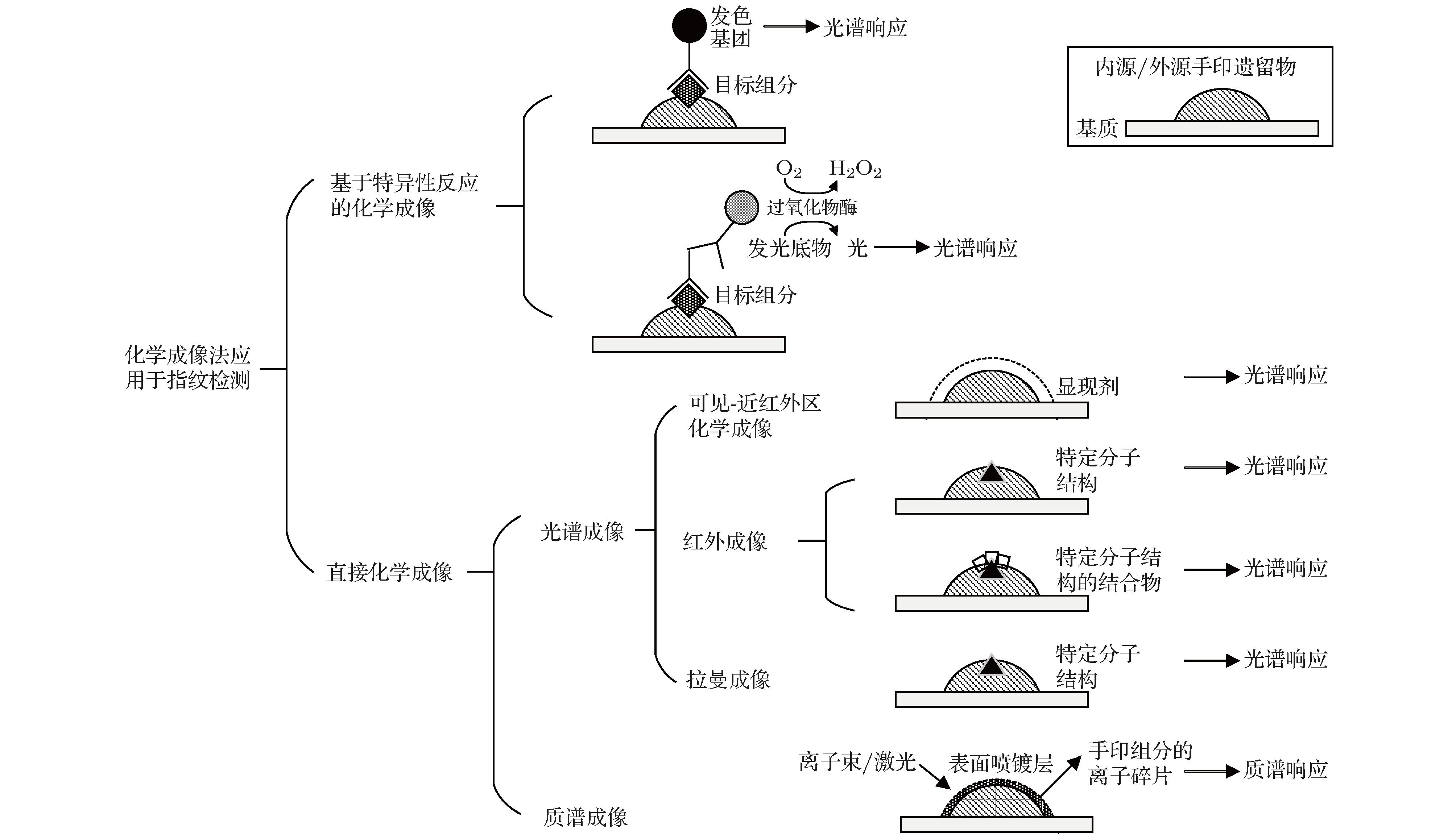
图 1 基于不同化学成像技术的指纹检验应用示例图
Figure 1. Application of various chemical imaging methods for fingerprint visualization and trace analysis.
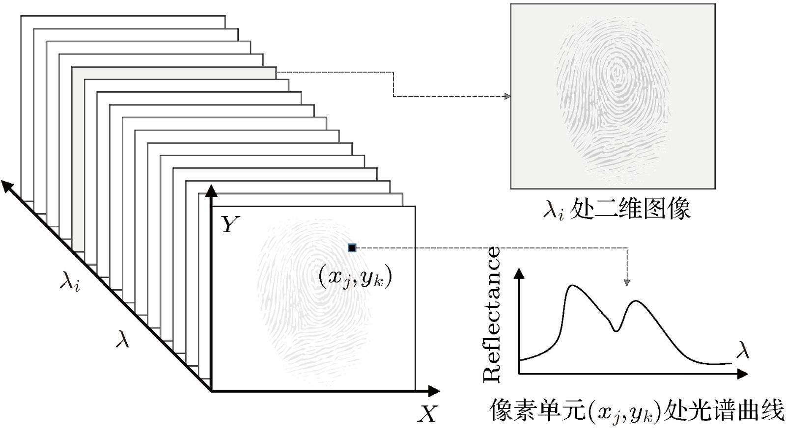
图 2 利用光谱成像技术获得基于光学参量值的数据立方
Figure 2. Hypercube of the trace sample obtained from hyperspectral imaging.
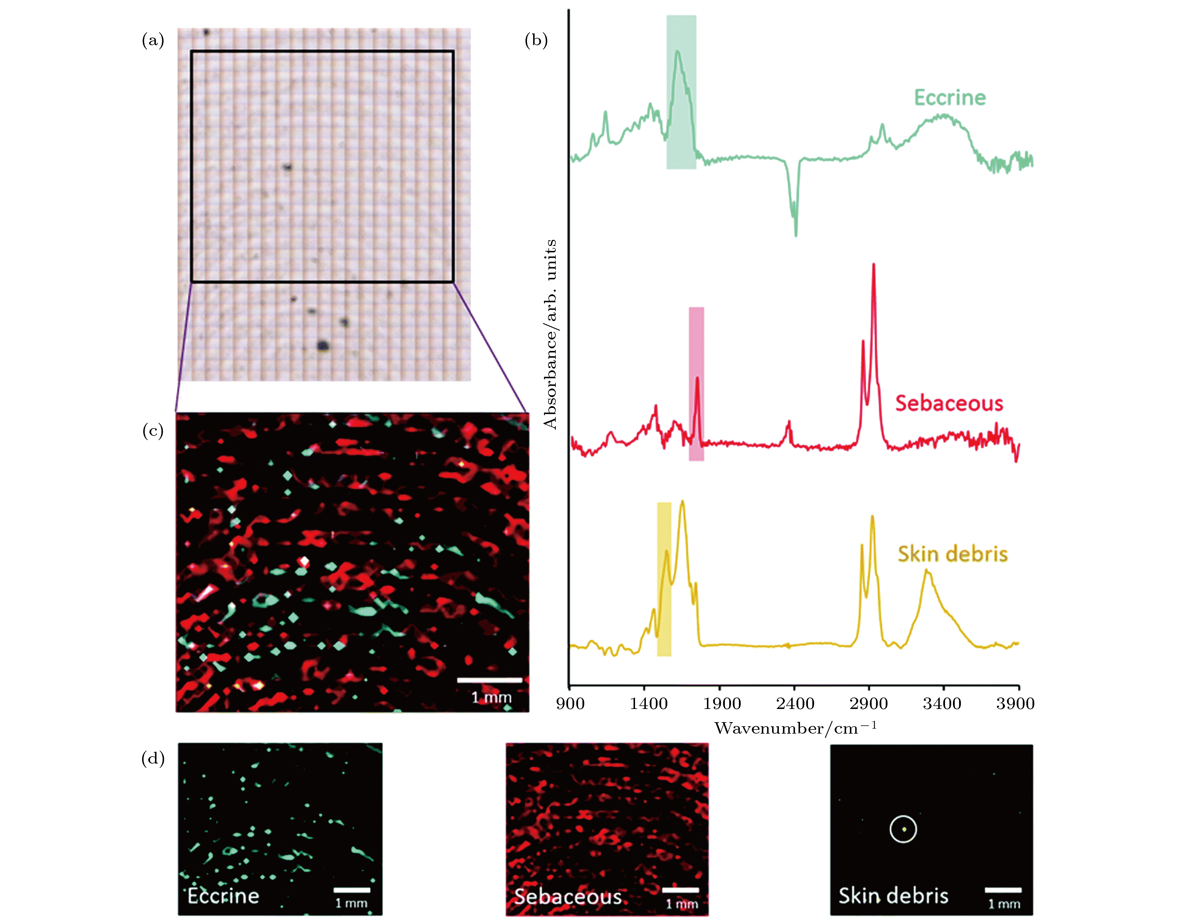
图 3 基于焦平面阵列红外光谱的指纹成像[29] (a)检测区域的亮场光学图像; (b)汗腺、皮脂腺分泌物和皮肤脱落物的FT-IR光谱; (c)指印物质组分的空间分布图像; (d)基于汗腺分泌物的O—H键弯曲振动吸收带(1520—1719 cm–1)、皮脂腺分泌物的C=O键吸收带(1713—1773 cm–1)和和皮肤脱落物的酰胺II带(1507—1548 cm–1)产生的指印物质空间分布图像
Figure 3. Fingerprint image investigated with FT-IR focal plane array imaging[29]: (a) Bright field optical image of the area investigated with FT-IR focal plane array imaging; (b) FT-IR spectra of eccrine, sebaceoussecretions and skin debris obtained using the conventional FT-IR spectroscopy; (c) the composite distribution map; (d) individual false colour images were generated by integrating over the O—H bending band for the eccrine material (1520−1719 cm–1), the C=O band for the sebaceous material (1713−1773 cm–1) and the amide II band (1507−1548 cm–1) for skin debris.
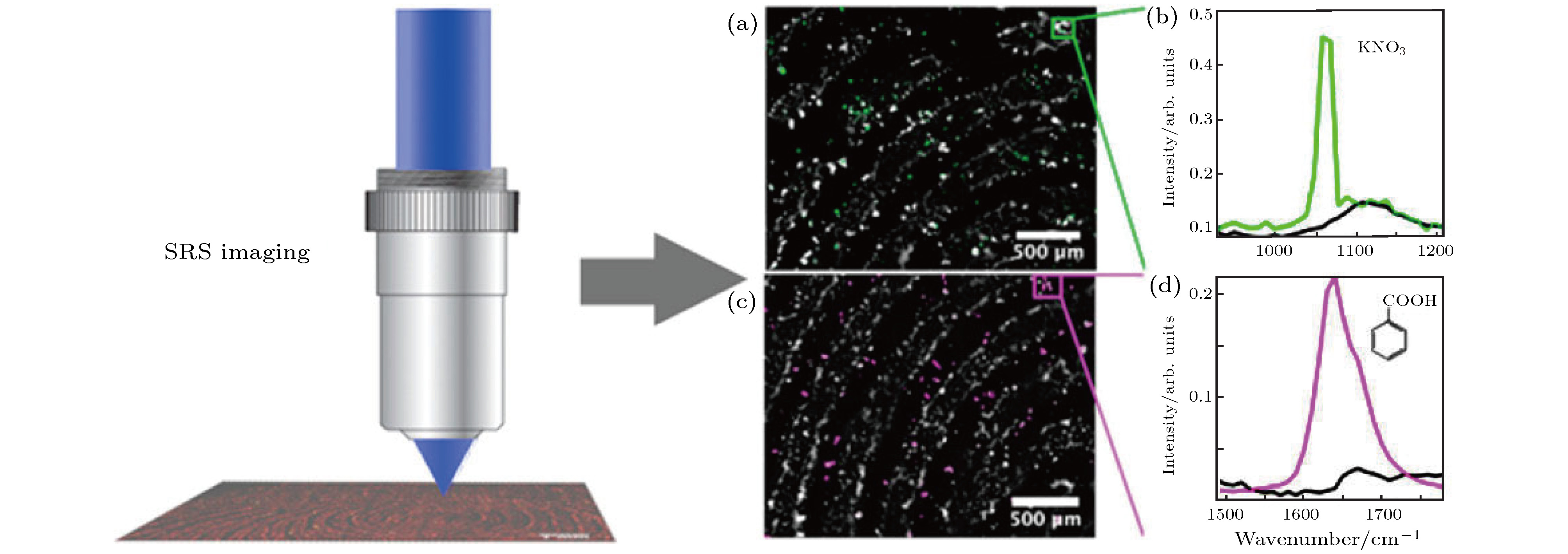
图 4 利用受激拉曼散射显微成像实现潜指纹的无标记化学成像[39] (a) 2850 cm–1和1067 cm–1波段融合图像; (b)潜指纹中KNO3的受激拉曼光谱; (c) 2850 cm–1和1639 cm–1波段融合图像; (d)苯甲酸的受激拉曼光谱
Figure 4. Label-free chemical imaging of latent fingerprints with stimulated Raman scattering microscopy[39]: (a) Image of merged channels from 2850 cm–1 and 1067 cm–1; (b) SRS spectrum of pure KNO3 on the LFP; (c) image of merged channels from 2850 cm–1 and 1639 cm–1; (b) SRS spectrum of benzoic acid.

图 5 利用MALDI/TOF MSI检出伪麻黄碱和指纹图像[45] (a)总离子流图像; (b)质荷比166的提取离子图; (c)总离子流和提取离子的叠加图; (d)标记区域质谱数据; 潜指纹经粉末刷显处理
Figure 5. Pseudoephedrine residue detection and fingerprint imaging using MLDI/TOF MSI[45]: (a) Total ion current image; (b) extracted ion image for m/z 166; (c) superimposed image of TIC and extracted ion image; (d) the corresponding mass spectra of the highlighted areas. Fingerprints were developed with fingerprint powder.
表 1 不同化学成像法的特点与适用性
Table 1. Features and applicabilities of various chemical imaging methods.
方法 预处理 适用范围 与传统显现技术的兼容性 优缺点 基于特异性反应的化学成像 基于抗原-抗体特异性结合, 使目标手印物质组分带上发色标记 显现含特定目标组分的手印 磁性粉、茚三酮、茚二酮、物理显影液、超级胶处理后, 可进一步采用本方法显现/增强显现[14,15] 检测目标明确, 灵敏度高, 适用目标组分筛查; 构建反应剂繁琐, 不适用于未知组分的定性. 可见-短波近红外区化学成像 一般不需特殊处理; 往往与传统显现剂顺序使用 当手印物质/处理后的表面物质与背景基质在可见-近红外区有差异化吸收特性时, 实现手印的显现/增强显现 先用茚三酮、物理显影液、刷显粉末、荧光剂、超级胶处理, 再进一步采用本方法增强显现; 或先采用本方法, 不影响后续使用传统显现法 与传统方法兼容, 操作便捷, 非接触式检测; 难以挖掘手印物质组分信息 红外成像 一般不需特殊处理; 依据手印遗留条件, 或使用与手印组分特定分子结构的结合物放大分子信号; 或先转移手印物质到检测基质上再采集光谱信号 组分在近红外-中红外区有响应时(如油脂手印、某些沾附炸药、违禁药物的手印等情形), 实现手印的显现/增强显现 针对一般的汗液手印, 先用超级胶处理, 再进一步采用本方法实现增强显现 适用范围相对广泛, 可挖掘手印组分物质信息, 空间分辨率较高; 灵敏度受手印组分含量限制 拉曼成像 一般不需特殊处理; 依据手印遗留条件, 或先转移手印物质到检测基质上再采集光谱信号 组分具有拉曼光谱响应时(如某些沾附药物、爆炸物的手印等情形), 实现手印的显现/增强显现 先采用本方法, 不影响后续使用传统显现法 适用范围相对广泛, 可挖掘手印组分物质信息, 空间分辨率较高, 非接触式检测; 灵敏度受手印组分含量限制 质谱成像 需进行表面喷镀处理 基于各种内源、外源手印物质组分, 实现手印的显现/增强
显现采用刷显粉末、超级胶处理后的手印, 可进一步采用本方法实现增强显现[17] 适用范围相对广泛, 可挖掘手印组分物质信息, 灵敏度较高  DownLoad: CSV
-
[1] Su B 2016 Anal. Bioanal. Chem. 408 2781
Google Scholar
[2] van Dam A, van Beek F, Aalders M, van Leeuwen T, Lambrechts S 2016 Sci. Justice 56 143
Google Scholar
[3] Wei Q, Zhang M, Ogorevc B, Zhang X 2016 Analyst 141 6172
Google Scholar
[4] Bhargava R, Perlman R, Fernandez D, Levin I, Bartick E 2009 Anal. Bioanal. Chem. 394 2069
Google Scholar
[5] Heide S, Calavia P, Hardwick S, Hudson S, Wolff K, Russell D 2015 Forensic Sci. Int. 250 1
Google Scholar
[6] Huynh C, Halamek J 2016 Trends Anal. Chem. 82 328
Google Scholar
[7] Annemieke D, Aalders M, Kevin B, Hardy H, Leeuwen T, Lambrechts S 2013 Forensic Sci. Int. 232 173
Google Scholar
[8] He Y, Xu L, Zhu Y, Wei Q, Zhang M, Su B 2014 Angew. Chemi. Int. Ed. 53 12609
Google Scholar
[9] Leggett R, Leesmith E, Jickells S, Russell D 2007 Angew. Chemi. Int. Ed. 46 4100
Google Scholar
[10] van Dam A, Nes K, Aalders M, Leeuwen T, Lambrechts S 2014 Anal. Methods 6 1051
Google Scholar
[11] Lam R, Hofstetter O, Lennard C, Roux C, Spindler X 2016 Forensic Sci. Int. 264 168
Google Scholar
[12] Xu L, Zhou Z, Zhang C, He Y, Su B 2014 Chem. Commun. 50 9097
Google Scholar
[13] Zhang Y, Zhou W, Xue Y, Yang J, Liu D 2016 Anal. Chem. 88 12502
Google Scholar
[14] van Dam A, Aalders M, van Leeuwen T, Lambrechts S 2013 J. Forensic Sci. 58 999
Google Scholar
[15] van Dam A, Aalders M, Puit M, Gorre S, Irmak D, Leeuwen T, Lambrechts S 2014 Sci. Justice 54 356
Google Scholar
[16] Edelman G, Gaston E, van Leeuwen T, Cullen P, Aalders M 2012 Forensic Sci. Int. 223 28
Google Scholar
[17] O'Neill K, Hinners P, Lee Y 2018 J. Forensic Sci. 63 1854
Google Scholar
[18] 齐敏珺, 陈弈桦, 王新全 2016 刑事技术 41 299
Qi M J, Chen Y H, Wang X Q 2016 Forensic Sci. Technol. 41 299
[19] Maynard P, Jenkins J, Edey C, Payne G, Lennard C, Mcdonagh A, Roux C 2009 Aust. J. Forensic Sci. 41 43
Google Scholar
[20] Xie H, Wen Q, Huang H, Sun T, Li P, Li Y, Yu X, Wang Q 2015 RSC Adv. 5 79525
Google Scholar
[21] 鲍文, 丁志华, 王川, 梅胜涛 2013 物理学报 62 114202
Google Scholar
Bao W, Ding Z H, Wang C, Mei S T 2013 Acta Phys. Sin. 62 114202
Google Scholar
[22] Ossa M, Amigo J, Garcia-Ruiz C 2014 Forensic Sci. Int. 242 228
Google Scholar
[23] Banas A, Banas K, Breese B, Loke J, Lim S 2014 Anal. Bioanal. Chem. 406 4173
Google Scholar
[24] Crane N, Bartick E, Perlman R, Huffman S 2007 J. Forensic Sci. 52 48
Google Scholar
[25] Sonnex E, Almond M, Bond J 2016 J. Forensic Sci. 61 1100
Google Scholar
[26] Ricci C, Bleay S, Kazarian S 2007 Anal. Chem. 79 5771
Google Scholar
[27] Chan K, Kazarian S 2006 Analyst 131 126
Google Scholar
[28] Ewing A, Kazarian S 2017 Analyst 142 257
Google Scholar
[29] Dorakumbura B, Boseley R, Becker T, Martin D E, Richter A, Tobin M, van Bronswjik W, Vongsvivut J, Hackett M, Lewis S 2018 Analyst 143 3961
Google Scholar
[30] 马金栋, 吴浩煜, 路桥, 马挺, 时雷, 孙青, 毛庆和 2018 物理学报 67 094207
Google Scholar
Ma J D, Wu H Y, Lu Q, Ma T, Shi L, Sun Q, Mao Q H 2018 Acta Phys. Sin. 67 094207
Google Scholar
[31] Ricci C, Phiriyavityopas P, Curum N, Chan K, Jickells S, Kazarian S 2007 Appl. Spectrosc. 61 514
Google Scholar
[32] Girod A, Xiao L, Reedy B, Roux C, Weyermann C 2015 Forensic Sci. Int. 254 185
Google Scholar
[33] Day J, Edwards H, Dobrowski S, Voice A 2004 Spectrochim. Acta Part A 60 563
Google Scholar
[34] Day J, Edwards H, Dobrowski S, Voice A 2004 Spectrochim. Acta Part A 60 1725
Google Scholar
[35] Widjaja E 2009 Analyst 134 769
Google Scholar
[36] Emmons E, Tripathi A, Guicheteau J, Christesen S, Fountain A 2009 Appl. Spectrosc. 63 1197
Google Scholar
[37] Tripathi A, Emmons E, Wilcox P, Guicheteau J, Fountain A 2011 Appl. Spectrosc. 65 611
Google Scholar
[38] Song W, Mao Z, Liu X, Lu Y, Li Z, Zhao B, Lu L 2012 Nanoscale 4 2333
Google Scholar
[39] Figueroa B, Chen Y, Berry K, Francis A, Fu D 2017 Anal. Chem. 89 4468
Google Scholar
[40] Gode D, Volmer D 2013 Analyst 138 1289
Google Scholar
[41] Tang H, Lu W, Che C, Ng K 2010 Anal. Chem. 82 1589
Google Scholar
[42] Cheng Y, Zhang Y, Chau S, Lai K, Tang H, Ng K 2016 ACS Appl. Mater. Interfaces 8 29668
Google Scholar
[43] Hinners P, O'Neill K, Lee Y 2018 Sci. Rep. 8 5149
Google Scholar
[44] Kaplan-Sandquist K, LeBeau M, Miller M 2014 Forensic Sci. Int. 235 68
Google Scholar
[45] Kaplan-Sandquist K, LeBeau M, Miller M 2015 J. Forensic Sci. 60 611
Google Scholar
[46] Bradshaw R, Denison N, Francese S 2017 Analyst 142 1581
Google Scholar
[47] Scotcher K, Bradshaw R 2018 Sci. Rep. 8 8765
Google Scholar
[48] Dutkiewicz E, Urban P 2016 Philos. Trans. R. Soc. A 374 20150380
Google Scholar
[49] Bandey H, Bowman V, Bleay S 2014 Fingermark Visualisation Manual (Sandridge: CAST, Home Office)
[50] Becue A 2016 Anal. Methods 8 7983
Google Scholar
[51] Bailey M, Bright N, Croxton R, Francese S, Ferguson L, Hinder S, Jickells S, Jones B, Kazarian S, Ojeda J, Webb R, Wolstenholme R, Bleay S 2012 Anal. Chem. 84 8514
Google Scholar
[52] Yang J, Yoh J 2018 Microchem. J. 139 386
Google Scholar
[53] Pleik S, Spengler B, Bhandari D R, Luhn S, Schafer T, Urbach D, Kirsch D 2018 Analyst 143 1197
Google Scholar
[54] Amigo J, Babamoradi H, Elcoroaristizabal S 2015 Anal. Chim. Acta 896 34
Google Scholar
[55] Green T, Kuznetsov I, Willingham D, Naes B, Eiden G, Zhu Z, Chao W, Rocca J, Menoni C, Duffin A 2017 J. Anal. At. Spectrom. 32 1092
Google Scholar
[56] Zhou G, Cao Q, Kartner F, Chang G 2018 Opt. Lett. 43 2953
Google Scholar

 DownLoad:
Catalog
Metrics
- Abstract views: 8923
- PDF Downloads: 125
- Cited By: 0
