


-
In a swept source-optical coherence tomography system, the telecentric scanning mode gives rise to central saturation artifacts,partial structural loss, and low SNR (signal-to-noise ratio) area in the corneal image, which affects the accuracy of corneal contour extraction. In order to solve this problem, in this paper we propose an automatic extraction algorithm for corneal image of low quality. This algorithm divides the image into high and low SNR region according to the standard deviation distribution of the cornea image. For the high SNR region, we localize the peak point to extract the contour. For the low SNR region, image enhancement is achieved by the registration and superposition of successive frames, which provides reference contour points for low SNR areas. Then corneal contour localization is achieved by weighing the advantages and disadvantages of reference contour points and local line fitting results. Finally, global polynomial fitting is used to achieve the whole corneal contour information. Experiments on the optical eye model show that comparing with the existing algorithms, the accuracy of corneal contour extraction is improved by 4.9% on average.
-
Keywords:
- optical coherence tomography /
- swept-source optical coherence tomography /
- cornea /
- contour extraction
[1] Wang J F, Zheng W, Lin K, Huang Z W 2016 Opt. Lett. 41 3045
 Google Scholar
[2] Kumar A, Baumann B, Hafner J, Ginner L, Augustin M, Salas M, Pircher M, Leitgeb R, Prager S, Schmidterfurth U 2016 Biomed. Opt. Express 8 207
[3] Grulkowski I, Liu J J, Potsaid B, Jayaraman V, Jiang J, Fujimoto J G, Cable A E 2013 Opt. Lett. 38 673
Google Scholar
[4] Polans J, Cunefare D, Cole E, Keller B, Mettu P S, Cousins S W, Allingham M J, Izatt J A, Farsiu S 2017 Opt. Lett. 42 17
Google Scholar
[5] Camino A, Jia Y L, Liu G J, Wang J, Huang D 2017 Biomed. Opt. Express 8 3053
Google Scholar
[6] Kaluzny B J, Karnowski K, Szkulmowski M, Wojtkowski M, Gora M 2011 Biomed. Opt. Express 2 2709
Google Scholar
[7] Lawman S, Dong Y, Williams B M, Romano V, Kaye S, Harding S P, Willoughby C, Shen Y C, Zheng Y L 2016 Opt. Express 24 12395
Google Scholar
[8] Lawman S, Madden P W, Vito R, Romano V, Dong Y, Mason S, Williams B M, Kaye S B, Willoughby C E, Harding S P, Shen Y C 2017 Biomed. Opt. Express 8 5579
Google Scholar
[9] Wang L L, Xiong Q Z, Ge X, Bo E, Xie J, Liu X Y, Yu X J, Wang X H, Wang N S, Chen S, Wu X, Liu L B 2019 Opt. Express 27 1298
Google Scholar
[10] Williams D, Zheng Y L, Bao F J, Elsheikh A 2013 J. Biomed. Opt. 18 056003
[11] Li Y, Shekhar R, Huang D 2002 Proc. SPIE 4684 167
Google Scholar
[12] Li Y, Shekhar R, Huang D 2006 Ophthalmology 113 792
Google Scholar
[13] Shu P, Sun Y 2012 J. Innov. Opt. Heal. Sci. 5 9
[14] LaRocca F, Chiu S J, McNabb R P, Kuo A N, Izatt J A, Farsiu S 2011 Biomed. Opt. Express 2 1524
Google Scholar
[15] Keller B, Draelos M, Tang G, Farsiu S, Kuo A N, Hauser K, Izatt J A 2018 Biomed. Opt. Express 9 2716
Google Scholar
[16] Santos V A, Schmetterer L, Stegmann H, Pfister M, Messner A, Schmidinger G, Garhofer G, Werkmeister R M 2019 Biomed. Opt. Express 10 622
Google Scholar
[17] Otsu N 2007 IEEE. Trans. Syst. Man. Cybern. 9 62
[18] Doughty M J, Zaman M L 2000 Surv. Ophthalmol. 44 367
Google Scholar
[19] Gifford P, Ahmed A, Swarbrick H A 2011 Invest. Ophth. Vis. Sci. 52 3648
Google Scholar
[20] Atchison D A, Jones C E, Schmid K L, Nicola P, Pope J M, Strugnell W E, Riley R A 2004 Invest. Ophth. Vis. Sci. 45 3380
Google Scholar
[21] Wang Q, Liu W W, Wu Y L, Ma Y, Zhao G Q 2017 Clin. Exp. Optom. 100 250
[22] Saenzfrances F, Gonzalezpastor E, Borregosanz L, Jerezfidalgo M, Martinezdelacasa J, Mendezhernandez C, Santosbueso E, Fernandezvidal A, Garciasanchez J, Garciafeijoo J 2012 J. FR. Ophtalmol. 35 333
Google Scholar
[23] Read S A, Collins M J 2009 Optometry & Vision Science Official Publication of the American Academy of Optometry 86 170
[24] Zaki F, Wang Y H, Su H, Yuan X, Liu X 2017 Biomed. Opt. Express 8 2720
Google Scholar
[25] 张强, 那彦, 李建军 2006 应用光学 27 4
Zhang Q, Na Y, Li J J 2006 J. Appl. Opt. 27 4
-

图 1 角膜轮廓提取算法流程图
Figure 1. Flow chart of corneal contour extraction algorithm

图 2 角膜图像高、低信噪比区域划分 (a) 列间标准差平滑结果; (b) 角膜图像高、低信噪比区域划分结果
Figure 2. Division between high and low SNR (signal-to-noise ratio) regions of corneal image: (a) Smoothing result of standard deviation between columns; (b) division results of high and low SNR regions of corneal image.

图 3 高信噪比区域的轮廓提取结果 (a) 轮廓点初步提取结果; (b) 轮廓点精确提取结果
Figure 3. Contour extraction results of high SNR region: (a) Preliminary extraction result of contour points; (b) accurate extraction result of contour points.

图 4 角膜整体轮廓提取过程 (a) OTSU算法处理结果; (b) 中值滤波处理结果
Figure 4. Extraction process of the overall cornea contour: (a) Processing result by OTSU algorithm; (b) processing result by median filtering.

图 5 低信噪比区域角膜轮廓建模及提取结果 (a)低信噪比区域表面轮廓点建模结果; (b)低信噪比区域轮廓提取结果
Figure 5. Modeling and extraction results of corneal contour in low SNR region: (a) Modeling results of surface contour points in low SNR region; (b) contour extraction result of low SNR region.

图 6 角膜完整轮廓的提取过程 (a)角膜上下表面轮廓点提取结果; (b) 角膜轮廓拟合结果
Figure 6. Extraction process of the complete cornea contour: (a) Extraction results of the contour points in the upper and lower cornea surfaces; (b) fitting result of the cornea contour
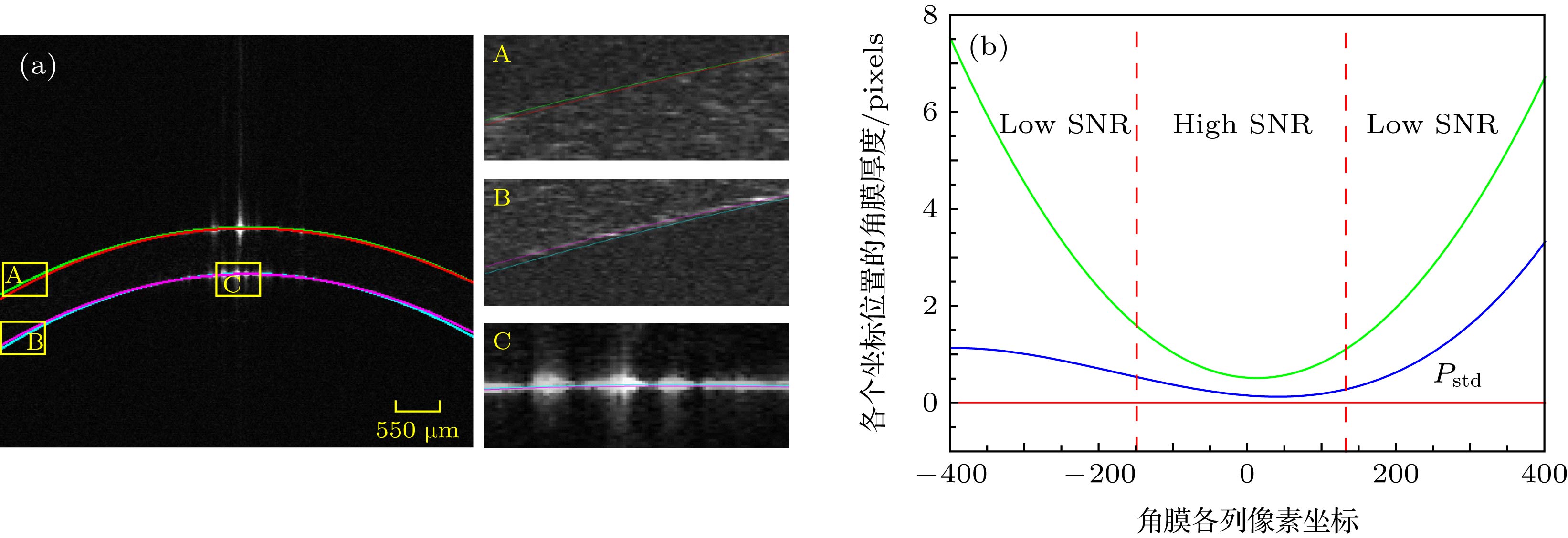
图 7 两种算法效果对比 (a)两种算法轮廓提取结果; (b) 两种算法角膜厚度平均计算结果
Figure 7. Comparison of the effects of the two algorithms: (a) Results of the contour extraction of the two algorithms; (b) results of the corneal thickness calculated by the two algorithms.
-
[1] Wang J F, Zheng W, Lin K, Huang Z W 2016 Opt. Lett. 41 3045
Google Scholar
[2] Kumar A, Baumann B, Hafner J, Ginner L, Augustin M, Salas M, Pircher M, Leitgeb R, Prager S, Schmidterfurth U 2016 Biomed. Opt. Express 8 207
[3] Grulkowski I, Liu J J, Potsaid B, Jayaraman V, Jiang J, Fujimoto J G, Cable A E 2013 Opt. Lett. 38 673
Google Scholar
[4] Polans J, Cunefare D, Cole E, Keller B, Mettu P S, Cousins S W, Allingham M J, Izatt J A, Farsiu S 2017 Opt. Lett. 42 17
Google Scholar
[5] Camino A, Jia Y L, Liu G J, Wang J, Huang D 2017 Biomed. Opt. Express 8 3053
Google Scholar
[6] Kaluzny B J, Karnowski K, Szkulmowski M, Wojtkowski M, Gora M 2011 Biomed. Opt. Express 2 2709
Google Scholar
[7] Lawman S, Dong Y, Williams B M, Romano V, Kaye S, Harding S P, Willoughby C, Shen Y C, Zheng Y L 2016 Opt. Express 24 12395
Google Scholar
[8] Lawman S, Madden P W, Vito R, Romano V, Dong Y, Mason S, Williams B M, Kaye S B, Willoughby C E, Harding S P, Shen Y C 2017 Biomed. Opt. Express 8 5579
Google Scholar
[9] Wang L L, Xiong Q Z, Ge X, Bo E, Xie J, Liu X Y, Yu X J, Wang X H, Wang N S, Chen S, Wu X, Liu L B 2019 Opt. Express 27 1298
Google Scholar
[10] Williams D, Zheng Y L, Bao F J, Elsheikh A 2013 J. Biomed. Opt. 18 056003
[11] Li Y, Shekhar R, Huang D 2002 Proc. SPIE 4684 167
Google Scholar
[12] Li Y, Shekhar R, Huang D 2006 Ophthalmology 113 792
Google Scholar
[13] Shu P, Sun Y 2012 J. Innov. Opt. Heal. Sci. 5 9
[14] LaRocca F, Chiu S J, McNabb R P, Kuo A N, Izatt J A, Farsiu S 2011 Biomed. Opt. Express 2 1524
Google Scholar
[15] Keller B, Draelos M, Tang G, Farsiu S, Kuo A N, Hauser K, Izatt J A 2018 Biomed. Opt. Express 9 2716
Google Scholar
[16] Santos V A, Schmetterer L, Stegmann H, Pfister M, Messner A, Schmidinger G, Garhofer G, Werkmeister R M 2019 Biomed. Opt. Express 10 622
Google Scholar
[17] Otsu N 2007 IEEE. Trans. Syst. Man. Cybern. 9 62
[18] Doughty M J, Zaman M L 2000 Surv. Ophthalmol. 44 367
Google Scholar
[19] Gifford P, Ahmed A, Swarbrick H A 2011 Invest. Ophth. Vis. Sci. 52 3648
Google Scholar
[20] Atchison D A, Jones C E, Schmid K L, Nicola P, Pope J M, Strugnell W E, Riley R A 2004 Invest. Ophth. Vis. Sci. 45 3380
Google Scholar
[21] Wang Q, Liu W W, Wu Y L, Ma Y, Zhao G Q 2017 Clin. Exp. Optom. 100 250
[22] Saenzfrances F, Gonzalezpastor E, Borregosanz L, Jerezfidalgo M, Martinezdelacasa J, Mendezhernandez C, Santosbueso E, Fernandezvidal A, Garciasanchez J, Garciafeijoo J 2012 J. FR. Ophtalmol. 35 333
Google Scholar
[23] Read S A, Collins M J 2009 Optometry & Vision Science Official Publication of the American Academy of Optometry 86 170
[24] Zaki F, Wang Y H, Su H, Yuan X, Liu X 2017 Biomed. Opt. Express 8 2720
Google Scholar
[25] 张强, 那彦, 李建军 2006 应用光学 27 4
Zhang Q, Na Y, Li J J 2006 J. Appl. Opt. 27 4

 DownLoad:
Catalog
Metrics
- Abstract views: 6031
- PDF Downloads: 54
- Cited By: 0
