


-
Three-dimensional (3D) ultrafast imaging is important for ultrasound technology development. The traditional 3D imaging method based on fully sampled two-dimensional (2D) matrix often requires a large number of electronic channels with high density which limits the aperture size and imaging resolution in application. Recently developed row-column addressing (RCA) matrix effectively reduces the number of electronic channels from N × N to N + N by addressing the row and column elements. The beamforming strategy designed for 3D ultrasound imaging was based on the coherent compounding of orthogonal plane waves (OPW). Such a multi-angle OPW compounding strategy achieves virtual transmit focusing in both directions by transmitting a set of plane waves in one direction and receiving along the orthogonal direction, which finally leads to an isotropic point spread function (PSF). In this paper, multi-angle OPW method was investigated for 3D blood flow imaging using an RCA matrix with 128 rows and 128 columns, centered at 6 MHz. The delay and sum (DAS) beamforming was developed for coherent OPW compounding, and the singular value decomposition (SVD) filtering method was used for separating the dynamic blood flow signals from the static tissue signals and low-amplitude noise. The Doppler velocity was computed by the autocorrelation method, and finally the 3D power Doppler and color Doppler imaging of the blood flow were realized. To evaluate the imaging quality and investigate the effect of different OPW tilting angles, quantitative analysis was carried out using multiple parameters, including –6 dB resolution measurements of the PSF, SNR of the power Doppler images and velocity distribution of the color Doppler. The –6 dB resolution is improved from 0.986 mm to 0.493 mm with the number of angles increasing from 5 to 33. With 17 plane wave angles, the SNR of the power Doppler image reaches 30 dB, and the average deviation between the velocity distribution along the diameter of the blood flow phantom and the actual value is about 26.0%. In conclusion, results show that the ultrafast 3D imaging method based on RCA matrix can obtain 3D B-mode, power Doppler and color Doppler images. Increasing the number of tilting angles and enlarging the angle range can significantly improve the imaging quality. The proposed method can be helpful for developing 3D ultrafast ultrasound Doppler imaging and functional ultrasound imaging based on neuro-vascular coupling.
-
Keywords:
- Ultrafast ultrasound /
- three-dimensional (3D) imaging /
- orthogonal plane wave (OPW) /
- row-column addressing (RCA) matrix /
- ultrasound Doppler
[1] Fenster A, Downey D B 1996 IEEE Eng. Med. Biol. 15 41
 Google Scholar
[2] Huang Q, Zeng Z 2017 BioMed Res. Int. 2017 1
Google Scholar
[3] 许凯亮, 付亚鹏, 闫少渊, 隋怡晖, 他得安, 王威琪 2023 声学学报 48 173
Google Scholar
Xu K L, Fu Y P, Yan S Y, Sui Y H, Ta D A, Wang W Q 2023 Acta Acustica 48 173
Google Scholar
[4] Brinkley J F, Moritz W E, Baker D W 1978 Ultrasound Med. Biol. 4 317
Google Scholar
[5] Baranger J, Demene C, Frerot A, Faure F, Delanoë C, Serroune H, Houdouin A, Mairesse J, Biran V, Baud O, Tanter M 2021 Nat. Commun. 12 1
Google Scholar
[6] Logan A S, Wong L L P, Chen A I H, Yeow J T W 2011 IEEE T. Ultrason. Ferr. 58 1266
Google Scholar
[7] Von Ramm O T, Smith S W 1990 J. Digit. Imaging 3 261
Google Scholar
[8] Von Ramm O T, Smith S W, Pavy H G 1991 IEEE T. Ultrason. Ferr. 38 109
Google Scholar
[9] Li P C, Huang J J 2002 IEEE T. Ultrason. Ferr. 49 1191
Google Scholar
[10] Eames M, Zhou S, Hossack J 2005 2005 IEEE International Ultrasonics Symposium(IUS) Rotterdam, The Netherlands, September 18–21, 2005 p2243
[11] Provost J, Papadacci C, Demene C, Gennisson J L, Tanter M, Pernot M 2015 IEEE T. Ultrason. Ferr. 62 1467
Google Scholar
[12] Papadacci C, Bunting E A, Konofagou E E 2017 IEEE Trans. Med. Imaging 36 357
Google Scholar
[13] Heiles B, Correia M, Hingot V, Pernot M, Provost J, Tanter M, Couture O 2019 IEEE Trans. Med. Imaging 38 2005
Google Scholar
[14] Hara K, Sakano J, Mori M, Tamano S, Sinomura R, Yamazaki K Proceedings. ISPSD '05. The 17th International Symposium on Power Semiconductor Devices and ICs Santa Barbara CA, USA, May 23–26, 2005 p359
[15] Matrone G, Savoia A S, Terenzi M, Caliano G, Quaglia F, Magenes G 2014 IEEE T. Ultrason. Ferr. 61 792
Google Scholar
[16] Ramalli A, Boni E, Savoia A S, Tortoli P 2015 IEEE T. Ultrason. Ferr. 62 1580
Google Scholar
[17] Diarra B, Robini M, Tortoli P, Cachard C, Liebgott H 2013 IEEE T. Biomed. Eng. 60 3093
Google Scholar
[18] Morton C E, Lockwood G R 2003 IEEE International Symposium on Ultrasonics(IUS) Honolulu, Hawaii, October 5–8, 2003 p968
[19] Seo C H, Yen J T 2009 IEEE T. Ultrason. Ferr. 56 837
Google Scholar
[20] Denarie B, Tangen T A, Ekroll I K, Rolim N, Torp H, Bjåstad T, Lovstakken L 2013 IEEE Trans. Med. Imaging 32 1265
Google Scholar
[21] Flesch M, Pernot M, Provost J, Ferin G, Nguyen-Dinh A, Tanter M, Deffieux T 2017 Phys. Med. Bio. 62 4571
Google Scholar
[22] Sauvage J, Porée J, Rabut C, Férin G, Flesch M, Rosinski B, Nguyen-Dinh A, Tanter M, Pernot M, Deffieux T 2020 IEEE T. Med. Imaging 39 1884
Google Scholar
[23] Deffieux T, Demené C, Tanter M 2021 Neuroscience 474 110
Google Scholar
[24] Montaldo G, Tanter M, Bercoff J, Benech N, Fink M 2009 IEEE T. Ultrason. Ferr. 56 489
Google Scholar
[25] Rasmussen M F, Christiansen T L, Thomsen E V, Jensen J A 2015 IEEE T. Ultrason. Ferr. 62 947
Google Scholar
[26] Xu K, Minonzio J G, Ta D, Hu B, Wang W, Laugier P 2016 I IEEE T. Ultrason. Ferr. 63 1514
Google Scholar
[27] Jensen J A 1996 Proceedings of the 10th Nordic-Baltic Conference on Biomedical Imaging Published in Medical & Biological Engineering & Computing Tempere, Finland, June 9–13, 1996 p351
[28] Jensen J A, Svendsen N B 1992 IEEE T. Ultrason. Ferr. 39 262
Google Scholar
[29] Taghavi I, Schou M, Panduro N S, Andersen B G, Tomov B G, Sørensen, C M, Stuart M B, Jensen J A 2022 2022 IEEE International Ultrasonics Symposium (IUS) Venice, Italy, October 10–13, 2022 p1
[30] Alfred C H, Lovstakken L 2010 IEEE T. Ultrason. Ferr. 57 1096
Google Scholar
[31] 郁钧瑾, 郭星奕, 隋怡晖, 宋剑平, 他得安, 梅永丰, 许凯亮 2022 物理学报 71 174302
Google Scholar
Yu J J, Guo X Y, Sui Y H, Song J P, Ta D A, Mei Y F, Xu K L 2022 Acta Phys. Sin. 71 174302
Google Scholar
[32] 臧佳琦, 许凯亮, 韩清见, 陆起涌, 梅永丰, 他得安 2021 物理学报 70 114304
Google Scholar
Zang J Q, Xu K L, Han Q J, Lu Q Y, Mei Y F, Ta D A 2021 Acta Phys. Sin. 70 114304
Google Scholar
[33] Sui Y, Yan S, Yu J, Song J, Ta D, Wang W, Xu K 2022 IEEE T. Ultrason. Ferr. 69 2425
Google Scholar
[34] Xu K, Guo X, Sui Y, Hingot V, Couture O, Ta D, Wang W 2021 IEEE International Ultrasonics Symposium (IUS) Xi'an, China, September 11–16, 2021 p1
-
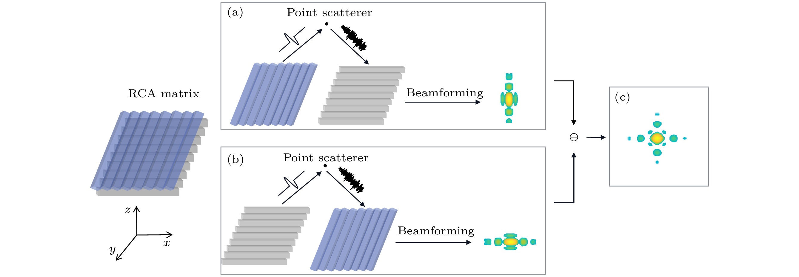
图 1 正交平面波复合 (a)行发射列接收; (b)列发射行接收; (c)各向同性的PSF图像
Figure 1. OPW compounding: (a) Row transmission and column reception; (b) column transmission and row reception; (c) coherent summation to obtain an isotropic PSF.
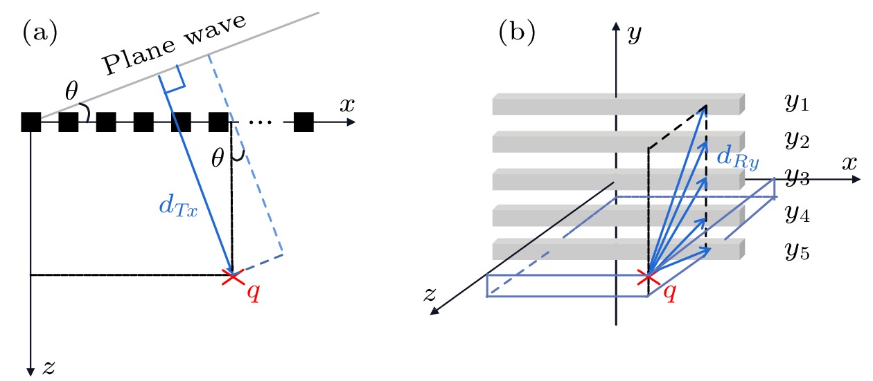
图 2 RCA阵列延时计算, 以行发射列接收为例 (a)发射距离; (b)接收距离
Figure 2. RCA matrix delay computation using row transmission and column reception as an example: (a) Forward distances; (b) back distances.
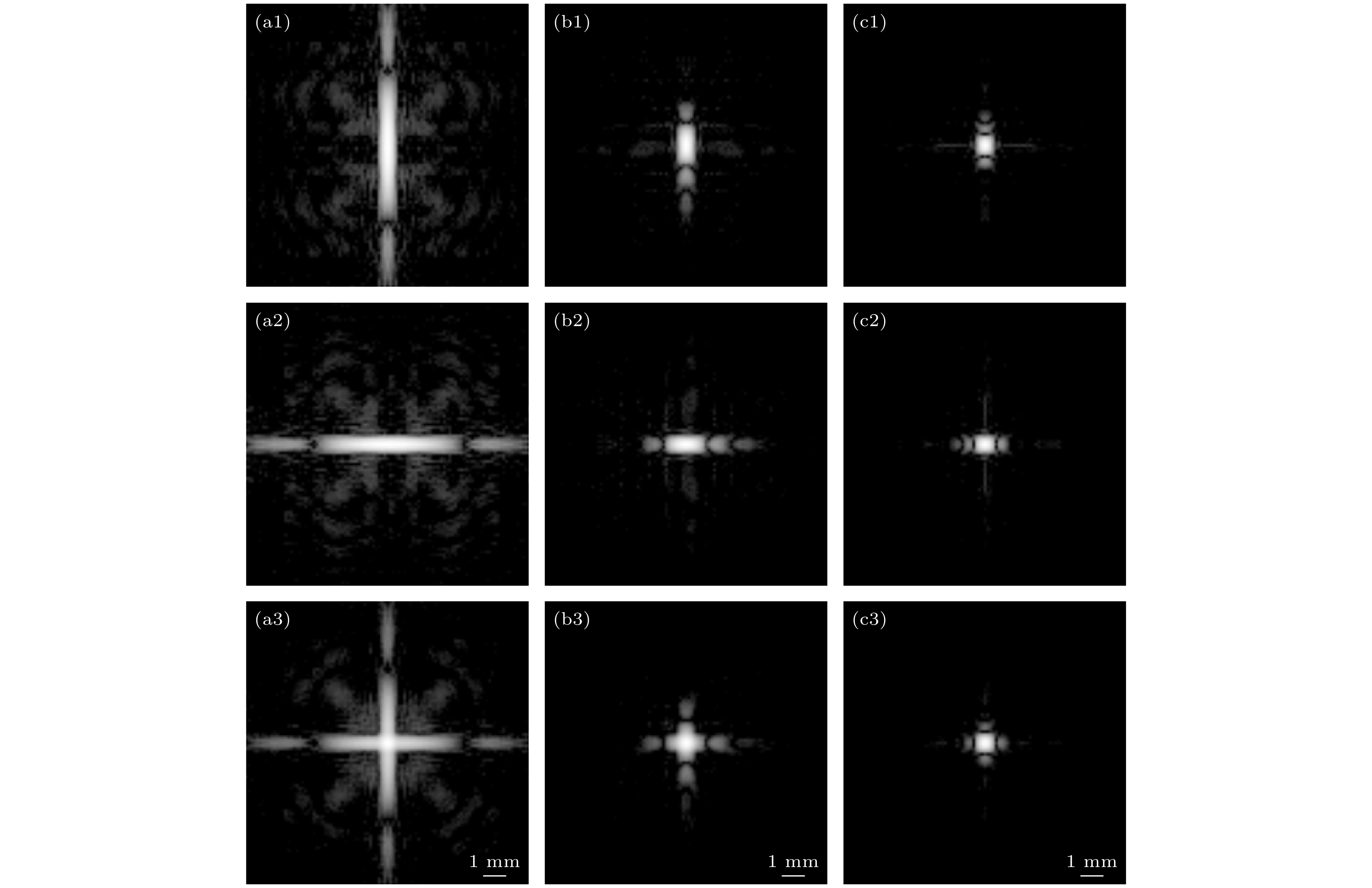
图 3 PSF图像(
$ \Delta \alpha = $ 1°), 角度数分别为: (a) 5; (b) 17; (c) 33 (1, 2为单次发射得到的PSF; 3为正交复合后的PSF)Figure 3. PSF results (
$ \Delta \alpha = $ 1°): (a) 5 angles; (b) 17 angles; (c) 33 angles (Images labeled 1 and 2 are the PSFs from single emission; images labeled 3 are the compounded PSFs).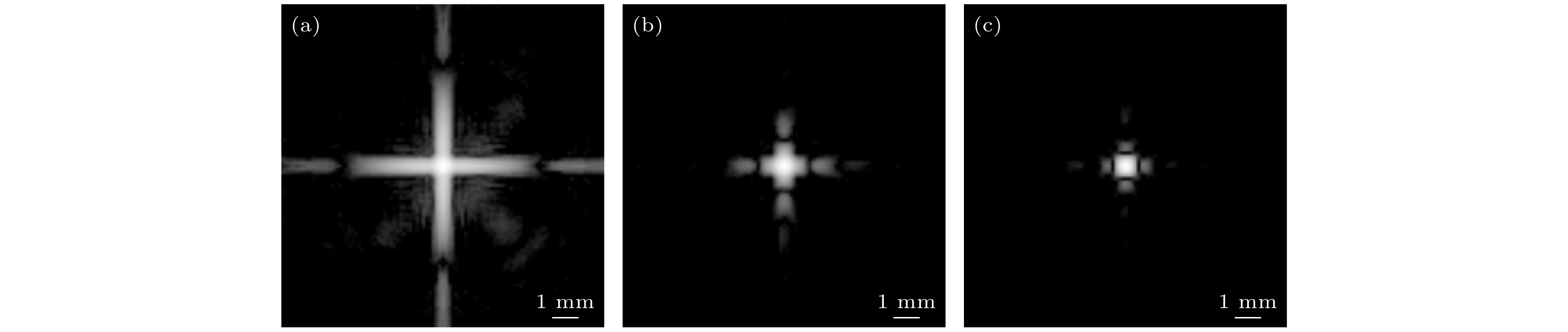
图 4
$ \Delta \alpha = $ 0.5°时的PSF图像 (a) 9个角度; (b) 33个角度; (c) 65个角度Figure 4. PSF results of
$ \Delta \alpha = $ 0.5°: (a) 9 angles; (b)33 angles; (c) 65 angles.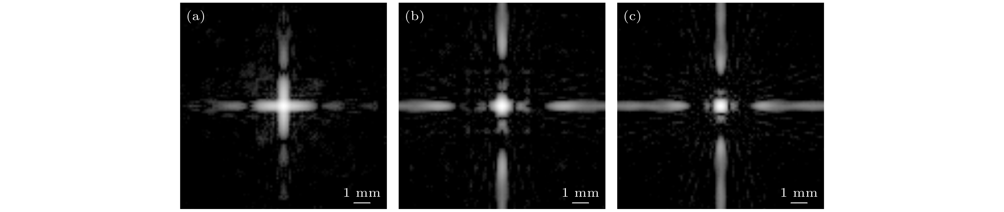
图 5 角度数为5时, 不同角度范围的PSF图像 (a) [–4°, 4°]; (b) [–8°, 8°]; (c) [–12°, 12°]
Figure 5. PSF results of 5 angles: (a) [–4°, 4°]; (b) [–8°, 8°]; (c) [–12°, 12°].
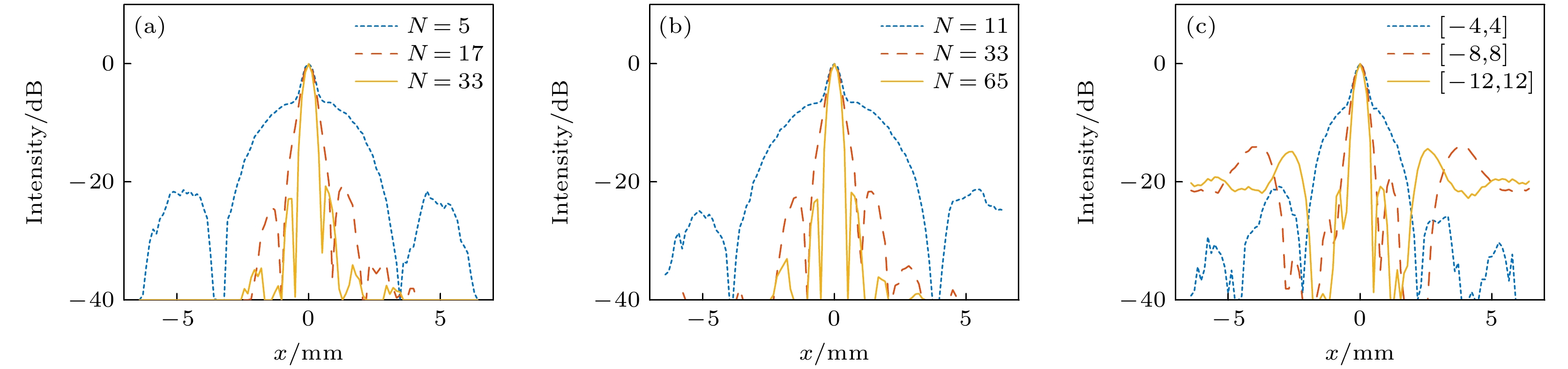
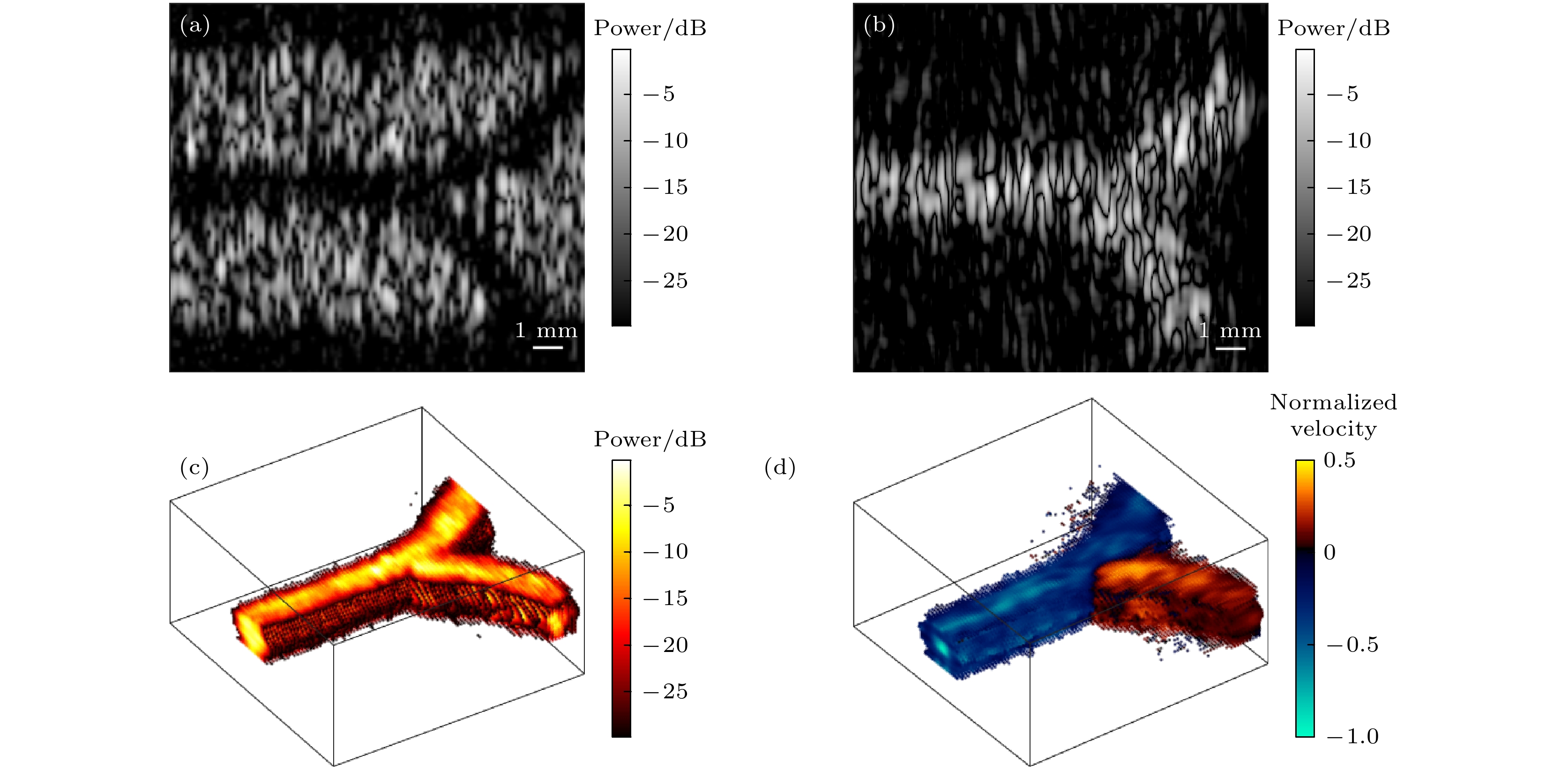
图 7 仿体血流成像结果 (a) SVD滤波前的二维B超图像; (b) SVD滤波后的二维B超图像; (c) 滤波后的三维功率多普勒图像; (d) 滤波后的三维彩色多普勒图像
Figure 7. Imaging results of the phantom blood flow: (a) B mode image before the clutter filtering; (b)B mode image after the clutter filtering; (c)3 D power Doppler image; (d)3 D color Doppler image.
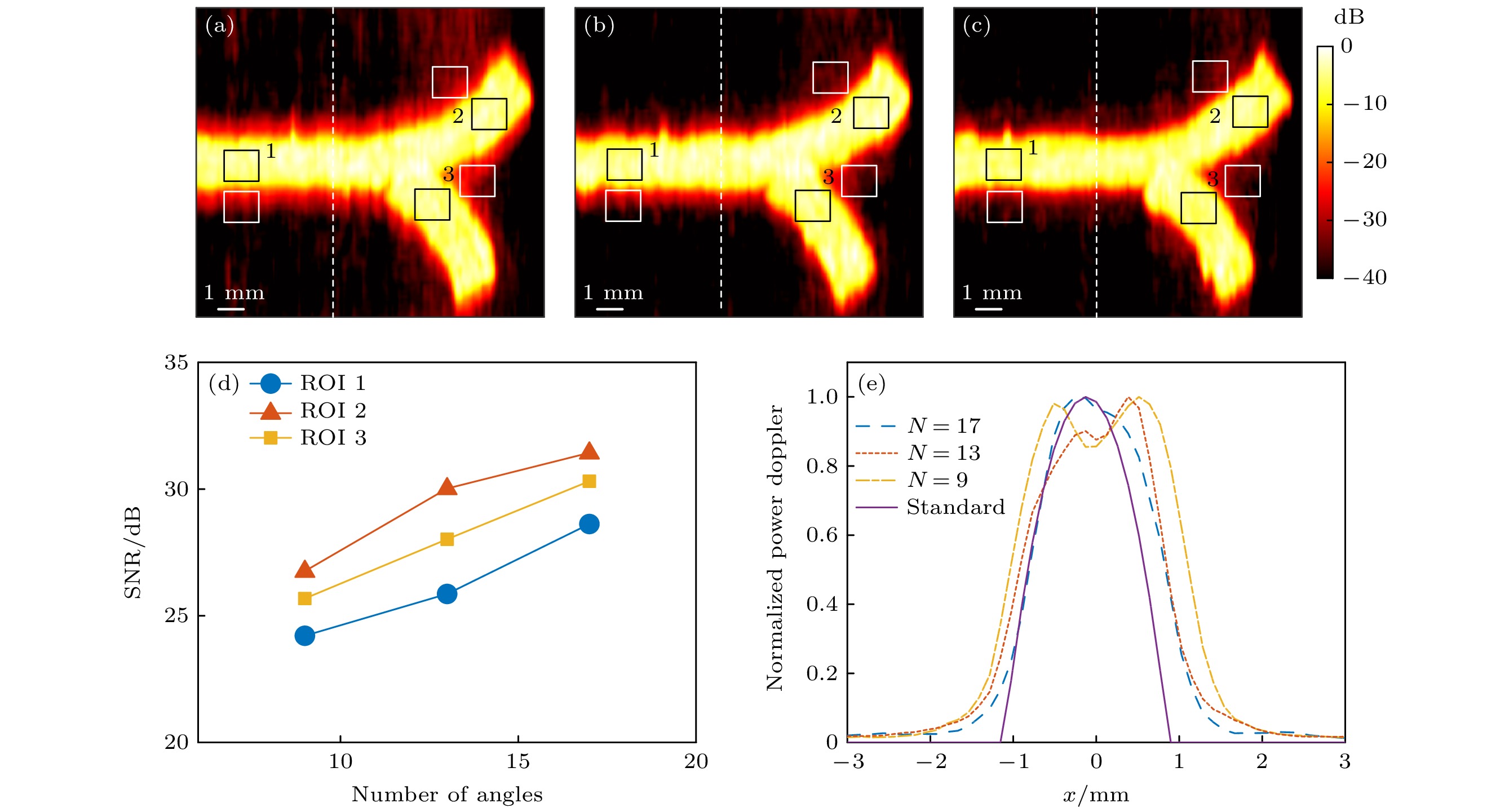
图 8 不同平面波复合角度下的功率多普勒图像及分析 (a) 9个角度; (b) 13个角度; (c) 17个角度; (d) SNR; (e) 沿虚线的功率多普勒能量分布
Figure 8. Power Doppler results with different numbers of steering angles: (a) 9 angles; (b) 13 angles; (c) 17 angles; (d) SNR; (e) power Doppler distribution along the dash line.
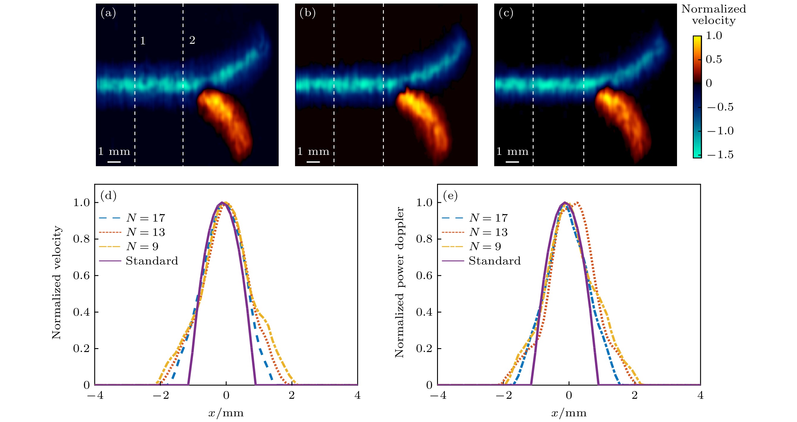
图 9 不同平面波复合角度下的彩色多普勒图像和速度分布情况 (a) 9个角度; (b) 13个角度; (c) 17个角度; (d) 沿虚线1的速度分布; (e) 沿虚线2的速度分布
Figure 9. Color Doppler image results with different numbers of steering angles: (a) 9 angles; (b) 13 angles; (c) 17 angles; (d) velocity distribution along the dash line 1; (e) velocity distribution along the dash line 2.
表 1 RCA阵列参数设置
Table 1. Parameters of the RCA matrix.
阵元数 128+128 中心频率 f0/MHz 6 声速 c/(m·s–1) 1540 波长 λ/μm 256.7 阵元中心间距/mm 0.2 阵元宽度/mm 0.175 阵列孔径/mm2 25.6$ \times $25.6  DownLoad: CSV
表 2 彩色多普勒图像速度分布的平均误差
Table 2. Average error of the velocity distribution of the color Doppler.
角度数N 9 13 17 平均误差1/% 48.73 40.48 25.03 平均误差2/% 43.55 49.70 26.86
DownLoad: CSV
-
[1] Fenster A, Downey D B 1996 IEEE Eng. Med. Biol. 15 41
Google Scholar
[2] Huang Q, Zeng Z 2017 BioMed Res. Int. 2017 1
Google Scholar
[3] 许凯亮, 付亚鹏, 闫少渊, 隋怡晖, 他得安, 王威琪 2023 声学学报 48 173
Google Scholar
Xu K L, Fu Y P, Yan S Y, Sui Y H, Ta D A, Wang W Q 2023 Acta Acustica 48 173
Google Scholar
[4] Brinkley J F, Moritz W E, Baker D W 1978 Ultrasound Med. Biol. 4 317
Google Scholar
[5] Baranger J, Demene C, Frerot A, Faure F, Delanoë C, Serroune H, Houdouin A, Mairesse J, Biran V, Baud O, Tanter M 2021 Nat. Commun. 12 1
Google Scholar
[6] Logan A S, Wong L L P, Chen A I H, Yeow J T W 2011 IEEE T. Ultrason. Ferr. 58 1266
Google Scholar
[7] Von Ramm O T, Smith S W 1990 J. Digit. Imaging 3 261
Google Scholar
[8] Von Ramm O T, Smith S W, Pavy H G 1991 IEEE T. Ultrason. Ferr. 38 109
Google Scholar
[9] Li P C, Huang J J 2002 IEEE T. Ultrason. Ferr. 49 1191
Google Scholar
[10] Eames M, Zhou S, Hossack J 2005 2005 IEEE International Ultrasonics Symposium(IUS) Rotterdam, The Netherlands, September 18–21, 2005 p2243
[11] Provost J, Papadacci C, Demene C, Gennisson J L, Tanter M, Pernot M 2015 IEEE T. Ultrason. Ferr. 62 1467
Google Scholar
[12] Papadacci C, Bunting E A, Konofagou E E 2017 IEEE Trans. Med. Imaging 36 357
Google Scholar
[13] Heiles B, Correia M, Hingot V, Pernot M, Provost J, Tanter M, Couture O 2019 IEEE Trans. Med. Imaging 38 2005
Google Scholar
[14] Hara K, Sakano J, Mori M, Tamano S, Sinomura R, Yamazaki K Proceedings. ISPSD '05. The 17th International Symposium on Power Semiconductor Devices and ICs Santa Barbara CA, USA, May 23–26, 2005 p359
[15] Matrone G, Savoia A S, Terenzi M, Caliano G, Quaglia F, Magenes G 2014 IEEE T. Ultrason. Ferr. 61 792
Google Scholar
[16] Ramalli A, Boni E, Savoia A S, Tortoli P 2015 IEEE T. Ultrason. Ferr. 62 1580
Google Scholar
[17] Diarra B, Robini M, Tortoli P, Cachard C, Liebgott H 2013 IEEE T. Biomed. Eng. 60 3093
Google Scholar
[18] Morton C E, Lockwood G R 2003 IEEE International Symposium on Ultrasonics(IUS) Honolulu, Hawaii, October 5–8, 2003 p968
[19] Seo C H, Yen J T 2009 IEEE T. Ultrason. Ferr. 56 837
Google Scholar
[20] Denarie B, Tangen T A, Ekroll I K, Rolim N, Torp H, Bjåstad T, Lovstakken L 2013 IEEE Trans. Med. Imaging 32 1265
Google Scholar
[21] Flesch M, Pernot M, Provost J, Ferin G, Nguyen-Dinh A, Tanter M, Deffieux T 2017 Phys. Med. Bio. 62 4571
Google Scholar
[22] Sauvage J, Porée J, Rabut C, Férin G, Flesch M, Rosinski B, Nguyen-Dinh A, Tanter M, Pernot M, Deffieux T 2020 IEEE T. Med. Imaging 39 1884
Google Scholar
[23] Deffieux T, Demené C, Tanter M 2021 Neuroscience 474 110
Google Scholar
[24] Montaldo G, Tanter M, Bercoff J, Benech N, Fink M 2009 IEEE T. Ultrason. Ferr. 56 489
Google Scholar
[25] Rasmussen M F, Christiansen T L, Thomsen E V, Jensen J A 2015 IEEE T. Ultrason. Ferr. 62 947
Google Scholar
[26] Xu K, Minonzio J G, Ta D, Hu B, Wang W, Laugier P 2016 I IEEE T. Ultrason. Ferr. 63 1514
Google Scholar
[27] Jensen J A 1996 Proceedings of the 10th Nordic-Baltic Conference on Biomedical Imaging Published in Medical & Biological Engineering & Computing Tempere, Finland, June 9–13, 1996 p351
[28] Jensen J A, Svendsen N B 1992 IEEE T. Ultrason. Ferr. 39 262
Google Scholar
[29] Taghavi I, Schou M, Panduro N S, Andersen B G, Tomov B G, Sørensen, C M, Stuart M B, Jensen J A 2022 2022 IEEE International Ultrasonics Symposium (IUS) Venice, Italy, October 10–13, 2022 p1
[30] Alfred C H, Lovstakken L 2010 IEEE T. Ultrason. Ferr. 57 1096
Google Scholar
[31] 郁钧瑾, 郭星奕, 隋怡晖, 宋剑平, 他得安, 梅永丰, 许凯亮 2022 物理学报 71 174302
Google Scholar
Yu J J, Guo X Y, Sui Y H, Song J P, Ta D A, Mei Y F, Xu K L 2022 Acta Phys. Sin. 71 174302
Google Scholar
[32] 臧佳琦, 许凯亮, 韩清见, 陆起涌, 梅永丰, 他得安 2021 物理学报 70 114304
Google Scholar
Zang J Q, Xu K L, Han Q J, Lu Q Y, Mei Y F, Ta D A 2021 Acta Phys. Sin. 70 114304
Google Scholar
[33] Sui Y, Yan S, Yu J, Song J, Ta D, Wang W, Xu K 2022 IEEE T. Ultrason. Ferr. 69 2425
Google Scholar
[34] Xu K, Guo X, Sui Y, Hingot V, Couture O, Ta D, Wang W 2021 IEEE International Ultrasonics Symposium (IUS) Xi'an, China, September 11–16, 2021 p1

 DownLoad:


Catalog
Metrics
- Abstract views: 3128
- PDF Downloads: 96
- Cited By: 0
