


-
Optical microscopy has the advantages of real-time, non-invasive, tomography, three-dimensional imaging and living imaging. However, its spatial resolution cannot exceed half wavelength due to the existence of optical diffraction limit, which limits the development of optical microscopy. The primary task of super-resolution imaging is to break the diffraction limit and improve the resolution of optical microscopy for study of subcellular structure. Many kinds of super-resolution imaging technologies have been reported, among which the stimulated emission depletion (STED) microscopy is the earliest imaging technology to break the optical diffraction limit at present. STED microscopy can achieve nanometer-scale spatial resolution by breaking the optical diffraction limit with pure optical methods and a clever optical design. However, the application of STED microscopy in biomedicine, especially in live cell imaging is limited by high illumination power of STED light. In this paper, a new type of STED probe has been developed. The spectral analysis results show that the peak of the excitation and emission spectrum of this probe is as far as 122 nm away from each other, which is very suitable for the study of STED super-resolution because of its long stokes redshift. After colocalization with commercial mitochondrial dyes, it was found that the probe had a higher localization coefficient with commercial dyes and could be well positioned on mitochondrial organelles. At the same time, it was found that strong mitochondrial signal could be detected with low-power excitation light (only 1 μW in the experiment), and can get higher resolution of 62 nm under the STED light with 39.5 mW. The result of measuring the transverse resolution obtained by STED light under different power shows that the saturated light power of the probe is 3.5 mW (1.1 MW·cm–2). Through the anti-bleaching testing, the probe still has a strong fluorescence intensity after more than 300 times of high power light irradiation, which indicates that the probe has a strong anti-bleaching property. Through a series of tests, this paper present a novel STED probe which has good mitochondrial targeting, excellent photobleaching-resistance, high resolution and low saturation power, which provides a new research tool for long-term live cell mitochondrial super-resolution imaging.
-
Keywords:
- stimulated emission depletion microscopy /
- super-resolution imaging /
- fluorescent probe /
- mitochondria /
- living cell imaging
[1] Webb R H 1996 Rep. Prog. Phys. 59 427
 Google Scholar
[2] 林丹樱, 屈军乐 2017 物理学报 66 148703
Google Scholar
Lin D Y, Qu J L 2017 Acta Phys. Sin. 66 148703
Google Scholar
[3] Hell S W 2003 Nat. Biotechnol. 21 1347
Google Scholar
[4] Yan W, Yang Y L, Tan Y, Chen X, Li Y, Qu J L, Ye T 2017 Photonics Res. 5 176
Google Scholar
[5] Wang L W, Yan W, Li R Z, Weng X Y, Zhang J, Yang Z G, Liu L W, Ye T, Qu J L 2018 Nanophotonics 7 1971
Google Scholar
[6] Huang B, Bates M, Zhuang X W 2009 Annu. Rev. Biochem. 78 993
Google Scholar
[7] Wang L W, Chen Y, Yan W, Weng X Y, Yang Z G, Ye T, Qu J L 2019 J. Biophotonics 12 e201800315
Google Scholar
[8] Wang L W, Chen B L, Yan W, Yang Z G, Peng X, Lin D Y, Weng X Y, Ye T, Qu J L 2018 Nanoscale 10 16252
Google Scholar
[9] Klar T A, Engel E, Hell S W 1994 Opt. Lett. 19 780
Google Scholar
[10] Hell S W, Jakobs S, Kastrup L 2003 Appl. Phys. A 77 859
Google Scholar
[11] Folling J, Bossi M, Bock H, Medda R, Wurm C A, Hein B, Jakobs S, Eggeling C 2008 Nat. Methods 5 943
Google Scholar
[12] Gustafsson M G 2000 J. Microsc. 198 82
Google Scholar
[13] Betzig E, Patterson G H, Sougrat R, Lindwasser O W, Olenych S, Bonifacino J S, Davidson M W, Schwartz J L, Hess H F 2006 Science 313 1642
Google Scholar
[14] Bates M, Huang B, Dempsey G T, Zhuang X W 2007 Science 317 793
Google Scholar
[15] Aloi A, Vilanova N, Albertazzi L, Voets I K 2016 Nanoscale 8 8712
Google Scholar
[16] Hell S W 2007 Science 316 1153
Google Scholar
[17] Hotta J, Fron E, Dedecker P, Janssen K P F, Li C, Mullen K, Harke B, Buckers J, Hell S W, Hofkens J 2010 J. Am. Chem. Soc. 132 5021
Google Scholar
[18] Vicidomini G, Schonle A, Ta H, Han K Y, Moneron G, Eggeling C, Hell S W 2013 PloS One 8 e54221
Google Scholar
[19] Liu Y J, Lu Y Q, Yang X S, Zheng X L, Wen S H, Wang F, Vidal X, Zhao J B, Liu D M, Zhou Z G, Ma C S, Zhou J, Peper J A, Xi P, Jin D Y 2017 Nature 543 229
Google Scholar
[20] Zhan Q Q, Liu H C, Wang B J, Wu Q S, Pu R, Zhou C, Huang B R, Peng X Y, Agren H, He S L 2017 Nat. Commun. 8 1058
Google Scholar
[21] Li D Y, Qin W, Xu B, Qian J 2017 Adv. Mater. 29 1703643
Google Scholar
[22] Ye S, Yan W, Zhao M J, Peng X, Song J, Qu J L 2018 Adv. Mater. 30 1800167
Google Scholar
[23] Martin O L, Hugo G S, Alexander S, James H C, Alice C N, Daniel M D, Chris D, Mark A N, Paul M W 2014 J. Biophotonics 7 29
Google Scholar
[24] Kuang C F, Li S, Liu W, Hao X, Gu X H, Wang Y F, Ge J H, Li H F, Liu X 2013 Sci. Rep. 3 1441
Google Scholar
[25] Schubbe S, Cavelius C, Schumann C, Koch M, Kraegeloh A 2010 Adv. Eng. Mater. 12 417
Google Scholar
[26] Gorlitz F, Hoyer P, Falk H J, Kastrup L, Engelhardt J, Hell S W 2014 Prog. Electromagn. Res. 147 57
Google Scholar
[27] Friedman J R, Nunnari J 2014 Nature 505 335
Google Scholar
[28] Desler C, Rasmussen L J 2012 Mitochondrion 12 472
Google Scholar
[29] 黄义梅, 杨洪钦, 陈江旭, 王瑜华, 郑莉琴, 谢树森 2012 中国激光 39 s104002
Google Scholar
Huang Y M, Yang H Q, Chen J X, Wang Y H, Zheng L Q, Xie S S 2012 Chin. J. Lasers 39 s104002
Google Scholar
[30] Jakobs S, Wurm C A 2014 Curr. Opin. Chem. Biol. 20 9
Google Scholar
[31] Hell S W 2009 Nat. Methods 6 24
Google Scholar
[32] Ha C E, Bhagavan N V 2013 Biochim. Biophys. Acta 1830 5486
Google Scholar
[33] Chen Q, Liu X, Zeng J, Cheng Z, Liu Z 2016 Biomater. 98 23
Google Scholar
[34] Samanta S, Halder S, Das G 2018 Anal. Chem. 90 7561
Google Scholar
[35] Samanta S, Huang M N, Lin F R, Das P, Chen B L, Yan W, Chen J J, Ji K, Liu L W, Qu J L, Yang Z G 2020 Anal. Chem. 92 1541
Google Scholar
[36] Kastrup L, Wildanger D, Rankin B, Hell S W 2010 STED Microscopy With Compact Light Sources, Nanoscopy and Multidimensional Optical Fluorescence Microscopy (Boca Raton: Chapmann and Hall/Crc Press) pp1–13
-
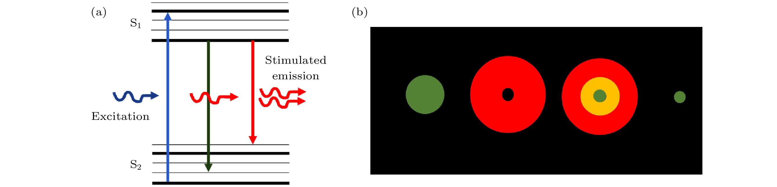
图 1 STED原理示意图 (a)受激辐射损耗能级图;(b)STED光斑示意图
Figure 1. Schematic diagram of STED: (a) Diagram of energy level for stimulated emission depletion; (b) schematic diagram of STED light spot.
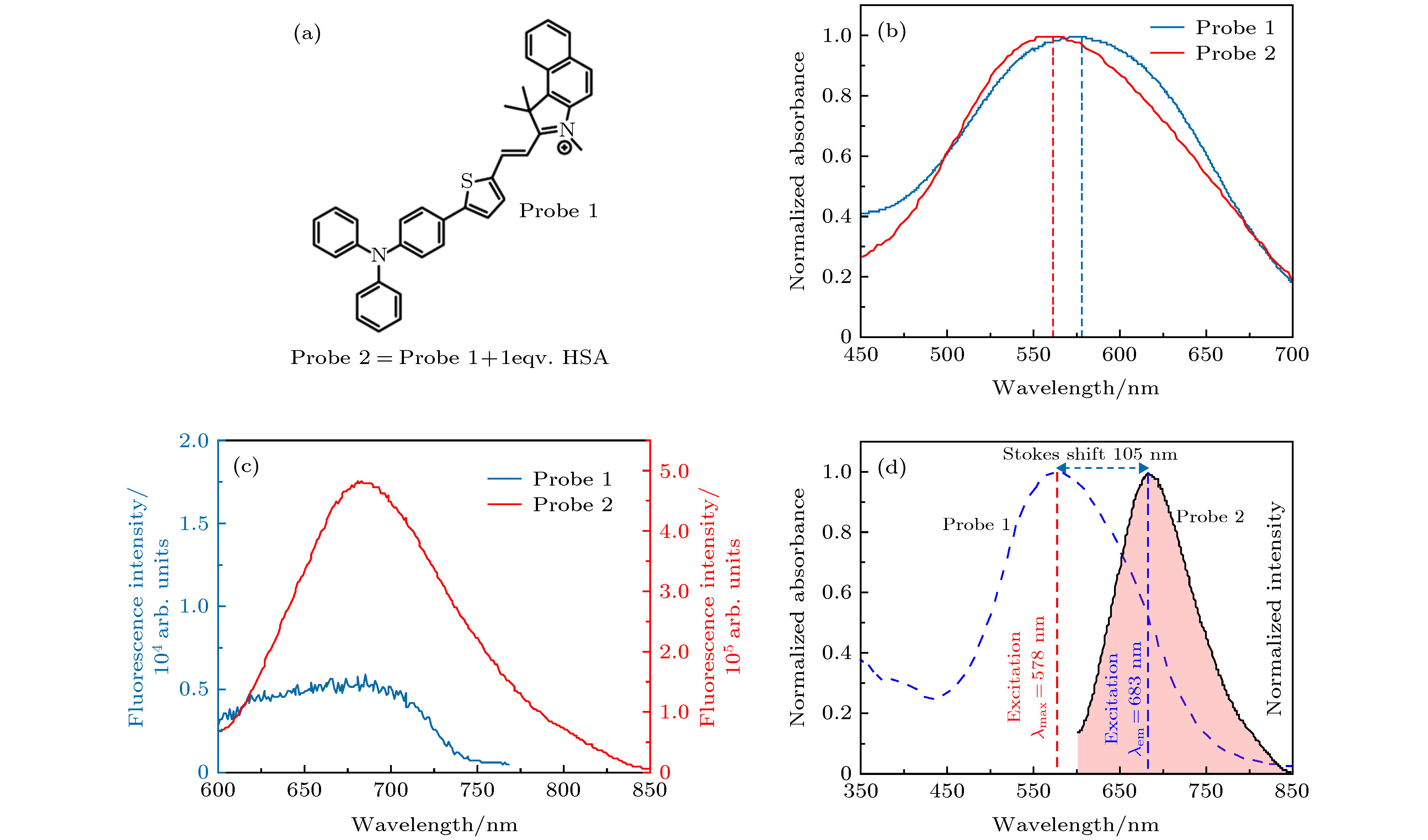
图 2 目标探针的化学结构和光谱表征 (a)探针1的化学结构; (b)探针1(蓝色)和2(红色)的激发谱; (c)探针1 (蓝色)和2 (红色)的发射谱; (d)探针1的激发谱(蓝色)和2的发射谱(红色)对比
Figure 2. Chemical structure and spectra characterization of target probe: (a) Chemical structure of probe 1; (b) excitation spectra of probe1 (blue)and 2 (red); (c) emission spectra of probe1 (blue) and 2 (red); (d) excitation spectrum of probe 1 (blue) and emission spectrum of probe 2 (red).
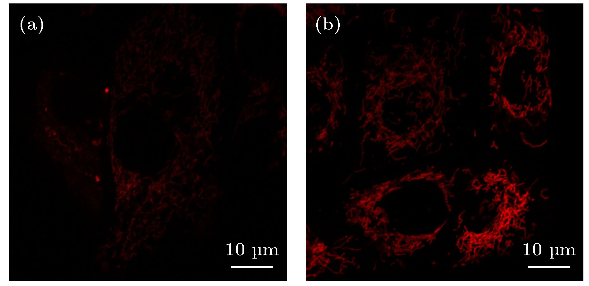
图 3 用探针1和探针2标记的HeLa细胞的共聚焦显微荧光图像, 比例尺为10 μm (a)探针1的荧光成像; (b)探针2的荧光成像
Figure 3. Confocal images of HeLa cells labeled with probe 1 and probe 2. Scale bar is 10 μm: (a) Confocal image of HeLa cells labeled with probe 1; (b) confocal image of HeLa cells labeled with probe2.
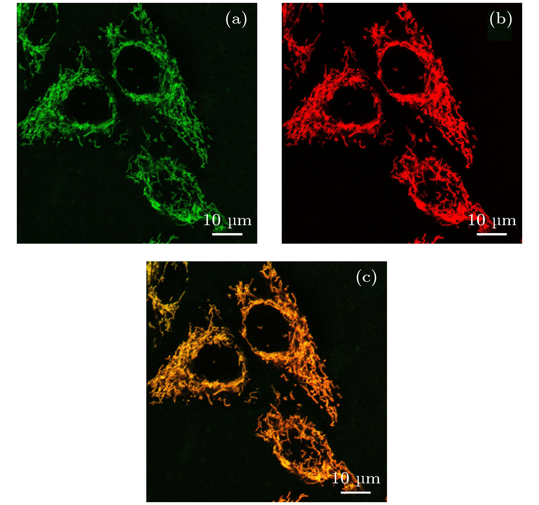
图 4 用Mito Tracker Green FM和探针2共处理的HeLa细胞的共定位图像, 比例尺为10 μm (a) Mito Tracker Green FM标记的细胞图像; (b)探针2标记的细胞图像; (c)图(a)和图(b)两者的重合
Figure 4. Co-localization images of Hela cells treated with Mito Tracker Green FM and Probe 2. Scale bar is 10 μm: (a) Image of Mito Tracker Green FM; (b) image of probe 2; (c) overlay of image (a) and (b).
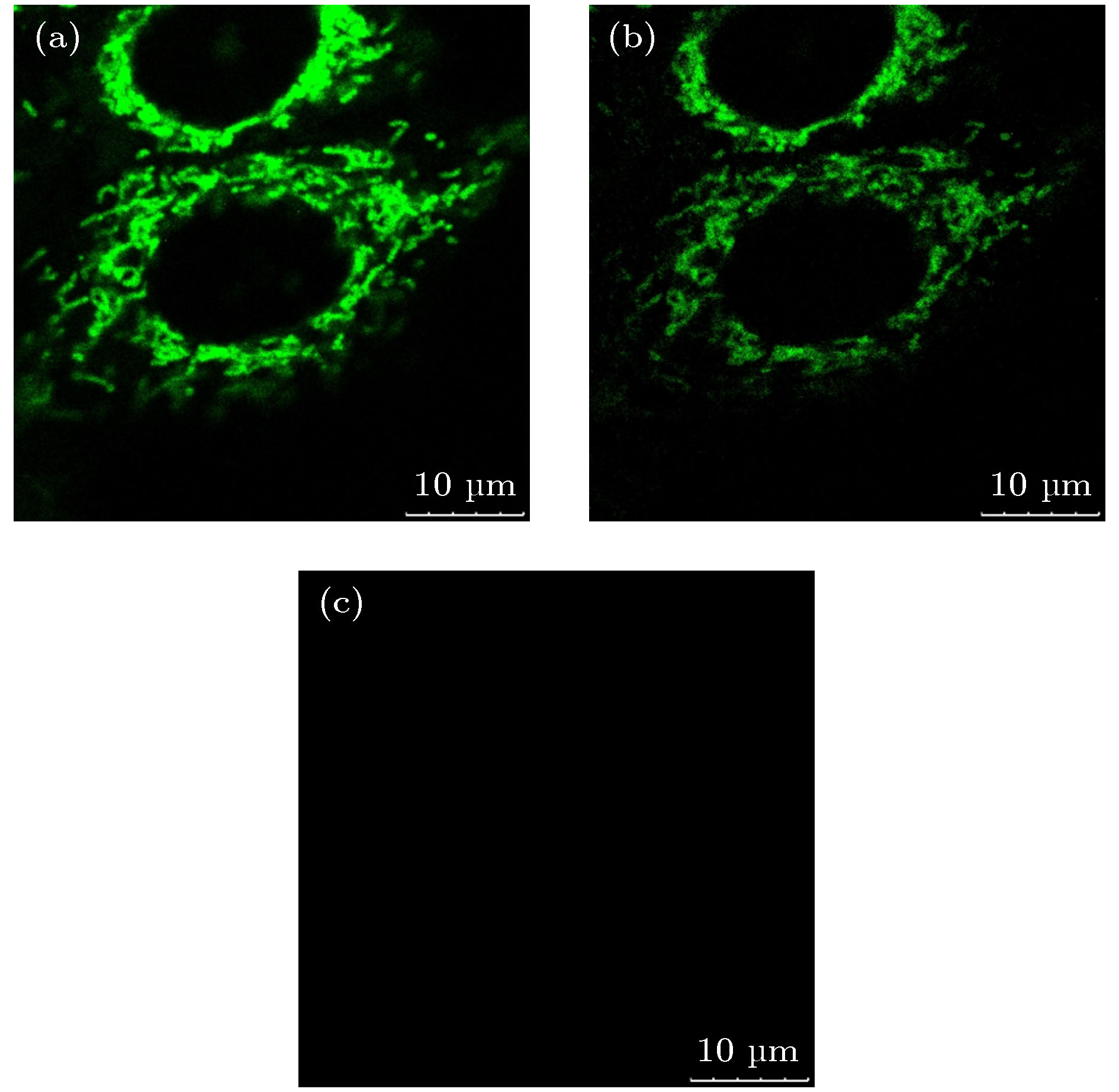
图 5 三种不同波长的光单独照射样品时的细胞图像 (a) 561 nm的光照射; (b) 660 nm的光照射; (c) 775 nm的光照射
Figure 5. Cell images illuminated by light of different wavelengths: (a) Illuminated by light of 561 nm; (b) illuminated by light of 660 nm; (c) illuminated by light of 775 nm.
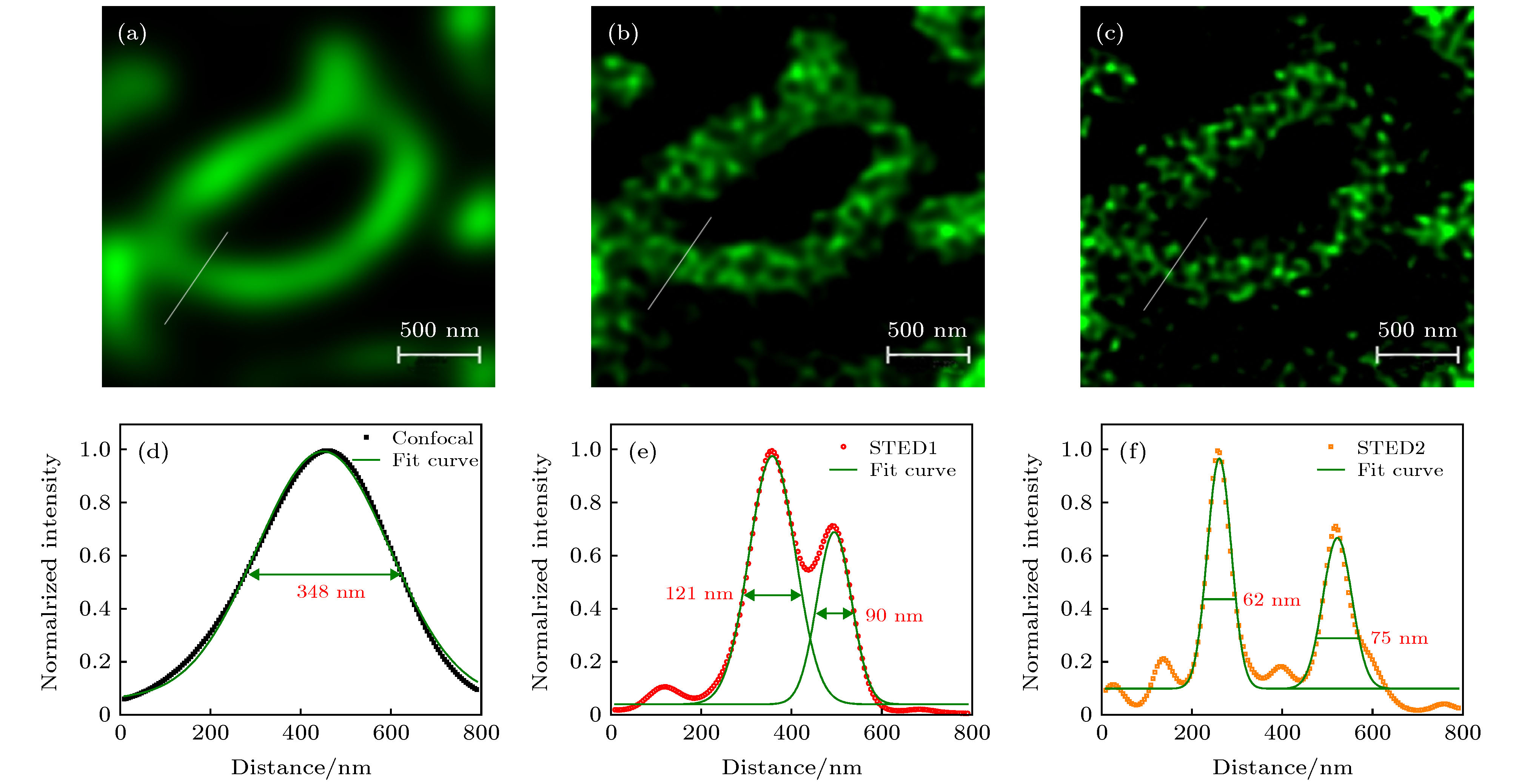
图 6 用探针2标记HeLa的共聚焦和STED图像, 比例尺为500 nm (a)共聚焦图像; (b)损耗光功率为19.75 mW时线粒体的STED图像; (c)损耗光功率为39.5 mW时的线粒体STED图像; (d)−(f)分别为图(a)−(c)中划线部分对应的信号曲线和分辨率
Figure 6. Confocal and STED images of HeLa cells labeled with probe 2. Scale bar is 500 nm: (a) Confocal image; (b) STED image of mitochondria obtained with 19.75 mW STED light; (c) STED image of mitochondria obtained with 39.5 mW STED light; (d)−(f) normalized signal intensity profiles along the lines in (a)−(c) respectively as well as the spatial resolutions.
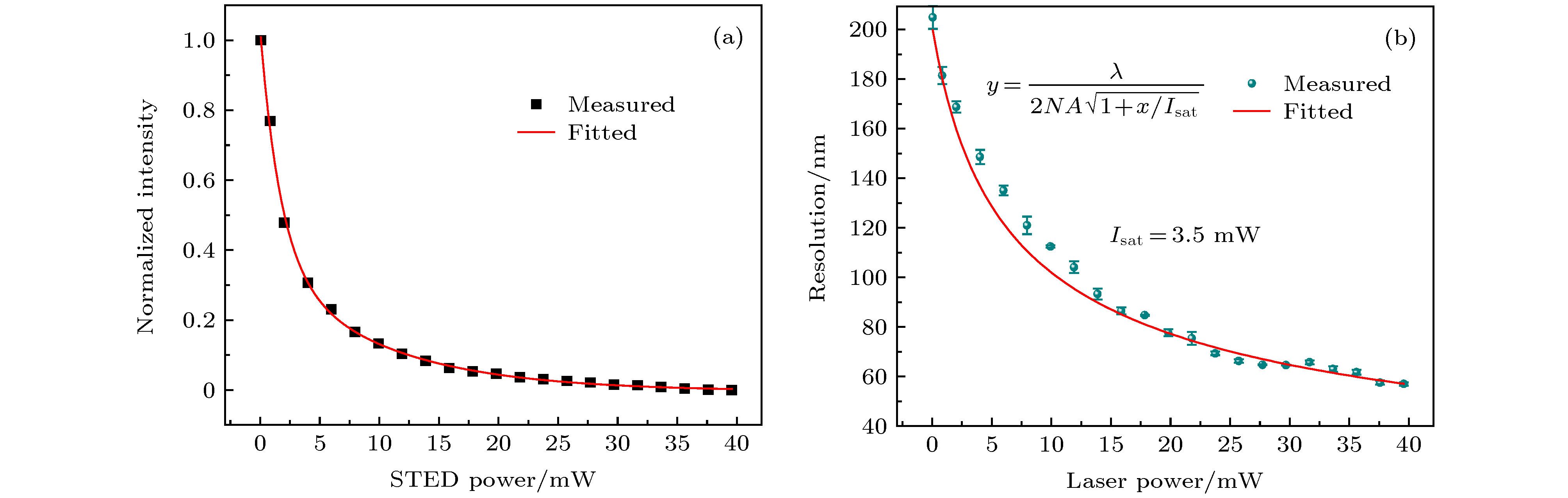
图 7 STED光功率对成像能力的影响 (a)探针2的受激辐射损耗效率; (b)增加损耗功率情况下获得的STED图像的分辨率
Figure 7. Effect of STED power on imaging performance: (a) Stimulated emission depletion efficiency of Probe 2; (b) resolution of STED images obtained at increased depletion power.
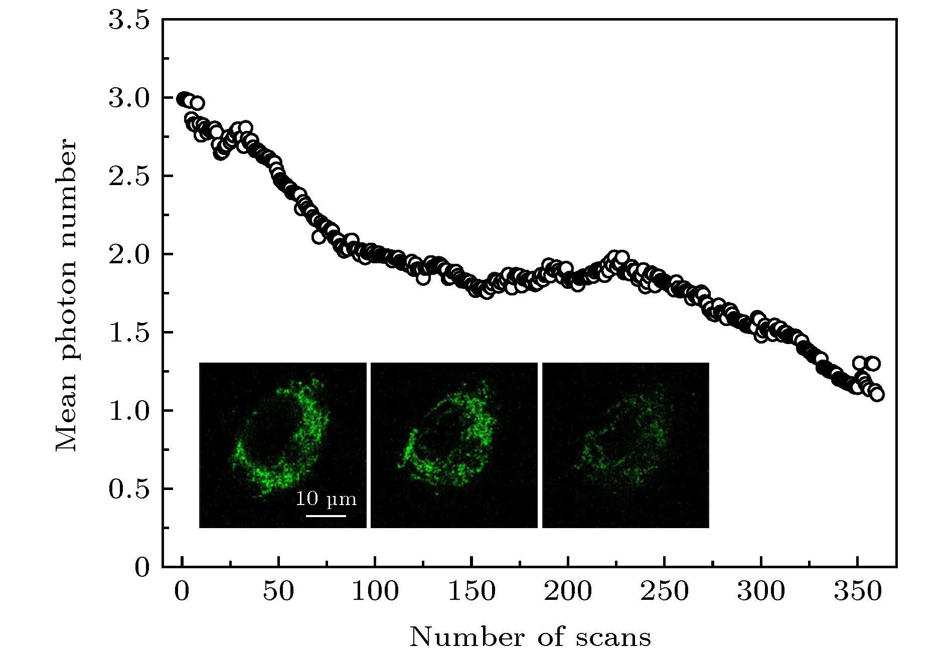
图 8 探针的抗光漂白测试结果. 内插图分别为对单个细胞第1次扫描、第180次扫描和第360次扫描后得到的图像, 比例尺为10 μm
Figure 8. Results of bleaching test. Inset pictures are the images of single cell obtained after the first scan, 180 th scan, and 360 th scan. Scale bar is 10 μm.
-
[1] Webb R H 1996 Rep. Prog. Phys. 59 427
Google Scholar
[2] 林丹樱, 屈军乐 2017 物理学报 66 148703
Google Scholar
Lin D Y, Qu J L 2017 Acta Phys. Sin. 66 148703
Google Scholar
[3] Hell S W 2003 Nat. Biotechnol. 21 1347
Google Scholar
[4] Yan W, Yang Y L, Tan Y, Chen X, Li Y, Qu J L, Ye T 2017 Photonics Res. 5 176
Google Scholar
[5] Wang L W, Yan W, Li R Z, Weng X Y, Zhang J, Yang Z G, Liu L W, Ye T, Qu J L 2018 Nanophotonics 7 1971
Google Scholar
[6] Huang B, Bates M, Zhuang X W 2009 Annu. Rev. Biochem. 78 993
Google Scholar
[7] Wang L W, Chen Y, Yan W, Weng X Y, Yang Z G, Ye T, Qu J L 2019 J. Biophotonics 12 e201800315
Google Scholar
[8] Wang L W, Chen B L, Yan W, Yang Z G, Peng X, Lin D Y, Weng X Y, Ye T, Qu J L 2018 Nanoscale 10 16252
Google Scholar
[9] Klar T A, Engel E, Hell S W 1994 Opt. Lett. 19 780
Google Scholar
[10] Hell S W, Jakobs S, Kastrup L 2003 Appl. Phys. A 77 859
Google Scholar
[11] Folling J, Bossi M, Bock H, Medda R, Wurm C A, Hein B, Jakobs S, Eggeling C 2008 Nat. Methods 5 943
Google Scholar
[12] Gustafsson M G 2000 J. Microsc. 198 82
Google Scholar
[13] Betzig E, Patterson G H, Sougrat R, Lindwasser O W, Olenych S, Bonifacino J S, Davidson M W, Schwartz J L, Hess H F 2006 Science 313 1642
Google Scholar
[14] Bates M, Huang B, Dempsey G T, Zhuang X W 2007 Science 317 793
Google Scholar
[15] Aloi A, Vilanova N, Albertazzi L, Voets I K 2016 Nanoscale 8 8712
Google Scholar
[16] Hell S W 2007 Science 316 1153
Google Scholar
[17] Hotta J, Fron E, Dedecker P, Janssen K P F, Li C, Mullen K, Harke B, Buckers J, Hell S W, Hofkens J 2010 J. Am. Chem. Soc. 132 5021
Google Scholar
[18] Vicidomini G, Schonle A, Ta H, Han K Y, Moneron G, Eggeling C, Hell S W 2013 PloS One 8 e54221
Google Scholar
[19] Liu Y J, Lu Y Q, Yang X S, Zheng X L, Wen S H, Wang F, Vidal X, Zhao J B, Liu D M, Zhou Z G, Ma C S, Zhou J, Peper J A, Xi P, Jin D Y 2017 Nature 543 229
Google Scholar
[20] Zhan Q Q, Liu H C, Wang B J, Wu Q S, Pu R, Zhou C, Huang B R, Peng X Y, Agren H, He S L 2017 Nat. Commun. 8 1058
Google Scholar
[21] Li D Y, Qin W, Xu B, Qian J 2017 Adv. Mater. 29 1703643
Google Scholar
[22] Ye S, Yan W, Zhao M J, Peng X, Song J, Qu J L 2018 Adv. Mater. 30 1800167
Google Scholar
[23] Martin O L, Hugo G S, Alexander S, James H C, Alice C N, Daniel M D, Chris D, Mark A N, Paul M W 2014 J. Biophotonics 7 29
Google Scholar
[24] Kuang C F, Li S, Liu W, Hao X, Gu X H, Wang Y F, Ge J H, Li H F, Liu X 2013 Sci. Rep. 3 1441
Google Scholar
[25] Schubbe S, Cavelius C, Schumann C, Koch M, Kraegeloh A 2010 Adv. Eng. Mater. 12 417
Google Scholar
[26] Gorlitz F, Hoyer P, Falk H J, Kastrup L, Engelhardt J, Hell S W 2014 Prog. Electromagn. Res. 147 57
Google Scholar
[27] Friedman J R, Nunnari J 2014 Nature 505 335
Google Scholar
[28] Desler C, Rasmussen L J 2012 Mitochondrion 12 472
Google Scholar
[29] 黄义梅, 杨洪钦, 陈江旭, 王瑜华, 郑莉琴, 谢树森 2012 中国激光 39 s104002
Google Scholar
Huang Y M, Yang H Q, Chen J X, Wang Y H, Zheng L Q, Xie S S 2012 Chin. J. Lasers 39 s104002
Google Scholar
[30] Jakobs S, Wurm C A 2014 Curr. Opin. Chem. Biol. 20 9
Google Scholar
[31] Hell S W 2009 Nat. Methods 6 24
Google Scholar
[32] Ha C E, Bhagavan N V 2013 Biochim. Biophys. Acta 1830 5486
Google Scholar
[33] Chen Q, Liu X, Zeng J, Cheng Z, Liu Z 2016 Biomater. 98 23
Google Scholar
[34] Samanta S, Halder S, Das G 2018 Anal. Chem. 90 7561
Google Scholar
[35] Samanta S, Huang M N, Lin F R, Das P, Chen B L, Yan W, Chen J J, Ji K, Liu L W, Qu J L, Yang Z G 2020 Anal. Chem. 92 1541
Google Scholar
[36] Kastrup L, Wildanger D, Rankin B, Hell S W 2010 STED Microscopy With Compact Light Sources, Nanoscopy and Multidimensional Optical Fluorescence Microscopy (Boca Raton: Chapmann and Hall/Crc Press) pp1–13

 DownLoad:
Catalog
Metrics
- Abstract views: 6364
- PDF Downloads: 108
- Cited By: 0
