


-
In spite of the success of fluorescence microscopes (such as stimulated emission depletion microscopy, stochastic optical reconstruction microscopy and photoactivated localization microscopy) in biomedical field, which have realized nanometer scale imaging resolution and promoted the great development of bio-medicine, the super-resolution imaging method for non-fluorescent sample is still scarce, and the resolution still has a big gap to nanometer scale. Among existing methods, structured illumination microscopy, PSF engineering, super-oscillatory lens and microsphere assisted nanoscopy are more mature and widely used. However, limited by the theory itself or engineering practice, the resolutions of these methods are hard to exceed 50 nm, which limits their applications in many fields. Enlightened by synthetic aperture technique, researchers have proposed spatial frequency shift super-resolution microscopy through shifting and combining the spatial frequency spectrum of imaging target, which is a promising super-resolution imaging scheme, for its resolution limit can be broken through continually. Currently, owing to the limitation of the refractive index of optical material, the wavelength of illumination evanescent wave is hard to shorten when this wave is generated at prism surface via total internal reflection, which determines the highest resolution of this spatial frequency shift super-resolution imaging system. Another deficiency of this scheme is the difference in imaging resolution among different directions, for the image has the highest resolution only in the direction along the wave vector of illumination evanescent wave; while, the image has the lowest resolution in the direction perpendicular to the wave vector, which is the same as that obtained by far-field illumination. In order to solve the above thorny questions, a new model of generating the evanescent wave is proposed, which can generates an omnidirectional evanescent wave with arbitrary wavelength based on the phase modulation of nano-structure, and solve the both problem in imaging system at the same time. To verify the our scheme, we set up a complete simulation model for spatial frequency shift imaging scheme, which includes three parts: the generation of evanescent wave and the interaction of the evanescent wave with the nano-structures at imaging target, which can be simulated with FDTD algorithm; the propagation of light field from near-field to far-field region, from the sample surface to the focal plane of objective lens, which can be calculated with angular spectrum theory; the propagation of light field from the focal place to the image plane, which can be worked out with Chirp-Z transform. Firstly, with this complete simulation model, we compare the resolution of microscopy illuminated by evanescent wave with that by propagating wave. The experimental results verify the super-resolution imaging ability of evanescent wave illumination and the influence of prism refractive index. The higher the refractive index, the shorter the wavelength of evanescent wave is and the higher the resolution of spatial frequency shift imaging system. Secondly, we demonstrate the resolution difference in a series of directions with a three-bar imaging target rotated to different directions. The result shows that the highest imaging resolution occurs in the direction of illumination evanescent wave vector, and the lowest resolution appears in the direction perpendicular to the wave vector. Finally, we simulate the evanescent wave generated by nano-strcuture and demonstrate its properties of wavelength and vector direction. When applied to near-field illumination super-resolution imaging, the omnidirectional evanescent wave solves the both problems in the model of total internal reflection from the prism surface. Therefore, the advantages of our scheme are higher imaging resolution and faster imaging speed, no need for multi-direction and multiple imaging, and also image post-processing. In this study, a new spatial frequency shift super-resolution imaging method is proposed, which lays a theoretical foundation for its applications. -
Keywords:
- microscopy /
- super-resolution imaging /
- near-field illumination /
- evanescent wave
[1] Kner P, Chhun B, Griffis E, Winoto L, Gustafsson M 2009 Nat. Methods 6 339
 Google Scholar
[2] Shao L, Kner P, Gustafsson M 2011 Nat. Methods 8 1044
Google Scholar
[3] Rogers E, Lindberg J, Zheludev N 2015 Nat. Mater. 11 432
Google Scholar
[4] Qin F, Huang K, Wu J J, Jiao J, Luo X, Qiu C W, Hong M H 2015 Sci. Rep. 5 09977
Google Scholar
[5] Hajj B, Beheiry E, Dahan M 2016 Biomed. Opt. Express 7 726
Google Scholar
[6] Izeddin I, Beheiry M, Andilla J, Ciepielewski D, Darzacq X, Dahan M 2012 Opt. Express 20 4957
Google Scholar
[7] Wang Z B, Guo W, Li L, Luk’yanchuk B, Khan A, Liu Z, Chen Z C, Hong M H 2011 Nat. Commun. 2 218
Google Scholar
[8] Zhou S, Deng Y, Zhou W, Yu M, Urbach H, Wu Y 2017 Appl. Phys. B 123 236
Google Scholar
[9] Kwon S, Park J, Kim K, Cho Y, Lee M 2022 Light-Sci. Appl. 11 32
Google Scholar
[10] Yan Y Z, Li L, Feng C, Guo W, Lee S J, Hong M H 2014 ACS Nano 8 1809
Google Scholar
[11] Lü G, Li Q, Chen Y, Feng H, Xu Z, Mu J 2019 Opt. Rev. 26 664
Google Scholar
[12] Gao W P, Yuan Y, Wang X R, Ma L, Zhao Z S, Yuan H 2021 Opt. Express 29 11869
Google Scholar
[13] Zheng G, Horstmeyer R, Yang C 2015 Nat. Photonics 9 621
Google Scholar
[14] Lee D J, Weiner A M 2014 Opt. Express 22 9380
Google Scholar
[15] Alekseyev L, Narimanov E, Khurgin J 2012 Opt. Express 19 22350
Google Scholar
[16] Kim M, Choi Y W, Fang Y C Sung Y J, Dasari R R, Feld M S, Choi W 2011 Opt. Lett. 36 148
Google Scholar
[17] Hao X, Liu X, Kuang C F 2013 Appl. Phys. Lett. 102 013104
Google Scholar
[18] Hao X, Kuang C F, Li Y H, Liu X 2013 Opt. Lett. 38 2455
Google Scholar
[19] Liu X, Kuang C F, Hao X, Pang C L, Xu P, Li H, Liu Y, Yu C, Xu Y, Nan D, Shen W, Fang Y, He L, Liu X, Yang Q 2017 Phys. Rev. Lett. 118 076101
Google Scholar
[20] Pang C L, Li J X, Tang M W, Wang J P, Mela I, Ströhl F, Hecker L, Shen W D, Liu Q L, Liu X W, Wang Y N, Zhang H, Xu M, Zhang X H, Liu X, Yang Q, Kaminski C 2019 Adv. Funct. Mater. 29 1900188
Google Scholar
[21] 郝翔, 杨青, 匡翠方, 刘旭 2021 光学学报 41 0111001
Google Scholar
Hao X, Yang Q, Kang C F, Liu X 2021 Acta Opt. Sin. 41 0111001
Google Scholar
[22] Ling J Z, Wang Y C, Liu X, Wang X R 2021 Opt. Lett. 46 1265
Google Scholar
-
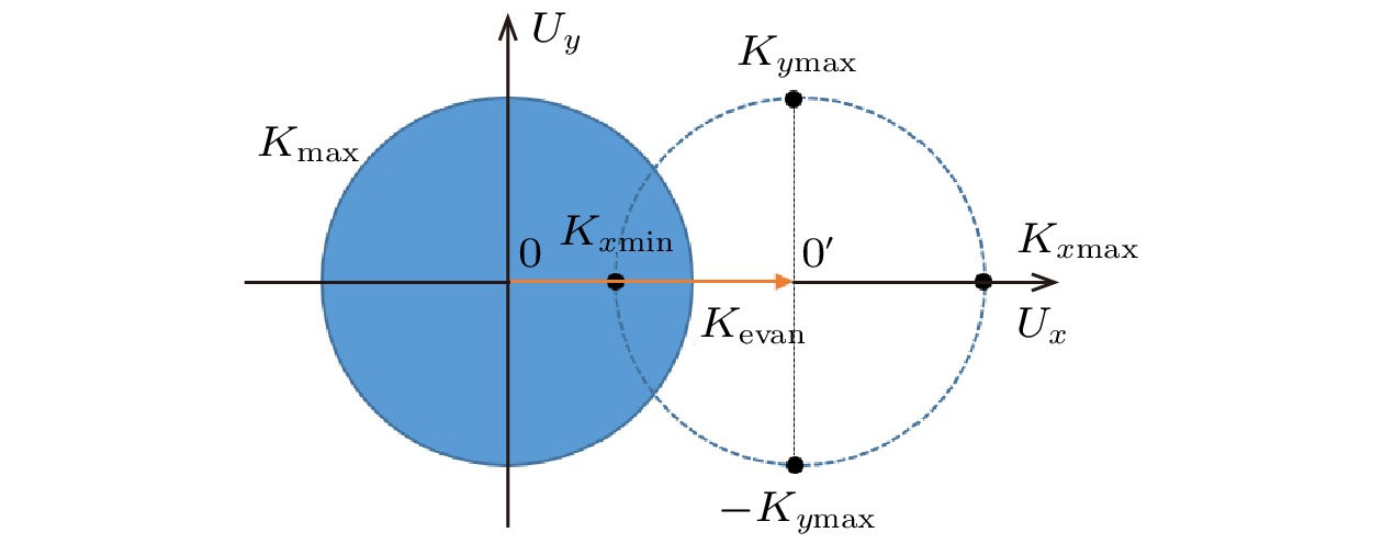
图 1 空间移频成像中的频谱移动示意图
Figure 1. Spatial frequency shifting in imaging system.

图 2 两种倏逝波的产生方案 (a) 基于棱镜全反射; (b) 基于微纳结构的倏逝波生成
Figure 2. Two methods for the generation of evanescent wave: (a) Scheme based on the total internal reflection at upper surface of prism; (b) scheme based on the transmission wave from nano-structures.
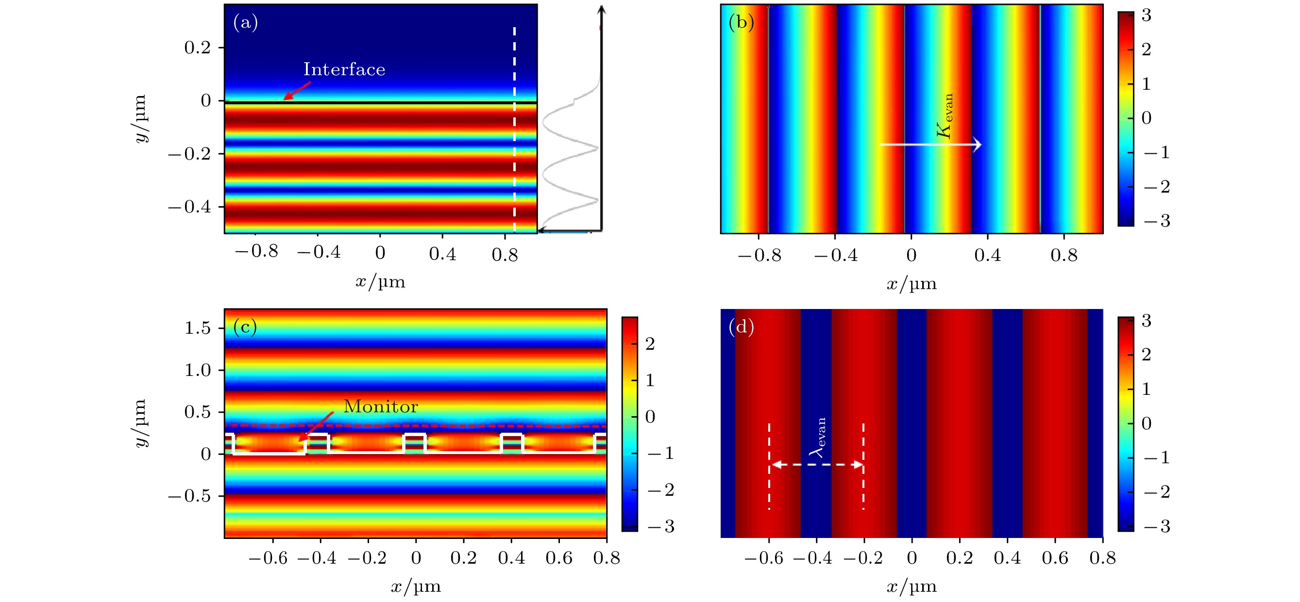
图 3 两种方案中倏逝波的特性比较 (a) 棱镜全反射时横截面上的光强分布; (b) 棱镜上表面的相位分布; (c) 微纳结构附近的相位分布; (d)微纳结构上方探测器位置的相位分布
Figure 3. Comparison of evanescent wave generated by above two methods: (a) Light intensity distribution around the interface of prism when total internal reflection occurs; (b) light phase distribution at the upper interface of prism; (c) light phase distribution around the nano-structure; (d) phase distribution at the monitor closely above the nano-structure
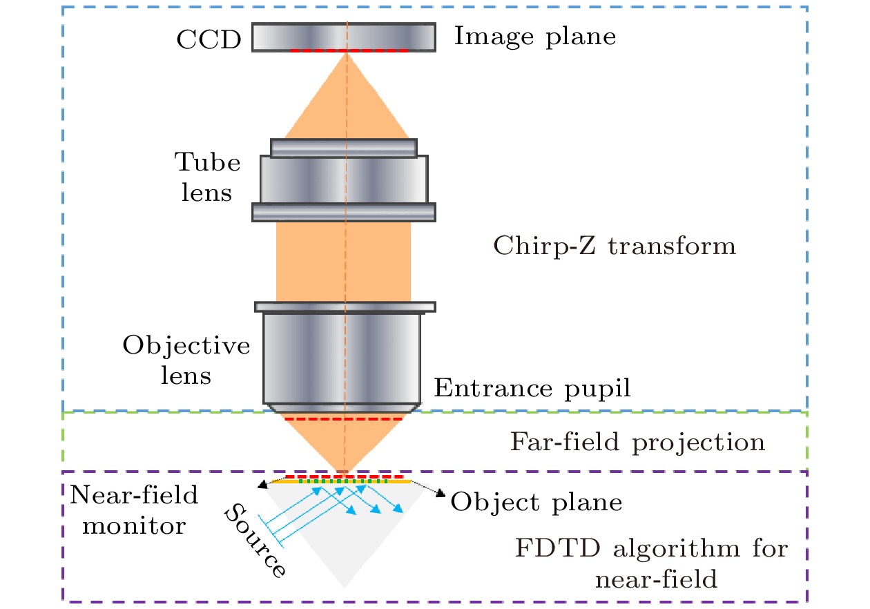
图 4 SFSSRI仿真模型示意图
Figure 4. Schematic diagram of the simulation model for SFSSRI.
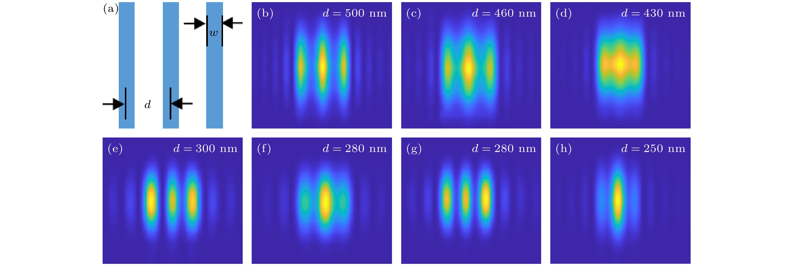
图 5 远场照明与近场照明时的成像分辨率比较 (a) 成像目标及参数; (b)—(d)远场照明的成像结果; (e), (f) 棱镜折射率n = 1.5时的近场照明成像结果; (g), (h) 棱镜折射率n = 1.8时的近场照明成像结果
Figure 5. Imaging resolution contrast between far-field illumination and near-field evanescent wave illumination: (a) Imaging target and its parameters; (b)–(d) imaging results obtained by far-field illumination; (e), (f) imaging results of near-field illumination when the refractive index of prism n = 1.5; (g), (h) imaging results of near-field illumination when the refractive index of prism n = 1.8

图 6 SFSSRI中的分辨率方向差异性 (a) 成像样品的方位示意图; (b)—(g) 不同方位角时样品的成像结果
Figure 6. Directional differences in imaging resolution of spatial frequency shift super-resolution imaging: (a) Imaging target and its direction; (b)–(g) imaging results obtained at different azimuth angles.

图 7 微纳结构及其产生的倏逝波 (a) 微纳结构示意图; (b) 微纳结构表面倏逝波的相位分布
Figure 7. Directional differences in imaging resolution of spatial frequency shifting super-resolution imaging method: (a) Sketch of the nano-structure; (b) phase distribution of the evanescent wave generated above the upper surface of nano-structure.

图 8 不同倏逝波的成像性能比较 (a), (b) 成像样品的结构示意图; (c), (d) 利用棱镜全反射所生成的倏逝波照明的成像结果; (e), (f) 利用微纳结构生成的倏逝波照明的成像结果
Figure 8. Comparison of imaging characteristic under different evanescent wave illumination: (a), (b) Sketch of the imaging targets; (c), (d) imaging results obtained under the evanescent wave illumination generated by the total internal reflection at prism surface; (e), (f) imaging results obtained under the evanescent wave illumination generated by nano-structures.
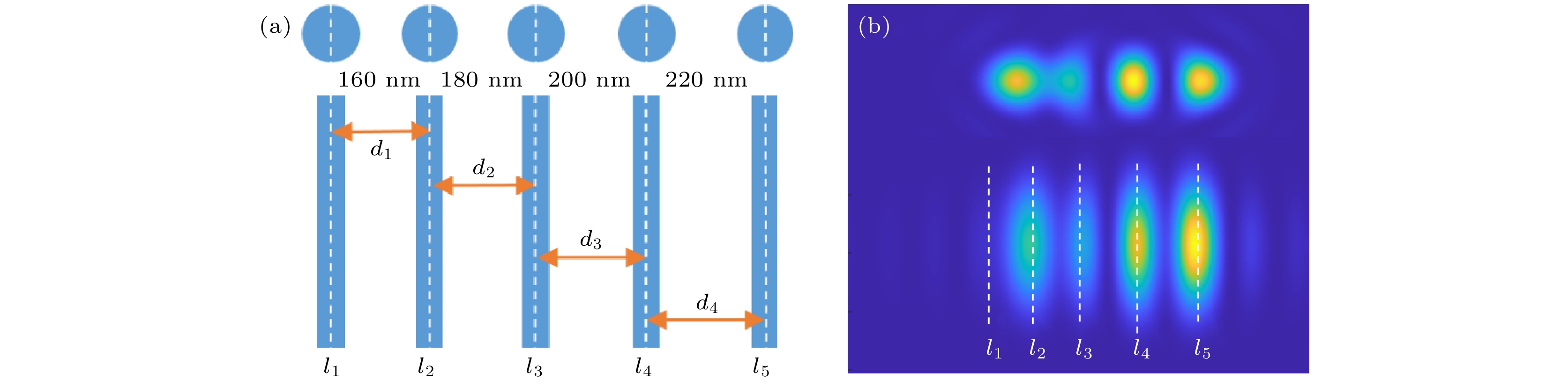
图 9 超高成像分辨率的实现 (a) 成像样品的结构示意图; (b) 倏逝波照明的成像结果
Figure 9. Realization of ultra-high imaging resolution: (a) Parameters of the imaging targets; (b) imaging results under evanescent wave illumination.
-
[1] Kner P, Chhun B, Griffis E, Winoto L, Gustafsson M 2009 Nat. Methods 6 339
Google Scholar
[2] Shao L, Kner P, Gustafsson M 2011 Nat. Methods 8 1044
Google Scholar
[3] Rogers E, Lindberg J, Zheludev N 2015 Nat. Mater. 11 432
Google Scholar
[4] Qin F, Huang K, Wu J J, Jiao J, Luo X, Qiu C W, Hong M H 2015 Sci. Rep. 5 09977
Google Scholar
[5] Hajj B, Beheiry E, Dahan M 2016 Biomed. Opt. Express 7 726
Google Scholar
[6] Izeddin I, Beheiry M, Andilla J, Ciepielewski D, Darzacq X, Dahan M 2012 Opt. Express 20 4957
Google Scholar
[7] Wang Z B, Guo W, Li L, Luk’yanchuk B, Khan A, Liu Z, Chen Z C, Hong M H 2011 Nat. Commun. 2 218
Google Scholar
[8] Zhou S, Deng Y, Zhou W, Yu M, Urbach H, Wu Y 2017 Appl. Phys. B 123 236
Google Scholar
[9] Kwon S, Park J, Kim K, Cho Y, Lee M 2022 Light-Sci. Appl. 11 32
Google Scholar
[10] Yan Y Z, Li L, Feng C, Guo W, Lee S J, Hong M H 2014 ACS Nano 8 1809
Google Scholar
[11] Lü G, Li Q, Chen Y, Feng H, Xu Z, Mu J 2019 Opt. Rev. 26 664
Google Scholar
[12] Gao W P, Yuan Y, Wang X R, Ma L, Zhao Z S, Yuan H 2021 Opt. Express 29 11869
Google Scholar
[13] Zheng G, Horstmeyer R, Yang C 2015 Nat. Photonics 9 621
Google Scholar
[14] Lee D J, Weiner A M 2014 Opt. Express 22 9380
Google Scholar
[15] Alekseyev L, Narimanov E, Khurgin J 2012 Opt. Express 19 22350
Google Scholar
[16] Kim M, Choi Y W, Fang Y C Sung Y J, Dasari R R, Feld M S, Choi W 2011 Opt. Lett. 36 148
Google Scholar
[17] Hao X, Liu X, Kuang C F 2013 Appl. Phys. Lett. 102 013104
Google Scholar
[18] Hao X, Kuang C F, Li Y H, Liu X 2013 Opt. Lett. 38 2455
Google Scholar
[19] Liu X, Kuang C F, Hao X, Pang C L, Xu P, Li H, Liu Y, Yu C, Xu Y, Nan D, Shen W, Fang Y, He L, Liu X, Yang Q 2017 Phys. Rev. Lett. 118 076101
Google Scholar
[20] Pang C L, Li J X, Tang M W, Wang J P, Mela I, Ströhl F, Hecker L, Shen W D, Liu Q L, Liu X W, Wang Y N, Zhang H, Xu M, Zhang X H, Liu X, Yang Q, Kaminski C 2019 Adv. Funct. Mater. 29 1900188
Google Scholar
[21] 郝翔, 杨青, 匡翠方, 刘旭 2021 光学学报 41 0111001
Google Scholar
Hao X, Yang Q, Kang C F, Liu X 2021 Acta Opt. Sin. 41 0111001
Google Scholar
[22] Ling J Z, Wang Y C, Liu X, Wang X R 2021 Opt. Lett. 46 1265
Google Scholar

 DownLoad:
Catalog
Metrics
- Abstract views: 1180
- PDF Downloads: 69
- Cited By: 0
