


-
High-resolution microscopy has opened the door to the exploration of the micro-world, while femtosecond laser has provided a measurement method for detecting ultrafast physical/chemical phenomena. Combination of these two techniques can produce new microscopic techniques with both ultra-high spatial resolution and ultra-fast temporal resolution, and thus has great importance in exploring new scientific phenomena and mechanisms on an extremely small spatial scale and temporal scale. This paper reviews the basic principles and properties of main microscopic techniques with ultra-high temporal resolution and spatial resolution, and introduces the latest research progress of their applications in various fields such as characterizing optoelectronic materials and devices, monitoring femtosecond laser micromachining, and detecting surface plasmon excitation dynamics. In order to conduct these researches systematically, we group these techniques based on time dimension and space dimension, including the near-field multi-pulse imaging techniques, the far-field multi-pulse imaging techniques, and the far-field single-pulse imaging techniques. In Section 2, we introduce the principles and characteristics of the ultra-high spatiotemporally resolved microscopic techniques. The near-field multi-pulse spatiotemporally microscopic techniques based on nano-probe are described in Subsection 2.1, in which is shown the combination of common near-field imaging techniques such as atomic force microscopy (AFM), near-field scanning optical microscopy (NSOM), scanning tunneling microscope (STM), and the ultra-fast temporal detection of pump-probe technique. In Subsection 2.2, we introduce the far-field multi-pulse spatiotemporal microscopic techniques. In contrast to near-field cases, the far-field spatiotemporal microscopic techniques have lower spatial resolution but possess more advantages of being non-invasive and non-contact, wider field of view, and faster imaging speed. In Subsection 2.3 we introduce the far-field single-pulse spatiotemporal microscopic techniques, in which is used a single ultrafast light pulse to capture dynamic processes at different moments in time, thereby enabling real-time imaging of ultrafast phenomena. In Section 3 , the advances in the application of the ultra-high spatiotemporal resolved microscopic techniques are introduced in many frontier areas, including the monitoring of femtosecond laser micromachining in Subsection 3.1, the detection of optoelectronic materials/devices in Subsection 3.2, and the characterization of surface plasmon dynamics in Subsection 3.3. Finally, in Section 4, we summarize the features of all above-mentioned spatiotemporal microscopic techniques in a table, including the spatial resolution and temporal resolution, advantages and disadvantages of each technique, and we also provide an outlook on future development trend in this research field. Looking forward to the future, ultra-high spatiotemporally resolved microscopy will develop rapidly toward the goal of "smaller, faster, smarter and more extensive". Its development not only promotes the research of the microscopy technology, but also provides a powerful tool for various practical applications such as precision machining, two-dimensional material dynamics, optoelectronic device design and characterization.
[1] Born M, Wolf E 2013 Principles of Optics: Electromagnetic Theory of Propagation, Interference and Diffraction of Light (Oxford: Pergamon Press)
[2] Hell S W 2003 Nat. Biotechnol. 21 1347
 Google Scholar
[3] Rust M J, Bates M, Zhuang X 2006 Nat. Methods 3 793
Google Scholar
[4] Betzig E, Patterson G H, Sougrat R, Lindwasser O W, Olenych S, Bonifacino J S, Davidson M W, Lippincott-Schwartz J, Hess H F 2006 Science 313 1642
Google Scholar
[5] Gustafsson M G 2000 J. Microsc. 198 82
Google Scholar
[6] Dertinger T, Colyer R, Iyer G, Weiss S, Enderlein J 2009 Proc. Natl. Acad. Sci. U. S. A. 106 22287
Google Scholar
[7] Balzarotti F, Eilers Y, Gwosch K C, Gynnå A H, Westphal V, Stefani F D, Elf J, Hell S W 2017 Science 355 606
Google Scholar
[8] Vigoureux J M, Courjon D 1992 Appl. Opt. 31 3170
Google Scholar
[9] Betzig E, Chichester R J 1993 Science 262 1422
Google Scholar
[10] Mohammed A, Abdullah A 2018 Proceedings of the 2018 International Conference on Hydraulics and Pneumatics— HERVEX Băile Govora, Romania, 2018 pp7–9
[11] Egerton R F 2008 Rep. Prog. Phys. 72 016502
[12] Pennycook S J, Nellist P D 2011 Scanning transmission electron microscopy: imaging and analysis (Springer Science & Business Media)
[13] Adachi S, Ishii H, Kanai T, Ishii N, Kosuge A, Watanabe S 2007 Opt. Lett. 32 2487
Google Scholar
[14] Ji S Y, Yang L, Hu Y L, Ni J C, Du W Q, Li J W, Zhao G, Wu D, Chu J R 2017 Small 13 1701190
Google Scholar
[15] Lao Z X, Hu Y L, Zhang C C, Yang L, Li J W, Chu J R, Wu D 2015 ACS Nano 9 12060
Google Scholar
[16] Bargheer M, Zhavoronkov N, Woerner M, Elsaesser T 2006 ChemPhysChem 7 783
Google Scholar
[17] Chapman H N, Fromme P, Barty A, White T A, Kirian R A, Aquila A, Hunter M S, Schulz J, DePonte D P, Weierstall U 2011 Nature 470 73
Google Scholar
[18] Holldack K, Khan S, Mitzner R, Quast T 2006 Phys. Rev. Lett. 96 054801
Google Scholar
[19] Van Tilborg J, Schroeder C, Filip C, Tóth C, Geddes C, Fubiani G, Huber R, Kaindl R, Esarey E, Leemans W 2006 Phys. Rev. Lett. 96 014801
Google Scholar
[20] Point G, Brelet Y, Houard A, Jukna V, Milián C, Carbonnel J, Liu Y, Couairon A, Mysyrowicz A 2014 Phys. Rev. Lett. 112 223902
Google Scholar
[21] Panagiotopoulos P, Whalen P, Kolesik M, Moloney J V 2015 Nat. Photonics 9 543
Google Scholar
[22] Krausz F, Ivanov M 2009 Rev. Mod. Phys. 81 163
Google Scholar
[23] Vozzi C, Calegari F, Ferrari F, Lucchini M, De Silvestri S, Svelto O, Sansone G, Stagira S, Nisoli M 2009 Laser Phys. Lett. 6 259
Google Scholar
[24] Morimoto Y, Baum P 2018 Nat. Phys. 14 252
Google Scholar
[25] Dudley J M, Finot C, Richardson D J, Millot G 2007 Nat. Phys. 3 597
Google Scholar
[26] Kravtsov V, Ulbricht R, Atkin J M, Raschke M B 2016 Nat. Nanotechnol. 11 459
Google Scholar
[27] Kim S, Jin J, Kim Y J, Park I Y, Kim Y, Kim S W 2008 Nature 453 757
Google Scholar
[28] Park I Y, Kim S, Choi J, Lee D H, Kim Y J, Kling M F, Stockman M I, Kim S W 2011 Nat. Photonics 5 677
Google Scholar
[29] Zhang Y Q, Shen J F, Min C J, Jin Y F, Jiang Y Q, Liu J, Zhu S W, Sheng Y L, Zayats A V, Yuan X C 2018 Nano Lett. 18 5538
Google Scholar
[30] Jiang Y, Narushima T, Okamoto H 2010 Nat. Phys. 6 1005
Google Scholar
[31] Van Dao L, Lincoln C, Lowe M, Hannaford P 2005 Femtosecond Laser Spectroscopy (Boston: Springer) pp197– 224
[32] Douhal A, Lahmani F, Zewail A H 1996 Chem. Phys. 207 477
Google Scholar
[33] Yamanouchi K 2002 Science 295 1659
Google Scholar
[34] Peterman E J, Monshouwer R, van Stokkum I H, van Grondelle R, van Amerongen H 1997 Chem. Phys. Lett. 264 279
Google Scholar
[35] Keller E L, Brandt N C, Cassabaum A A, Frontiera R R 2015 Analyst 140 4922
Google Scholar
[36] 李智, 张家森, 杨景, 龚旗煌 2007 物理学报 56 3630
Google Scholar
Li Z, Zhang J S, Yang J, Gong Q H 2007 Acta Phys. Sin. 56 3630
Google Scholar
[37] Sternbach A J, Latini S, Chae S, Hübener H, De Giovannini U, Shao Y, Xiong L, Sun Z, Shi N, Kissin P 2020 Nat. Commun. 11 3567
Google Scholar
[38] Huber M A, Mooshammer F, Plankl M, Viti L, Sandner F, Kastner L Z, Frank T, Fabian J, Vitiello M S, Cocker T L 2017 Nat. Nanotechnol. 12 207
Google Scholar
[39] Xue M F, Li M, Huang Y S, Chen R K, Li Y L, Wang J Y, Xing Y J, Chen J J, Yan H G, Xu H Q 2020 Adv. Mater. 32 2004120
Google Scholar
[40] Binnig G, Quate C F, Gerber C 1986 Phys. Rev. Lett. 56 930
Google Scholar
[41] Jahng J, Brocious J, Fishman D A, Huang F, Li X W, Tamma V A, Wickramasinghe H K, Potma E O 2014 Phys. Rev. B 90 155417
Google Scholar
[42] Jahng J, Brocious J, Fishman D A, Yampolsky S, Nowak D, Huang F, Apkarian V A, Wickramasinghe H K, Potma E O 2015 Appl. Phys. Lett. 106 083113
Google Scholar
[43] Binnig G, Rohrer H, Gerber C, Weibel E 1982 Phys. Rev. Lett. 49 57
Google Scholar
[44] Yang B, Chen G, Ghafoor A, Zhang Y F, Zhang Y, Zhang Y, Luo Y, Yang J L, Sandoghdar V, Aizpurua J 2020 Nat. Photonics 14 693
Google Scholar
[45] Takeuchi O, Aoyama M, Oshima R, Okada Y, Oigawa H, Sano N, Shigekawa H, Morita R, Yamashita M 2004 Appl. Phys. Lett. 85 3268
Google Scholar
[46] Terada Y, Yoshida S, Takeuchi O, Shigekawa H 2010 Nat. Photonics 4 869
Google Scholar
[47] Sonnenfeld R, Hansma P K 1986 Science 232 211
Google Scholar
[48] Maire G, Giovannini H, Talneau A, Chaumet P C, Belkebir K, Sentenac A 2018 Opt. Lett. 43 2173
Google Scholar
[49] Wang Z B, Guo W, Li L, Luk'Yanchuk B, Khan A, Liu Z, Chen Z C, Hong M H 2011 Nat. Commun. 2 218
Google Scholar
[50] Lu D, Liu Z W 2012 Nat. Commun. 3 1205
Google Scholar
[51] Hao X, Liu X, Kuang C F, Li Y H, Ku Y L, Zhang H J, Li H F, Tong L M 2013 Appl. Phys. Lett. 102 013104
Google Scholar
[52] Fang N, Lee H, Sun C, Zhang X 2005 Science 308 534
Google Scholar
[53] Darafsheh A, Limberopoulos N I, Derov J S, Walker Jr D E, Astratov V N 2014 Appl. Phys. Lett. 104 061117
Google Scholar
[54] Domke M, Rapp S, Schmidt M, Huber H P 2012 Opt. Express 20 10330
Google Scholar
[55] Zhou K, Jia X, Jia T Q, Cheng K, Cao K Q, Zhang S A, Feng D H, Sun Z R 2017 J. Appl. Phys. 121 104301
Google Scholar
[56] Hirano Y, Matsuda A, Hiraoka Y 2015 Microscopy 64 237
Google Scholar
[57] Li D, Shao L, Chen B C, Zhang X, Zhang M S, Moses B, Milkie D E, Beach J R, Hammer III J A, Pasham M 2015 Science 349 aab3500
Google Scholar
[58] Guo Y T, Li D, Zhang S W, Yang Y R, Liu J J, Wang X Y, Liu C, Milkie D E, Moore R P, Tulu U S 2018 Cell 175 1430
Google Scholar
[59] Massaro E S, Hill A H, Grumstrup E M 2016 ACS Photonics 3 501
Google Scholar
[60] Xu J, Min C J, Zhang Y Q, Ni J L, Cao G W, Wei Q Y, Yang J J, Yuan X C 2022 Photonics Res. 10 1900
Google Scholar
[61] Su X Y, Zhang Q C 2010 Opt. Lasers Eng. 48 191
Google Scholar
[62] Su X, Chen W J 2001 Opt. Lasers Eng. 35 263
Google Scholar
[63] Tao T Y, Chen Q, Da J, Feng S J, Hu Y, Zuo C 2016 Opt. Express 24 20253
Google Scholar
[64] Yao P C, Gai S Y, Chen Y C, Chen W L, Da F P 2021 Opt. Lasers Eng. 143 106623
Google Scholar
[65] Chen F, Brown G M, Song M M 2000 Opt. Eng. 39 10
Google Scholar
[66] Schafer S 2017 光学与光电技术 15 1
Schafer S 2017 Optics & Optoelectronic Technology 15 1
[67] Barwick B, Flannigan D J, Zewail A H 2009 Nature 462 902
Google Scholar
[68] Zheng D G, Huang S Y, Zhu C H, Xu P, Li Z A, Wang H, Li J, Tian H F, Yang H X, Li J Q 2021 Nano Lett. 21 10238
Google Scholar
[69] Fu X W, Sun Z P, Ji S Z, Liu F, Feng M, Yoo B-K, Zhu Y M 2022 Nano Lett. 22 2009
Google Scholar
[70] Könenkamp R, Word R C, Rempfer G, Dixon T, Almaraz L, Jones T 2010 Ultramicroscopy 110 899
Google Scholar
[71] Dabrowski M, Dai Y N, Petek H 2020 Chem. Rev. 120 6247
Google Scholar
[72] Sun Q, Zu S, Misawa H 2020 J. Chem. Phys. 153 120902
Google Scholar
[73] Huber B, Pres S, Wittmann E, Dietrich L, Lüttig J, Fersch D, Krauss E, Friedrich D, Kern J, Lisinetskii V 2019 Rev. Sci. Instrum. 90 113103
Google Scholar
[74] 李耀龙, 刘运全, 龚旗煌 2021 光子学报 50 0850201
Google Scholar
Li Y L, Liu Y Q, Gong Q H 2021 Acta Photonica Sin. 50 0850201
Google Scholar
[75] Ogawa S, Nagano H, Petek H, Heberle A 1997 Phys. Rev. Lett. 78 1339
Google Scholar
[76] Fecher G H, Schmidt O, Hwu Y, Schönhense G 2002 J. Electron Spectrosc. Relat. Phenomena 126 77
Google Scholar
[77] Rotermund H H 1997 Surf. Sci. Rep. 29 265
Google Scholar
[78] Kubo A, Onda K, Petek H, Sun Z J, Jung Y S, Kim H K 2005 Nano Lett. 5 1123
Google Scholar
[79] Man M K, Margiolakis A, Deckoff-Jones S, Harada T, Wong E L, Krishna M B M, Madéo J, Winchester A, Lei S D, Vajtai R 2017 Nat. Nanotechnol. 12 36
Google Scholar
[80] Drezet A, Hohenau A, Koller D, Stepanov A, Ditlbacher H, Steinberger B, Aussenegg F R, Leitner A, Krenn J R 2008 Mater. Sci. Eng. , B 149 220
Google Scholar
[81] Hohenau A, Krenn J, Drezet A, Mollet O, Huant S, Genet C, Stein B, Ebbesen T 2011 Opt. Express 19 25749
Google Scholar
[82] Drezet A, Genet C 2013 Phys. Rev. Lett. 110 213901
Google Scholar
[83] Gorodetski Y, Chervy T, Wang S, Hutchison J A, Drezet A, Genet C, Ebbesen T W 2016 Optica 3 48
Google Scholar
[84] Gao L, Liang J Y, Li C Y, Wang L H V 2014 Nature 516 74
Google Scholar
[85] Li Z Y, Zgadzaj R, Wang X M, Chang Y-Y, Downer M C 2014 Nat. Commun. 5 3085
Google Scholar
[86] Liang J Y, Zhu L R, Wang L H V 2018 Light-Sci. Appl. 7 42
Google Scholar
[87] Zhu Y L, Zeng X K, Cai Y, Lu X W, Zhu Q F, Zeng L W, He T C, Li J Z, Yang Y, Zheng M J 2021 Opt. Express 29 27298
Google Scholar
[88] Yao Y H, He Y L, Qi D L, Cao F Y, Yao J L, Ding P P, Jin C Z, Wu X Y, Deng L Z, Jia T Q, Huang F, Liang J Y, Sun Z R, Zhang S A 2021 ACS Photonics 8 738
Google Scholar
[89] Nakagawa K, Iwasaki A, Oishi Y, Horisaki R, Tsukamoto A, Nakamura A, Hirosawa K, Liao H G, Ushida T, Goda K 2014 Nat. Photonics 8 695
Google Scholar
[90] Tamamitsu M, Nakagawa K, Horisaki R, Iwasaki A, Oishi Y, Tsukamoto A, Kannari F, Sakuma I, Goda K 2015 Opt. Lett. 40 633
Google Scholar
[91] Zeng X K, Zheng S Q, Cai Y, Lin Q G, Liang J Y, Lu X W, Li J Z, Xie W X, Xu S X 2020 Adv. Photonics 2 056002
[92] Downer M, Fork R L, Shank C V 1985 JOSA B 2 595
Google Scholar
[93] Garcia-Lechuga M, Puerto D, Fuentes-Edfuf Y, Solis J, Siegel J 2016 ACS Photonics 3 1961
Google Scholar
[94] Liu J K, Jia X, Wu W S, Cheng K, Feng D H, Zhang S A, Sun Z R, Jia T Q 2018 Opt. Express 26 6302
Google Scholar
[95] Pan C J, Jiang L, Sun J Y, Wang Q S, Wang F F, Wang K, Lu Y F, Wang Y L, Qu L T, Cui T H 2020 Light Sci. Appl. 9 80
Google Scholar
[96] Fang R R, Vorobyev A, Guo C L 2017 Light-Sci. Appl. 6 e16256
[97] Spellauge M, Doñate-Buendía C, Barcikowski S, Gökce B, Huber H P 2022 Light-Sci. Appl. 11 68
Google Scholar
[98] Emmony D, Howson R, Willis L 1973 Appl. Phys. Lett. 23 598
Google Scholar
[99] Huang M, Zhao F L, Cheng Y, Xu N S, Xu Z Z 2009 ACS Nano 3 4062
Google Scholar
[100] Bonse J, Rosenfeld A, Krüger J 2009 J. Appl. Phys. 106 104910
Google Scholar
[101] Splendiani A, Sun L, Zhang Y B, Li T S, Kim J, Chim C Y, Galli G, Wang F 2010 Nano Lett. 10 1271
Google Scholar
[102] Zeng H L, Dai J F, Yao W, Xiao D, Cui X D 2012 Nat. Nanotechnol. 7 490
Google Scholar
[103] Bernardi M, Palummo M, Grossman J C 2013 Nano Lett. 13 3664
Google Scholar
[104] Baugher B W, Churchill H O, Yang Y F, Jarillo-Herrero P 2014 Nat. Nanotechnol. 9 262
Google Scholar
[105] Yuan H T, Bahramy M S, Morimoto K, Wu S F, Nomura K, Yang B J, Shimotani H, Suzuki R, Toh M, Kloc C 2013 Nat. Phys. 9 563
Google Scholar
[106] Furchi M M, Pospischil A, Libisch F, Burgdörfer J, Mueller T 2014 Nano Lett. 14 4785
Google Scholar
[107] Wang L, Xu C, Li M Y, Li L J, Loh Z H 2018 Nano Lett. 18 5172
Google Scholar
[108] Wittenbecher L, Viñas Boström E, Vogelsang J, Lehman S, Dick K A, Verdozzi C, Zigmantas D, Mikkelsen A 2021 ACS Nano 15 1133
Google Scholar
[109] Garg M, Martin-Jimenez A, Pisarra M, Luo Y, Martín F, Kern K 2022 Nat. Photonics 16 196
Google Scholar
[110] Barnes W L, Dereux A, Ebbesen T W 2003 Nature 424 824
Google Scholar
[111] Wang Y L, Min C J, Zhang Y Q, Yuan X C 2022 Opto-Electron. Adv. 5 210047
Google Scholar
[112] Nishiyama Y, Imura K, Okamoto H 2015 Nano Lett. 15 7657
Google Scholar
[113] Zu S, Sun Q, Cao E, Oshikiri T, Misawa H 2021 Nano Lett. 21 4780
Google Scholar
[114] Spektor G, Kilbane D, Mahro A, Frank B, Ristok S, Gal L, Kahl P, Podbiel D, Mathias S, Giessen H 2017 Science 355 1187
Google Scholar
[115] Zhao Z L, Lang P, Qin Y L, Ji B Y, Song X W, Lin J Q 2020 Opt. Express 28 19023
Google Scholar
[116] Yan Q C, Cao E, Sun Q, Ao Y T, Hu X Y, Shi X, Gong Q H, Misawa H 2021 Nano Lett. 21 9270
Google Scholar
-
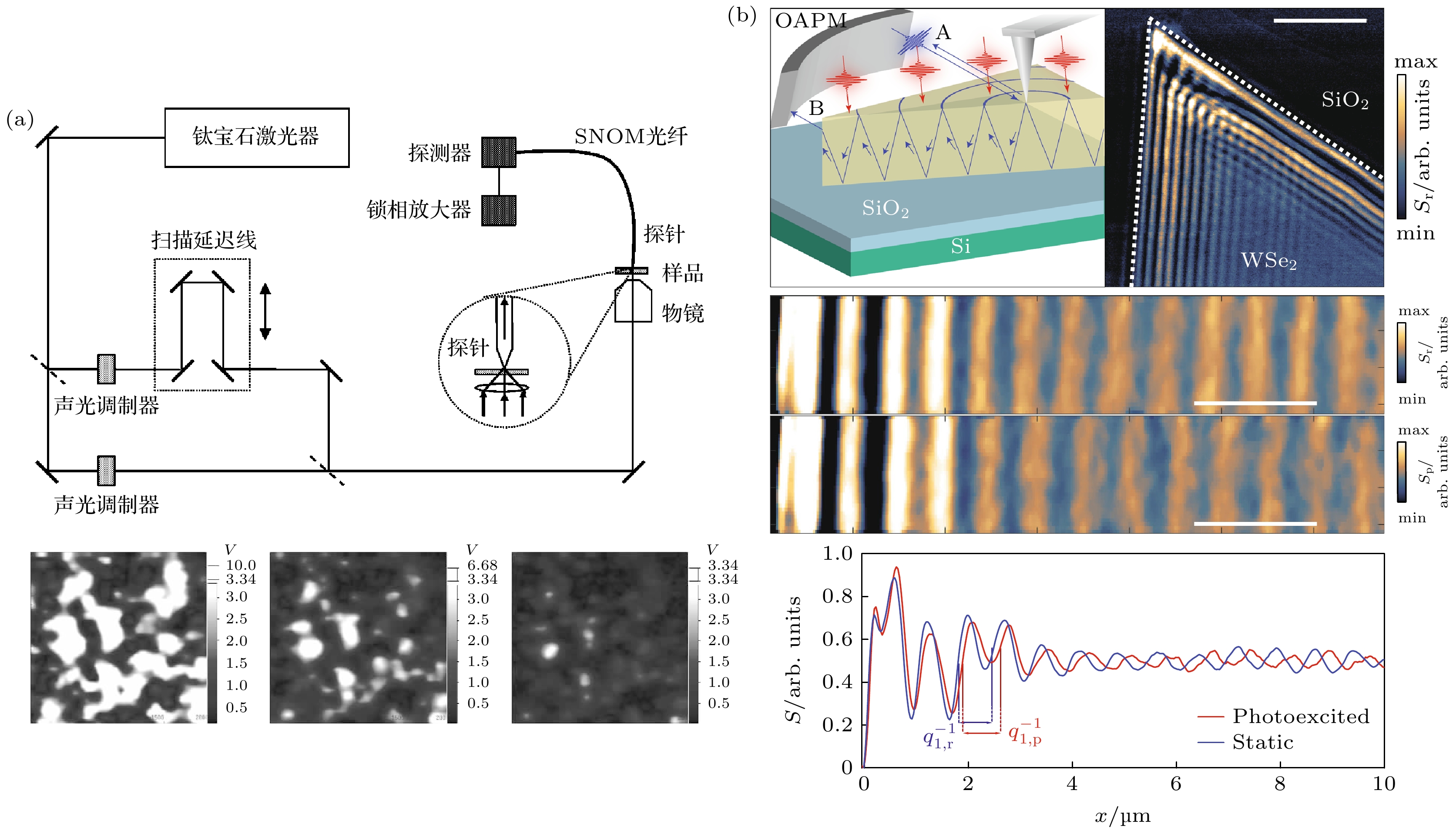
图 1 (a) 飞秒近场扫描显微成像系统示意图以及延迟时间0, 500 fs和2 ps时的近场泵浦-探测信号[36]; (b) 利用tr-NSOM对WSe2进行的时间分辨纳米尺度探测实验[37]
Figure 1. (a) Schematic representation of the femtosecond near-field scanning microscopy imaging system and the near-field pump-probe signal at the time delay of 0, 500 fs and 2 ps[36]; (b) time resolved infrared nano-imaging experiments on WSe2 by using tr-NSOM[37].

图 2 (a) tr-PiFM系统示意图[42]; 在两个不同时间延迟(–5.9和0.7 ps)下AFM所记录的(b), (c)形貌图和(d), (e)光诱导力图像[42]; (f)比较远场检测到的泵浦-探测信号(橙色实线)和光诱导力信号(黑色圆点)[42]
Figure 2. (a) Schematic of the tr-PiFM system[42]; topography image (b), (c) and optical force image (d), (e) are simultaneously recorded at negative time delay 5.9 ps (or positive time delay 0.7 ps) [42]; (f) comparison between the far-field detected pump-probe signal (orange solid line) and the tr-PiFM signal (black circle dot)[42].

图 3 (a) 超快FMW成像系统示意图, 经过脉冲整形系统后的飞秒脉冲通过刻蚀在金针尖上的光栅激发SPP并聚焦到针尖的顶点[26]; (b) 硅-金膜表面SPP热点S1, S2, S3位置对应的FWM扫描图像[26]; (c) 每个热点对应的AFM形貌图像[26]; (d) 金膜中SPP热点动力学的飞秒FWM纳米成像, 对应热点在不同延迟时间下的相对强度变化[26]
Figure 3. (a) Schematic of the ultrafast FMW imaging experiment, the femtosecond pulse after the pulse shaping system excited the SPP through a grating etched on the gold tip and focused to the vertex of the tip[26]; (b) near-field FWM image of a Si-Au step, showing SPP “hotspots” S1, S2 and S3[26]; (c) simultaneously acquired AFM topography[26]; (d) FWM nanoimages of the SPP “hotspots” dynamics of a Si-Au surface, corresponding to different inter-pulse delay, demonstrating evolution of the relative intensities in spots S1, S2 and S3[26].
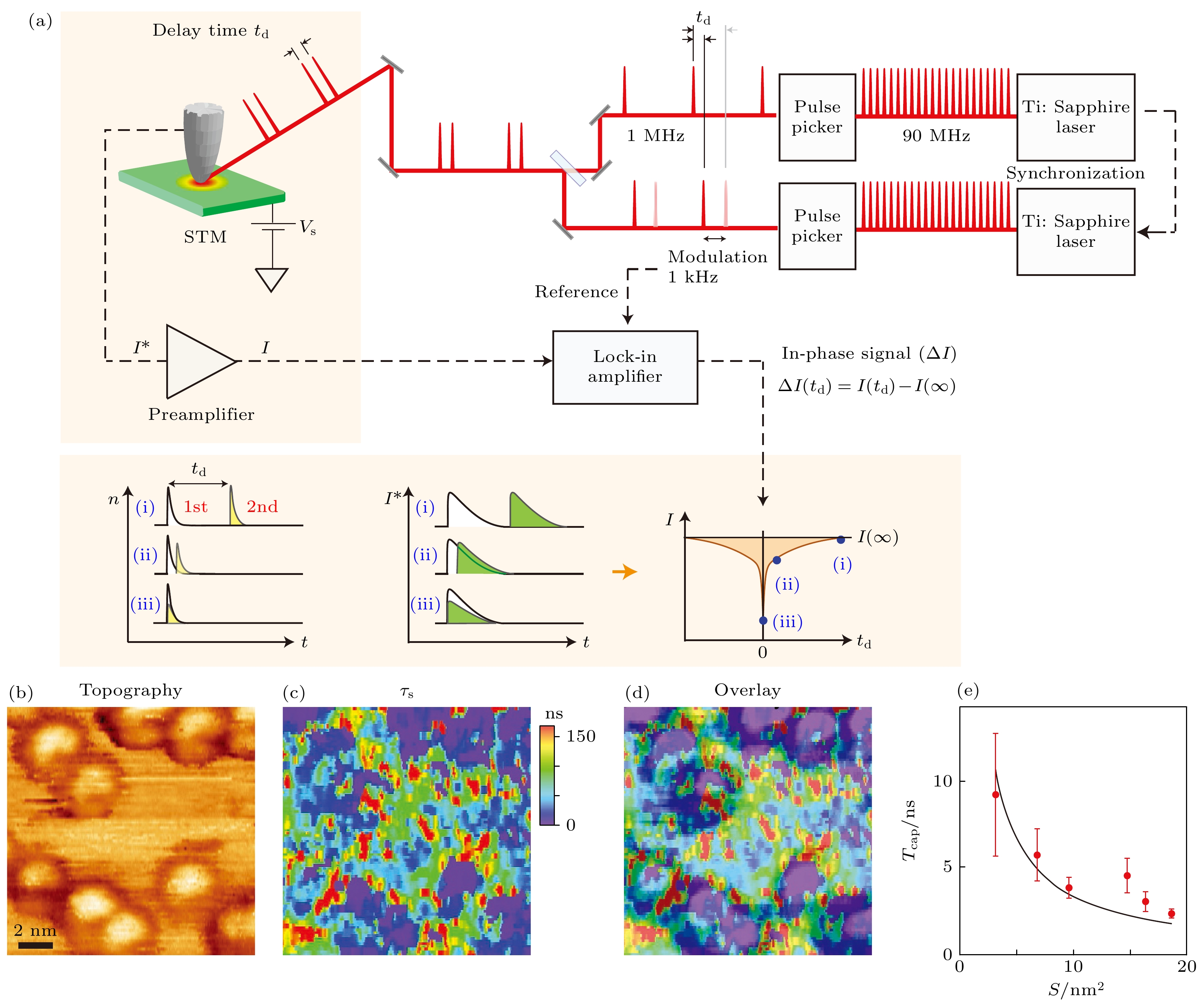
图 4 (a) Terada等[46]所提出的时间分辨STM工作原理图; (b) 实空间上GaAs样品表面Co纳米颗粒的STM形貌图[46]; (c) 图(b)中不同位置的空穴捕获率测量结果[46]; (d) 图(b)与图(c)的图象叠加[46]; (e) 空穴捕获率与Co纳米颗粒尺寸的依赖关系[46]
Figure 4. (a) Schematic of the time-resolved STM proposed by Yasuhiko Terada[46]; (b) STM topography of Co nanoparticles on the surface of GaAs samples in real space[46]; (c) hole capture rate measurements at different locations in Fig. (b) [46]; (d) superposition of Fig. (b) and Fig. (c) [46]; (e) size dependence of hole capture rate[46].

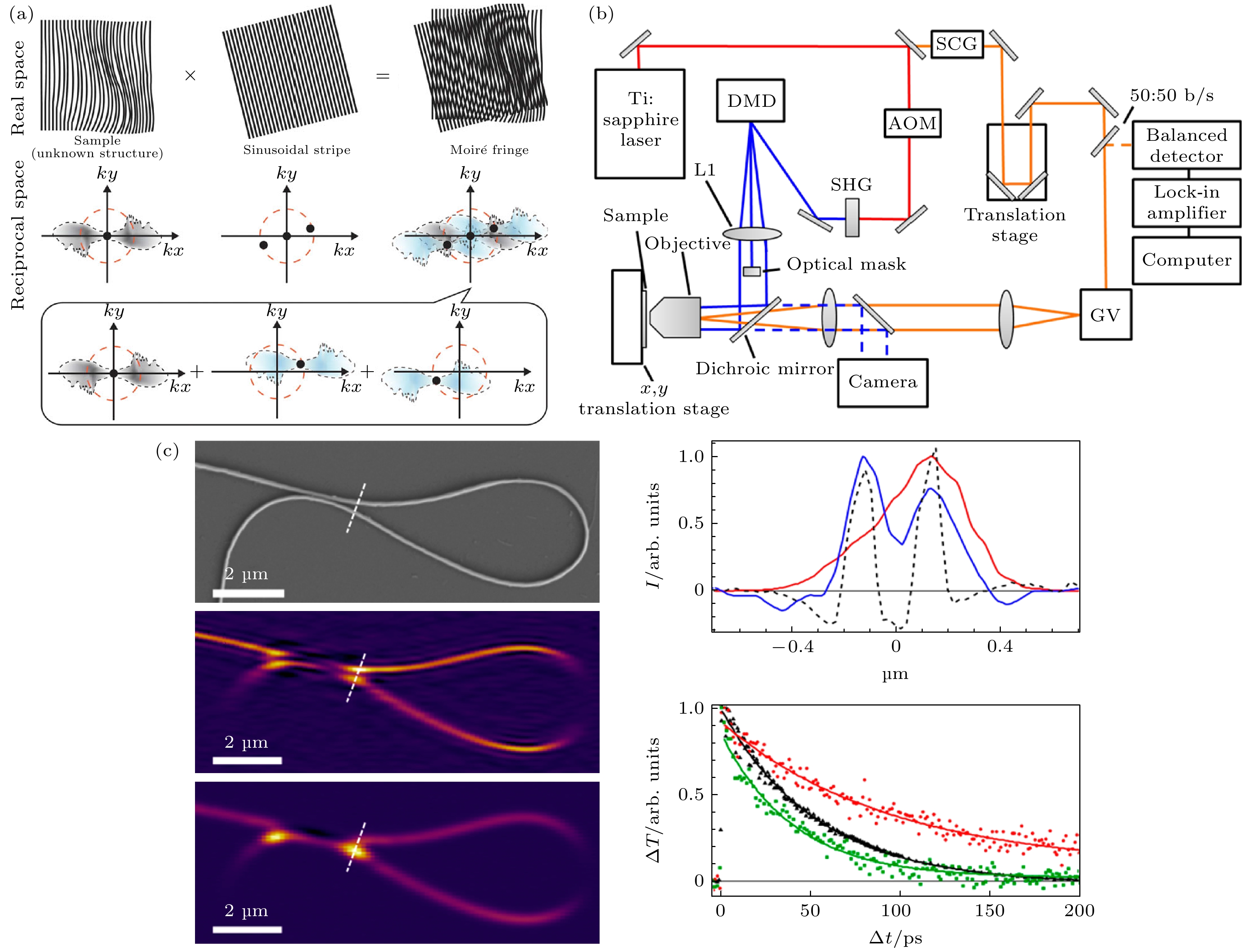
图 6 (a) 莫尔现象及SIM实现超分辨原理[56]; (b) 基于DMD的结构光照明泵浦-探测成像系统光路图; (c) 左侧分别为硅纳米线样品的SEM图像、SPPM成像、传统光学显微成像, 右侧为SPPM成像与传统光学显微成像分辨率对比、SPPM泵浦-探测获得样品不同位置的载流子弛豫过程[59]
Figure 6. (a) Moire phenomenon and the SIM principle of super-resolution[56]; (b) schematic of DMD-based structured pump–probe microscope[59]; (c) the left side: SEM image, SPPM image, and image by conventional optical microscopy of silicon nanowire samples respectively; the right side: the carrier relaxation process at different positions of SPPM imaging and conventional optical microscopy imaging resolution and SPPM pump-probe[59].
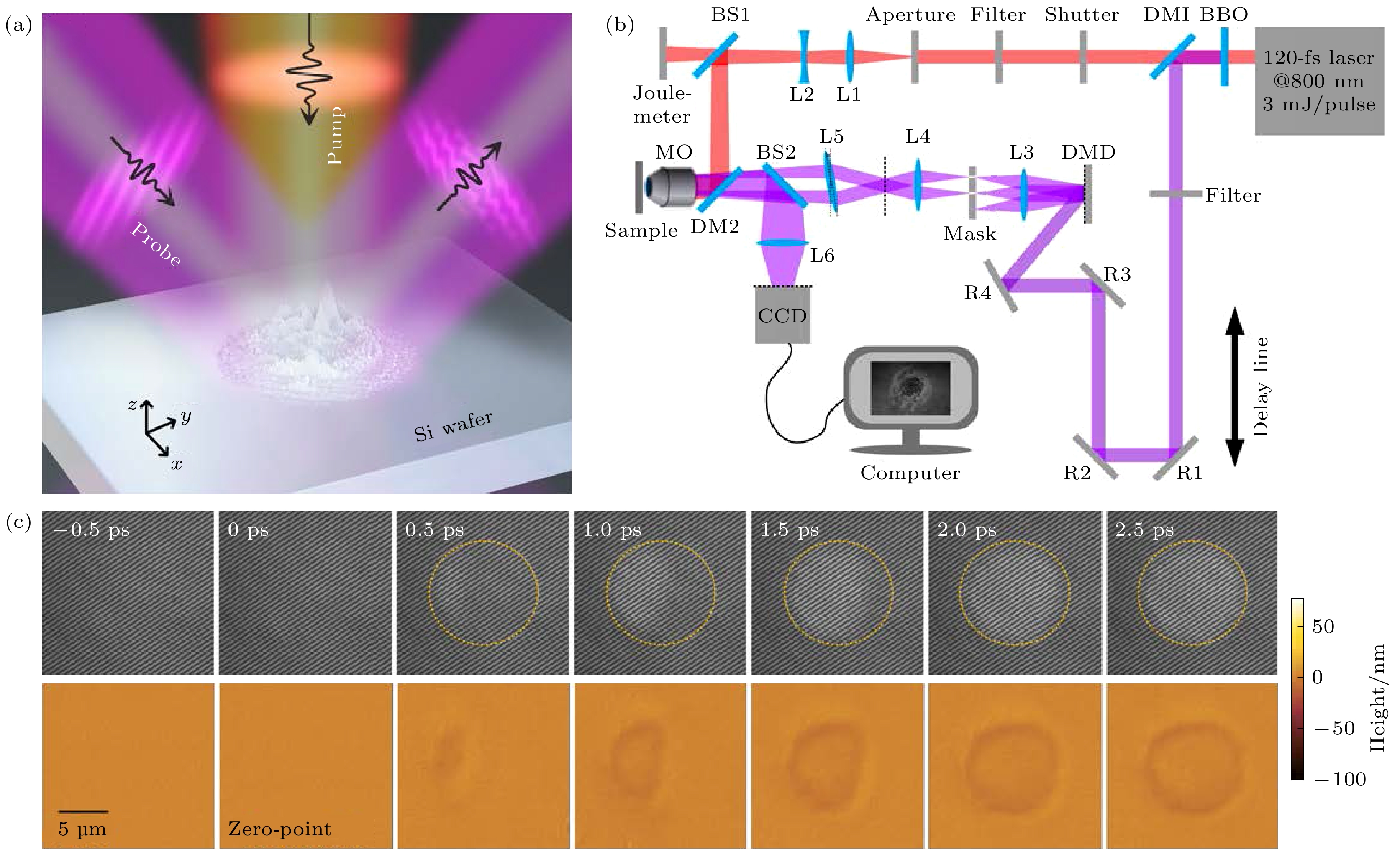
图 7 (a) SPSLM系统示意图及光路图, 其中红色和紫色分别代表泵浦光和探测光[60]; (b)单个泵浦光脉冲烧蚀硅表面从0到2.5 ps采集到的原始图像及重建得到的表面形貌演化图[60]
Figure 7. (a) Schematic of the SPSLM, where color red and color violet indicate pump light and probe light respectively[60]; (b) raw images and the reconstructed surface evolution height maps collected from 0 to 2.5 ps[60].
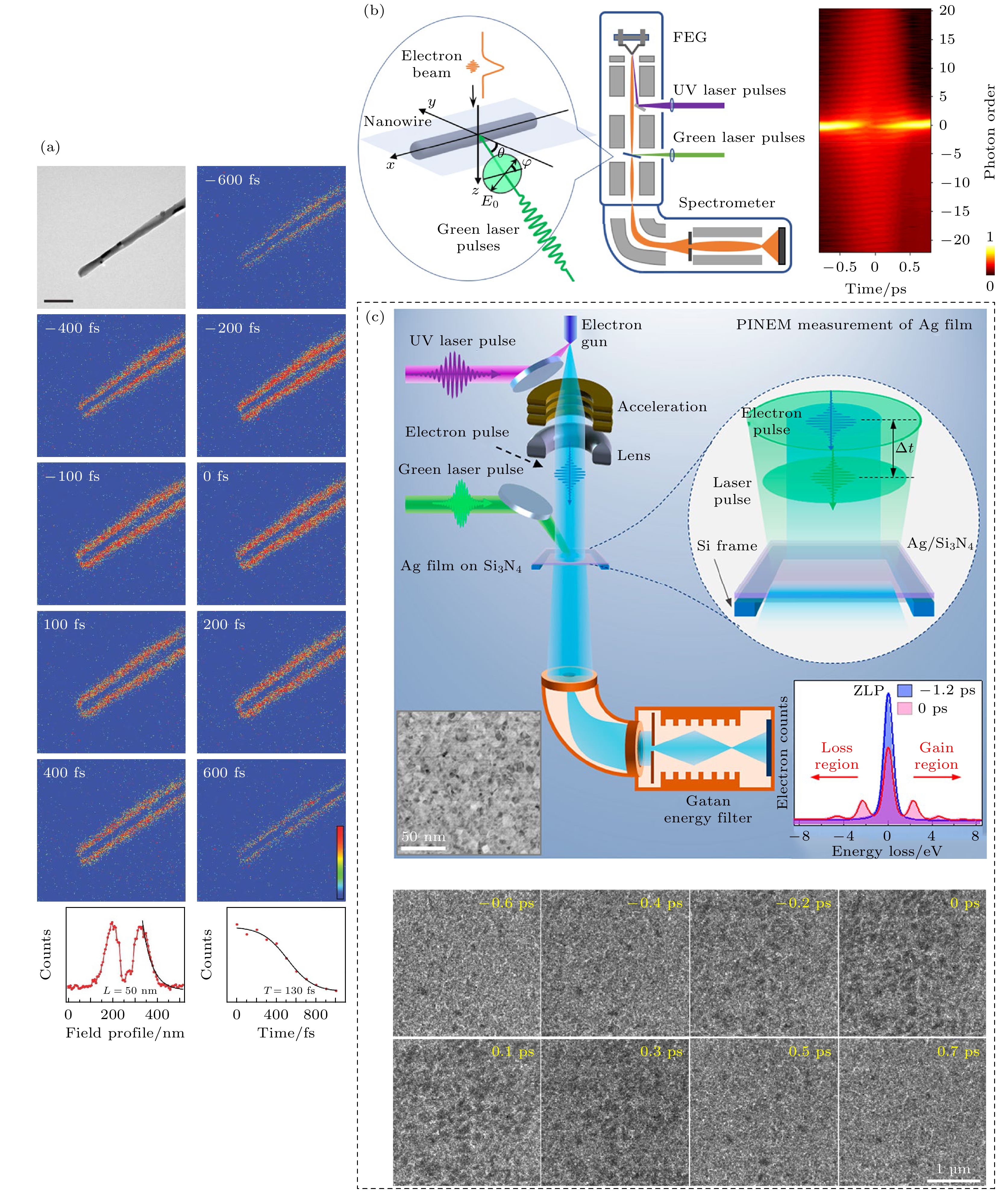
图 8 (a) 单个碳纳米管的PINEM时间分辨图像. 图片左上角的时间表示电子波包与飞秒脉冲到达纳米管的延迟时间, 颜色表示飞秒脉冲在纳米结构表面周围产生的场强度分布及其随时间的衰减[67]; (b) PINEM系统示意图(左)和采集的银纳米线在不同时间延迟下的电子能量损失谱(EELS)(右)[68]; (c) PINEM系统示意图及银膜表面等离激元的时间分辨能量过滤图像[69]
Figure 8. (a) PINEM result of an individual nanotube, the time in the upper left corner of the blue figures represents the delay time of the electron wave packet reaching the nanotube, and the color reveals the field intensity distribution around the surface of the nanostructure and its decay over time[67]; (b) schematic illustration of the PINEM apparatus (left) and electron energy-loss spectroscopy (EELS) data for pulsed-electrons interacting with the plasmon near-field (right)[68]; (c) schematic diagram of the system and energy-filtered time-resolved images of the surface plasmons on the silver film[69].
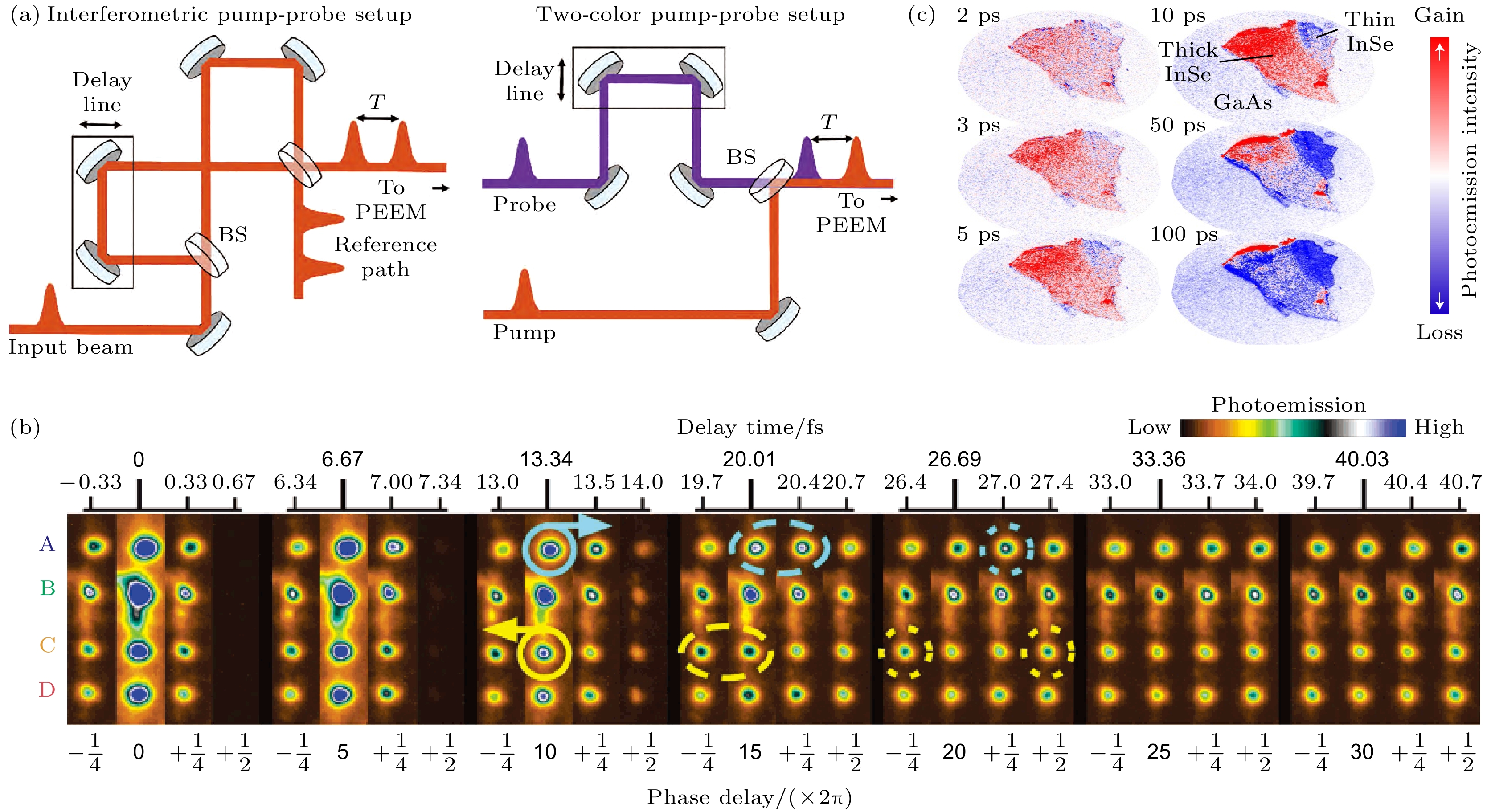
图 9 (a) tr-PEEM两种泵浦-探测光路类型[74]; (b) Ag光栅上的四个局部SPP的ITR-PEEM结果. 泵浦和探测脉冲之间的延迟时间(
${\tau _{\rm{d}}}$ ) 从–0.33 fs增加到40.69 fs, 步长为0.33 fs[78]; (c)通过tr-PEEM拍摄了在光激发后, InSe/GaAs异质结构中的电子随时间的积累(红色)和耗尽(蓝色)等转移过程[79]Figure 9. (a) Two pump-probe schematic of tr-PEEM[74]; (b) ITR-PEEM of the four localized plasmons on the silver grating framed. The delay time between the pump and probe pulses (
${\tau _{\rm{d}}}$ ) is advanced from –0.33 to 40.69 fs with an increment step of 0.33 fs[78]; (c) electron transport over time in the InSe/GaAs heterostructure showing the initial accumulation (red) and eventual recombination (blue) after photoexcitation[79].
图 10 (a) 通过飞秒LRM成像SPP波包运动的实现系统光路图[83]; (b) 延迟时间为190 fs时CMOS相机采集到的干涉图像[83]; (c) 不同时刻下(200, 230, 260 fs)实验中获得的泄露模干涉图与理论值(绿色虚线)的比较, 用于表征SPP波包运动[83]
Figure 10. (a) Implementation of femtosecond LRM imaging SPP wave packet[83]; (b) LRM image recorded on the CMOS camera at the output of the interferometer for a time delay of 190 fs[83]; (c) SPP wave packet motion of experimental time-resolved interferograms obtained by LRM cross-cut images for the different time delays of 200, 230 and 260 fs, compared with the theoretical values (green dashed line)[83].

图 11 (a) T-CUP系统示意图. 其中黑色虚线框为条纹相机中的条纹管的详细图示[86]; (b)使用T-CUP系统得到的飞秒脉冲时间聚焦的时空演化[86]
Figure 11. (a) Schematic of the T-CUP system, where the black dashed box is a detailed representation of the striped tube in the striped camera[86]; (b) spatiotemporal evolution of the femtosecond pulse time-focus obtained using the T-CUP system[86].
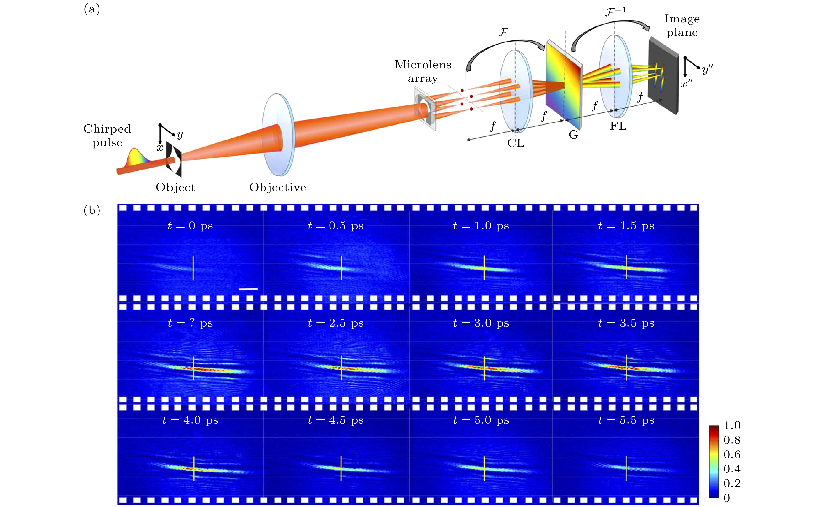
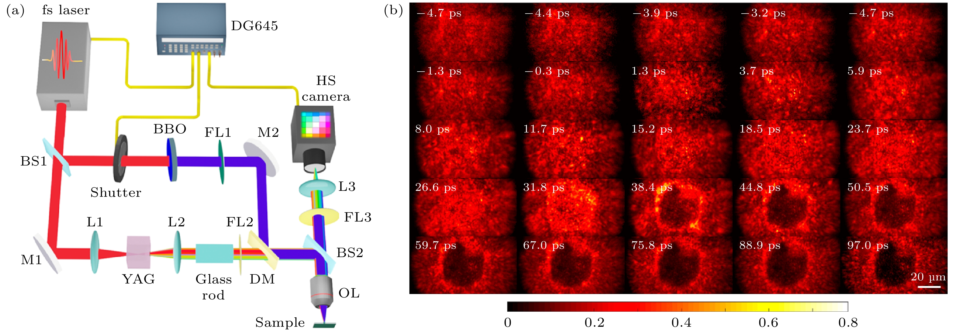
图 13 (a) 用于飞秒激光烧蚀测量的CSMUP系统[88]; (b) CSMUP在400 nm飞秒激光照射下对硅烧蚀的单脉冲不同时刻动力学成像结果[88]
Figure 13. (a) System configuration of CSMUP for the femtosecond laser ablation measurement [88]; (b) single-shot laser ablation dynamics measurement of silicon under a 400 nm femtosecond laser exposure captured with CSMUP[88].


图 16 (a) 在1.0 J/cm2能量密度的泵浦脉冲后, 锌表面在不同延迟时间的CCD图像[96]; (b) 空气和水中的皮秒激光诱导烧蚀动力学[97]; (c) 泵浦功率为0.29 J/cm2下, 第二和第三个脉冲叠加后硅表面的超快形貌演变, 并将最终结果与原子力显微镜(AFM)进行比较[60]
Figure 16. (a) CCD images of the Zn surface impacted by pump pulse at different delay times with energy of 1.0 J/cm2[96]; (b) picosecond laser-induced ablation in air and water[97]; (c) ultrafast topography evolution on the Si surface impacted by 2nd and 3rd pulses with energy of 0.29 J/cm2, and final results compared with AFM[60].
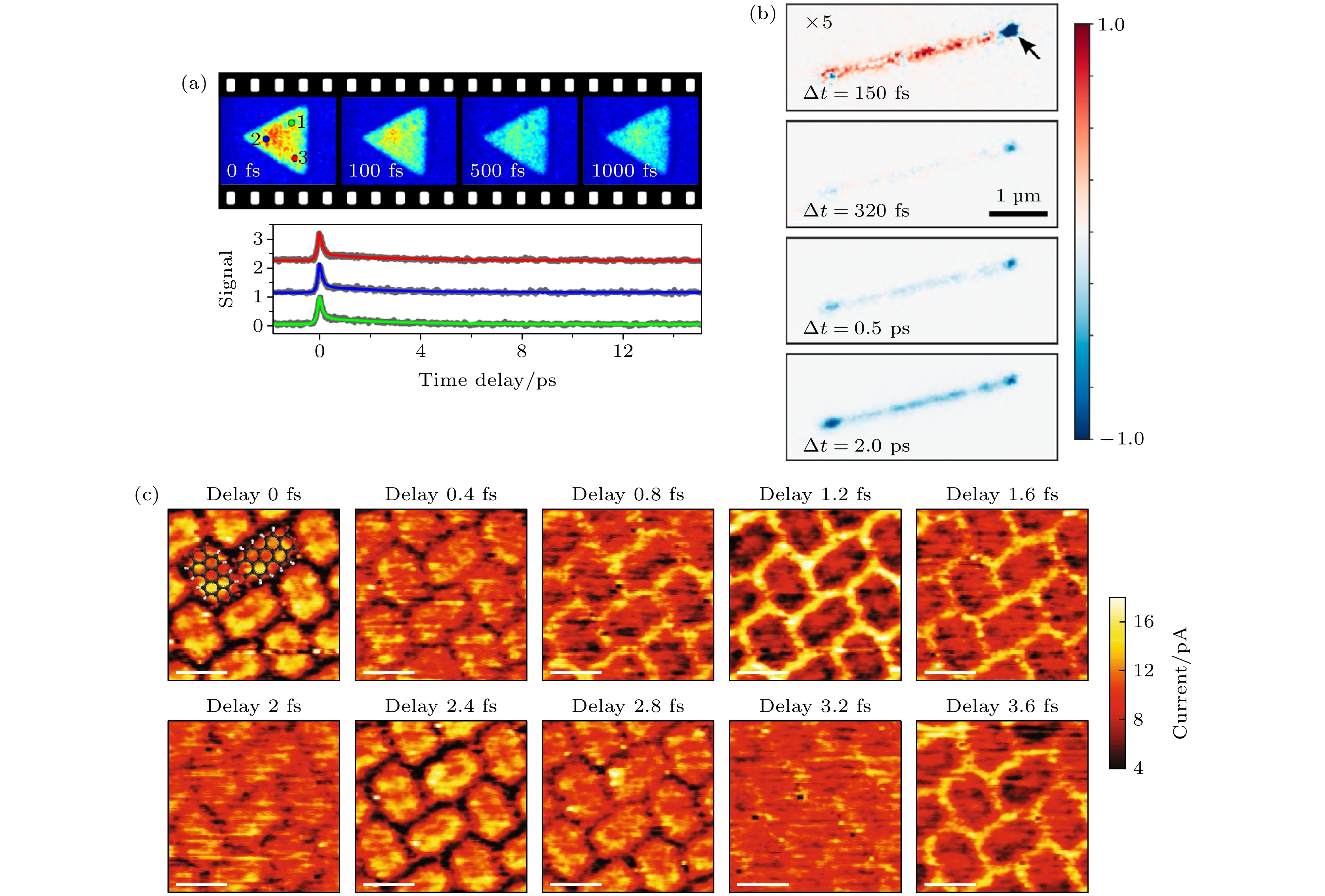
图 17 (a) WSe2薄片的时间分辨成像以及三个不同位点处信号强度随延迟时间变化曲线[107]; (b) 单个InAs纳米线中的热电子动力学. 在不同泵浦-探测延迟时间下拍摄的tr-PEEM图像[108]; (c) 苝四羧酸二酐分子(PTCDA)量子相干干涉的时空分辨成像[109]
Figure 17. (a) Time-resolved imaging of WSe2 flake and Representative time traces obtained for single pixels located at three different positions[107]; (b) hot electron dynamics in a single InAs nanowire. Time-resolved PEEM images taken at different pump-probe delay time[108]; (c) space-time-resolved imaging of quantum coherent interference of PTCDA[109].

图 18 (a) 金纳米棒的瞬态近场图像中沿着棒轴的线轮廓的时间变化[112]; (b) LCP和RCP光反对称模式和对称模式的激发下的光电发射强度随时间演变图及0 fs下的PEEM扫描图像[113]; (c) tr-PEEM观测SPP携带OAM实验示意及观测结果, SPP涡旋场在约2.67 fs的单个光学周期内演化状态的快照序列图[114]; (d) ITR-PEEM对从银膜矩形沟槽发射的飞秒SPP的时空演化成像[115]; (e) 金纳米链的拓扑边态动力学过程[116]
Figure 18. (a) Time variation of the line profiles along the rod axis in transient near-field images of nanorod[112]; (b) PEEM-measured (solid lines) and theoretically fitted (dashed lines) photoemission intensity curves against the delay time between the two pulses for LCP and RCP light, corresponding to excitation of the antisymmetric mode and symmetric mode, respectively, as indicated by the PEEM images with a delay time of 0 fs in the insets[113]; (c) schematic experimental methodology and experimental tr-PEEM snapshot sequence of the rotating field of a plasmonic vortex in the revolution stage within a single optical cycle of ~2.67 fs[114]; (d) spatial-temporal evolution imaging of the femtosecond SPP emitted from the rectangular grooves of the silver membranes by ITR-PEEM[115]; (e) topological edge state dynamic processes of gold nanocarticles[116].

图 19 超高时空分辨显微成像技术分辨率指标对比, 包括近场多脉冲显微成像技术(红色框)、远场多脉冲显微成像技术(蓝色框)以及远场单脉冲显微成像(黄色框)
Figure 19. Resolution of the ultra-high spatiotemporal resolved imaging techniques, including the near-field multi-pulse spatiotemporal microscopic techniques (red box), the far-field multi-pulse spatiotemporal microscopic techniques (blue) and the far-field single -pulse spatiotemporal microscopic techniques (yellow box).
表 1 超高时空分辨成像技术指标及优缺点对比
Table 1. Technical indexes, advantages and disadvantages of ultra-high spatiotemporal resolution imaging.
技术手段 空间分辨率 时间分辨率 优点 缺点 近场
多脉冲超快NSOM[37] 20 nm 亚fs 可实现空间超分辨 系统复杂, 视场小, 成像速度慢 超快四波混频AFM[26] 50 nm 10 fs 可实现空间超分辨, 可以得到样品表面形貌信息 系统复杂, 视场小, 成像速度慢, 需要激发非线性效应 超快PiFM[42] 10 nm 200 fs 可实现空间超分辨, 可以得到样品表面形貌信息 系统复杂, 视场小, 成像速度慢 超快STM[46] 0.1 nm 亚fs 可实现空间超分辨, 空间分辨率最高 系统复杂, 视场小, 成像速度慢, 只适用导电样品 远场
多脉冲高NA系统[55] 接近衍射极限 fs 量级 速度快, 大视场 无法实现空间超分辨 SPPM[59] 114 nm fs量级 可实现空间超分辨 视场小, 需要多步相移, 成像速度慢 SPSLM[60] 478 nm(横向);
22 nm (纵向)256 fs 单帧成像, 大视场, 有三维成像能力 无法实现空间超分辨 PINEM[67] 小于0.7 nm 10 fs 可实现空间超分辨 电子显微镜系统复杂, 设备昂贵, 样品要求高 超快PEEM[71] 10 nm 10 fs 可实现空间超分辨 电子显微镜系统复杂, 设备昂贵, 样品要求高, 空间分辨率受材料影响 LRM[83] 接近衍射极限 10 fs 可获得SPP传播相速度和群速度信息 目前仅能对SPP成像 远场
单脉冲CUP[86] 1 μm 100 fs 帧数高, 成像速度快 压缩感知算法较复杂, 条纹相机较为昂贵 OPR[87] 11.1 μm 100 fs 重建算法简单、直接、稳定性好, 时间分辨率高 空间分辨率较低, 目前仅有微米量级 CSMUP[88] 833 nm 4 ps 较高的空间分辨率, 图像尺寸更大 时间分辨率依赖于高光谱相机光谱带, 时间分辨率较低 STAMP[89] 1 μm 227 fs 在显微和宏观成像领域都适用, 普适性强 帧数和时间分辨率存在依赖关系, 难以兼得 FINCOPA[91] 3 μm 50 fs 时空分辨率、帧数、帧间隔相互独立 空间分辨率较低  DownLoad: CSV
-
[1] Born M, Wolf E 2013 Principles of Optics: Electromagnetic Theory of Propagation, Interference and Diffraction of Light (Oxford: Pergamon Press)
[2] Hell S W 2003 Nat. Biotechnol. 21 1347
Google Scholar
[3] Rust M J, Bates M, Zhuang X 2006 Nat. Methods 3 793
Google Scholar
[4] Betzig E, Patterson G H, Sougrat R, Lindwasser O W, Olenych S, Bonifacino J S, Davidson M W, Lippincott-Schwartz J, Hess H F 2006 Science 313 1642
Google Scholar
[5] Gustafsson M G 2000 J. Microsc. 198 82
Google Scholar
[6] Dertinger T, Colyer R, Iyer G, Weiss S, Enderlein J 2009 Proc. Natl. Acad. Sci. U. S. A. 106 22287
Google Scholar
[7] Balzarotti F, Eilers Y, Gwosch K C, Gynnå A H, Westphal V, Stefani F D, Elf J, Hell S W 2017 Science 355 606
Google Scholar
[8] Vigoureux J M, Courjon D 1992 Appl. Opt. 31 3170
Google Scholar
[9] Betzig E, Chichester R J 1993 Science 262 1422
Google Scholar
[10] Mohammed A, Abdullah A 2018 Proceedings of the 2018 International Conference on Hydraulics and Pneumatics— HERVEX Băile Govora, Romania, 2018 pp7–9
[11] Egerton R F 2008 Rep. Prog. Phys. 72 016502
[12] Pennycook S J, Nellist P D 2011 Scanning transmission electron microscopy: imaging and analysis (Springer Science & Business Media)
[13] Adachi S, Ishii H, Kanai T, Ishii N, Kosuge A, Watanabe S 2007 Opt. Lett. 32 2487
Google Scholar
[14] Ji S Y, Yang L, Hu Y L, Ni J C, Du W Q, Li J W, Zhao G, Wu D, Chu J R 2017 Small 13 1701190
Google Scholar
[15] Lao Z X, Hu Y L, Zhang C C, Yang L, Li J W, Chu J R, Wu D 2015 ACS Nano 9 12060
Google Scholar
[16] Bargheer M, Zhavoronkov N, Woerner M, Elsaesser T 2006 ChemPhysChem 7 783
Google Scholar
[17] Chapman H N, Fromme P, Barty A, White T A, Kirian R A, Aquila A, Hunter M S, Schulz J, DePonte D P, Weierstall U 2011 Nature 470 73
Google Scholar
[18] Holldack K, Khan S, Mitzner R, Quast T 2006 Phys. Rev. Lett. 96 054801
Google Scholar
[19] Van Tilborg J, Schroeder C, Filip C, Tóth C, Geddes C, Fubiani G, Huber R, Kaindl R, Esarey E, Leemans W 2006 Phys. Rev. Lett. 96 014801
Google Scholar
[20] Point G, Brelet Y, Houard A, Jukna V, Milián C, Carbonnel J, Liu Y, Couairon A, Mysyrowicz A 2014 Phys. Rev. Lett. 112 223902
Google Scholar
[21] Panagiotopoulos P, Whalen P, Kolesik M, Moloney J V 2015 Nat. Photonics 9 543
Google Scholar
[22] Krausz F, Ivanov M 2009 Rev. Mod. Phys. 81 163
Google Scholar
[23] Vozzi C, Calegari F, Ferrari F, Lucchini M, De Silvestri S, Svelto O, Sansone G, Stagira S, Nisoli M 2009 Laser Phys. Lett. 6 259
Google Scholar
[24] Morimoto Y, Baum P 2018 Nat. Phys. 14 252
Google Scholar
[25] Dudley J M, Finot C, Richardson D J, Millot G 2007 Nat. Phys. 3 597
Google Scholar
[26] Kravtsov V, Ulbricht R, Atkin J M, Raschke M B 2016 Nat. Nanotechnol. 11 459
Google Scholar
[27] Kim S, Jin J, Kim Y J, Park I Y, Kim Y, Kim S W 2008 Nature 453 757
Google Scholar
[28] Park I Y, Kim S, Choi J, Lee D H, Kim Y J, Kling M F, Stockman M I, Kim S W 2011 Nat. Photonics 5 677
Google Scholar
[29] Zhang Y Q, Shen J F, Min C J, Jin Y F, Jiang Y Q, Liu J, Zhu S W, Sheng Y L, Zayats A V, Yuan X C 2018 Nano Lett. 18 5538
Google Scholar
[30] Jiang Y, Narushima T, Okamoto H 2010 Nat. Phys. 6 1005
Google Scholar
[31] Van Dao L, Lincoln C, Lowe M, Hannaford P 2005 Femtosecond Laser Spectroscopy (Boston: Springer) pp197– 224
[32] Douhal A, Lahmani F, Zewail A H 1996 Chem. Phys. 207 477
Google Scholar
[33] Yamanouchi K 2002 Science 295 1659
Google Scholar
[34] Peterman E J, Monshouwer R, van Stokkum I H, van Grondelle R, van Amerongen H 1997 Chem. Phys. Lett. 264 279
Google Scholar
[35] Keller E L, Brandt N C, Cassabaum A A, Frontiera R R 2015 Analyst 140 4922
Google Scholar
[36] 李智, 张家森, 杨景, 龚旗煌 2007 物理学报 56 3630
Google Scholar
Li Z, Zhang J S, Yang J, Gong Q H 2007 Acta Phys. Sin. 56 3630
Google Scholar
[37] Sternbach A J, Latini S, Chae S, Hübener H, De Giovannini U, Shao Y, Xiong L, Sun Z, Shi N, Kissin P 2020 Nat. Commun. 11 3567
Google Scholar
[38] Huber M A, Mooshammer F, Plankl M, Viti L, Sandner F, Kastner L Z, Frank T, Fabian J, Vitiello M S, Cocker T L 2017 Nat. Nanotechnol. 12 207
Google Scholar
[39] Xue M F, Li M, Huang Y S, Chen R K, Li Y L, Wang J Y, Xing Y J, Chen J J, Yan H G, Xu H Q 2020 Adv. Mater. 32 2004120
Google Scholar
[40] Binnig G, Quate C F, Gerber C 1986 Phys. Rev. Lett. 56 930
Google Scholar
[41] Jahng J, Brocious J, Fishman D A, Huang F, Li X W, Tamma V A, Wickramasinghe H K, Potma E O 2014 Phys. Rev. B 90 155417
Google Scholar
[42] Jahng J, Brocious J, Fishman D A, Yampolsky S, Nowak D, Huang F, Apkarian V A, Wickramasinghe H K, Potma E O 2015 Appl. Phys. Lett. 106 083113
Google Scholar
[43] Binnig G, Rohrer H, Gerber C, Weibel E 1982 Phys. Rev. Lett. 49 57
Google Scholar
[44] Yang B, Chen G, Ghafoor A, Zhang Y F, Zhang Y, Zhang Y, Luo Y, Yang J L, Sandoghdar V, Aizpurua J 2020 Nat. Photonics 14 693
Google Scholar
[45] Takeuchi O, Aoyama M, Oshima R, Okada Y, Oigawa H, Sano N, Shigekawa H, Morita R, Yamashita M 2004 Appl. Phys. Lett. 85 3268
Google Scholar
[46] Terada Y, Yoshida S, Takeuchi O, Shigekawa H 2010 Nat. Photonics 4 869
Google Scholar
[47] Sonnenfeld R, Hansma P K 1986 Science 232 211
Google Scholar
[48] Maire G, Giovannini H, Talneau A, Chaumet P C, Belkebir K, Sentenac A 2018 Opt. Lett. 43 2173
Google Scholar
[49] Wang Z B, Guo W, Li L, Luk'Yanchuk B, Khan A, Liu Z, Chen Z C, Hong M H 2011 Nat. Commun. 2 218
Google Scholar
[50] Lu D, Liu Z W 2012 Nat. Commun. 3 1205
Google Scholar
[51] Hao X, Liu X, Kuang C F, Li Y H, Ku Y L, Zhang H J, Li H F, Tong L M 2013 Appl. Phys. Lett. 102 013104
Google Scholar
[52] Fang N, Lee H, Sun C, Zhang X 2005 Science 308 534
Google Scholar
[53] Darafsheh A, Limberopoulos N I, Derov J S, Walker Jr D E, Astratov V N 2014 Appl. Phys. Lett. 104 061117
Google Scholar
[54] Domke M, Rapp S, Schmidt M, Huber H P 2012 Opt. Express 20 10330
Google Scholar
[55] Zhou K, Jia X, Jia T Q, Cheng K, Cao K Q, Zhang S A, Feng D H, Sun Z R 2017 J. Appl. Phys. 121 104301
Google Scholar
[56] Hirano Y, Matsuda A, Hiraoka Y 2015 Microscopy 64 237
Google Scholar
[57] Li D, Shao L, Chen B C, Zhang X, Zhang M S, Moses B, Milkie D E, Beach J R, Hammer III J A, Pasham M 2015 Science 349 aab3500
Google Scholar
[58] Guo Y T, Li D, Zhang S W, Yang Y R, Liu J J, Wang X Y, Liu C, Milkie D E, Moore R P, Tulu U S 2018 Cell 175 1430
Google Scholar
[59] Massaro E S, Hill A H, Grumstrup E M 2016 ACS Photonics 3 501
Google Scholar
[60] Xu J, Min C J, Zhang Y Q, Ni J L, Cao G W, Wei Q Y, Yang J J, Yuan X C 2022 Photonics Res. 10 1900
Google Scholar
[61] Su X Y, Zhang Q C 2010 Opt. Lasers Eng. 48 191
Google Scholar
[62] Su X, Chen W J 2001 Opt. Lasers Eng. 35 263
Google Scholar
[63] Tao T Y, Chen Q, Da J, Feng S J, Hu Y, Zuo C 2016 Opt. Express 24 20253
Google Scholar
[64] Yao P C, Gai S Y, Chen Y C, Chen W L, Da F P 2021 Opt. Lasers Eng. 143 106623
Google Scholar
[65] Chen F, Brown G M, Song M M 2000 Opt. Eng. 39 10
Google Scholar
[66] Schafer S 2017 光学与光电技术 15 1
Schafer S 2017 Optics & Optoelectronic Technology 15 1
[67] Barwick B, Flannigan D J, Zewail A H 2009 Nature 462 902
Google Scholar
[68] Zheng D G, Huang S Y, Zhu C H, Xu P, Li Z A, Wang H, Li J, Tian H F, Yang H X, Li J Q 2021 Nano Lett. 21 10238
Google Scholar
[69] Fu X W, Sun Z P, Ji S Z, Liu F, Feng M, Yoo B-K, Zhu Y M 2022 Nano Lett. 22 2009
Google Scholar
[70] Könenkamp R, Word R C, Rempfer G, Dixon T, Almaraz L, Jones T 2010 Ultramicroscopy 110 899
Google Scholar
[71] Dabrowski M, Dai Y N, Petek H 2020 Chem. Rev. 120 6247
Google Scholar
[72] Sun Q, Zu S, Misawa H 2020 J. Chem. Phys. 153 120902
Google Scholar
[73] Huber B, Pres S, Wittmann E, Dietrich L, Lüttig J, Fersch D, Krauss E, Friedrich D, Kern J, Lisinetskii V 2019 Rev. Sci. Instrum. 90 113103
Google Scholar
[74] 李耀龙, 刘运全, 龚旗煌 2021 光子学报 50 0850201
Google Scholar
Li Y L, Liu Y Q, Gong Q H 2021 Acta Photonica Sin. 50 0850201
Google Scholar
[75] Ogawa S, Nagano H, Petek H, Heberle A 1997 Phys. Rev. Lett. 78 1339
Google Scholar
[76] Fecher G H, Schmidt O, Hwu Y, Schönhense G 2002 J. Electron Spectrosc. Relat. Phenomena 126 77
Google Scholar
[77] Rotermund H H 1997 Surf. Sci. Rep. 29 265
Google Scholar
[78] Kubo A, Onda K, Petek H, Sun Z J, Jung Y S, Kim H K 2005 Nano Lett. 5 1123
Google Scholar
[79] Man M K, Margiolakis A, Deckoff-Jones S, Harada T, Wong E L, Krishna M B M, Madéo J, Winchester A, Lei S D, Vajtai R 2017 Nat. Nanotechnol. 12 36
Google Scholar
[80] Drezet A, Hohenau A, Koller D, Stepanov A, Ditlbacher H, Steinberger B, Aussenegg F R, Leitner A, Krenn J R 2008 Mater. Sci. Eng. , B 149 220
Google Scholar
[81] Hohenau A, Krenn J, Drezet A, Mollet O, Huant S, Genet C, Stein B, Ebbesen T 2011 Opt. Express 19 25749
Google Scholar
[82] Drezet A, Genet C 2013 Phys. Rev. Lett. 110 213901
Google Scholar
[83] Gorodetski Y, Chervy T, Wang S, Hutchison J A, Drezet A, Genet C, Ebbesen T W 2016 Optica 3 48
Google Scholar
[84] Gao L, Liang J Y, Li C Y, Wang L H V 2014 Nature 516 74
Google Scholar
[85] Li Z Y, Zgadzaj R, Wang X M, Chang Y-Y, Downer M C 2014 Nat. Commun. 5 3085
Google Scholar
[86] Liang J Y, Zhu L R, Wang L H V 2018 Light-Sci. Appl. 7 42
Google Scholar
[87] Zhu Y L, Zeng X K, Cai Y, Lu X W, Zhu Q F, Zeng L W, He T C, Li J Z, Yang Y, Zheng M J 2021 Opt. Express 29 27298
Google Scholar
[88] Yao Y H, He Y L, Qi D L, Cao F Y, Yao J L, Ding P P, Jin C Z, Wu X Y, Deng L Z, Jia T Q, Huang F, Liang J Y, Sun Z R, Zhang S A 2021 ACS Photonics 8 738
Google Scholar
[89] Nakagawa K, Iwasaki A, Oishi Y, Horisaki R, Tsukamoto A, Nakamura A, Hirosawa K, Liao H G, Ushida T, Goda K 2014 Nat. Photonics 8 695
Google Scholar
[90] Tamamitsu M, Nakagawa K, Horisaki R, Iwasaki A, Oishi Y, Tsukamoto A, Kannari F, Sakuma I, Goda K 2015 Opt. Lett. 40 633
Google Scholar
[91] Zeng X K, Zheng S Q, Cai Y, Lin Q G, Liang J Y, Lu X W, Li J Z, Xie W X, Xu S X 2020 Adv. Photonics 2 056002
[92] Downer M, Fork R L, Shank C V 1985 JOSA B 2 595
Google Scholar
[93] Garcia-Lechuga M, Puerto D, Fuentes-Edfuf Y, Solis J, Siegel J 2016 ACS Photonics 3 1961
Google Scholar
[94] Liu J K, Jia X, Wu W S, Cheng K, Feng D H, Zhang S A, Sun Z R, Jia T Q 2018 Opt. Express 26 6302
Google Scholar
[95] Pan C J, Jiang L, Sun J Y, Wang Q S, Wang F F, Wang K, Lu Y F, Wang Y L, Qu L T, Cui T H 2020 Light Sci. Appl. 9 80
Google Scholar
[96] Fang R R, Vorobyev A, Guo C L 2017 Light-Sci. Appl. 6 e16256
[97] Spellauge M, Doñate-Buendía C, Barcikowski S, Gökce B, Huber H P 2022 Light-Sci. Appl. 11 68
Google Scholar
[98] Emmony D, Howson R, Willis L 1973 Appl. Phys. Lett. 23 598
Google Scholar
[99] Huang M, Zhao F L, Cheng Y, Xu N S, Xu Z Z 2009 ACS Nano 3 4062
Google Scholar
[100] Bonse J, Rosenfeld A, Krüger J 2009 J. Appl. Phys. 106 104910
Google Scholar
[101] Splendiani A, Sun L, Zhang Y B, Li T S, Kim J, Chim C Y, Galli G, Wang F 2010 Nano Lett. 10 1271
Google Scholar
[102] Zeng H L, Dai J F, Yao W, Xiao D, Cui X D 2012 Nat. Nanotechnol. 7 490
Google Scholar
[103] Bernardi M, Palummo M, Grossman J C 2013 Nano Lett. 13 3664
Google Scholar
[104] Baugher B W, Churchill H O, Yang Y F, Jarillo-Herrero P 2014 Nat. Nanotechnol. 9 262
Google Scholar
[105] Yuan H T, Bahramy M S, Morimoto K, Wu S F, Nomura K, Yang B J, Shimotani H, Suzuki R, Toh M, Kloc C 2013 Nat. Phys. 9 563
Google Scholar
[106] Furchi M M, Pospischil A, Libisch F, Burgdörfer J, Mueller T 2014 Nano Lett. 14 4785
Google Scholar
[107] Wang L, Xu C, Li M Y, Li L J, Loh Z H 2018 Nano Lett. 18 5172
Google Scholar
[108] Wittenbecher L, Viñas Boström E, Vogelsang J, Lehman S, Dick K A, Verdozzi C, Zigmantas D, Mikkelsen A 2021 ACS Nano 15 1133
Google Scholar
[109] Garg M, Martin-Jimenez A, Pisarra M, Luo Y, Martín F, Kern K 2022 Nat. Photonics 16 196
Google Scholar
[110] Barnes W L, Dereux A, Ebbesen T W 2003 Nature 424 824
Google Scholar
[111] Wang Y L, Min C J, Zhang Y Q, Yuan X C 2022 Opto-Electron. Adv. 5 210047
Google Scholar
[112] Nishiyama Y, Imura K, Okamoto H 2015 Nano Lett. 15 7657
Google Scholar
[113] Zu S, Sun Q, Cao E, Oshikiri T, Misawa H 2021 Nano Lett. 21 4780
Google Scholar
[114] Spektor G, Kilbane D, Mahro A, Frank B, Ristok S, Gal L, Kahl P, Podbiel D, Mathias S, Giessen H 2017 Science 355 1187
Google Scholar
[115] Zhao Z L, Lang P, Qin Y L, Ji B Y, Song X W, Lin J Q 2020 Opt. Express 28 19023
Google Scholar
[116] Yan Q C, Cao E, Sun Q, Ao Y T, Hu X Y, Shi X, Gong Q H, Misawa H 2021 Nano Lett. 21 9270
Google Scholar

 DownLoad:


Catalog
Metrics
- Abstract views: 3696
- PDF Downloads: 240
- Cited By: 0
