


-
Controlling the emission characteristics of fluorescent substances and increasing the intensity of fluorescence emission are crucial for fluorescence detecting technology in single-molecule detection, biomedicine, and sensing applications. Since fluorescence emission is isotropic in nature, the collected fluorescence is only accounted for a small fraction of the total emitted fluorescence. In this paper, a composite structure composed of dielectric microsphere and metallic planar nanolayers is proposed to enhance the fluorescence far-field directional emission intensity and improve the fluorescence collection efficiency. The excitation process and the emission process of quantum dots (QDs) located between the dielectric microspheres and the gold layer are investigated by the finite difference time domain (FDTD) method. In the emission process, the emission of QDs in a homogeneous medium is isotropic. Therefore, we usually select several special polarizations in theoretical analysis state for research. In this paper, we first study the effect of the structure on the fluorescence emission enhancement of QDs when the QDs are in the x-, y-, and z-polarization state. Some results can be obtained as shown below. When the radiation direction of the QDs is perpendicular to the microsphere plane layered structure, the structure is coupled with the emitted fluorescence, thereby realizing the directional enhancement of the emitted fluorescence of the QDs, and the obvious fluorescence enhancement is obtained in the x- and y-polarization state. Therefore, in the research, we choose and investigate the dipole light source of x-polarization state. We mainly study the influence of microsphere radius, refractive index, and QDs position on the fluorescence directional enhancement. The QDs as a fluorescent material are coated in polymethyl methacrylate (PMMA) to control the distance from the gold layer to tune the fluorescence enhancement. The structure is based on the synergistic effect among plasmon coupling, whispering gallery mode and photonic nanojet, which enhances the far-field fluorescence of QDs by a factor of 230, and the fluorescence collection efficiency is as high as 70%. Comparing with the enhanced fluorescence of the dielectric microspheres and the gold sphere dimer composite structure, the distance between the gold sphere dimers is not easy to control, and the QDs should be placed at specific positions between the gold spheres. The structure we propose is more convenient to implement. In this paper, not only the emission enhancement process of QDs is studied in detail, but also the excitation process of QDs is investigated. Our proposed dielectric microsphere metal planar nanolayered structure can enhance the excitation of QDs in most areas, proving that our designed structure can effectively realize the excitation enhancement of QDs. The above results have very important applications in the fluorescence biological detection, imaging, and light-emitting devices.
-
Keywords:
- fluorescence enhancement /
- photonic nanojet /
- dielectric microsphere /
- biological detection
[1] Wang J, Sun C, Ji M, Wang B, Wang P, Zhou G, Dong B, Du W, Huang L, Wang H, Ren L 2021 Protein. Expr. Purif. 187 105952
 Google Scholar
[2] Zhou M, Cao J, Akers W J 2016 Methods Mol. Biol. 1444 45
Google Scholar
[3] Zhou L, Zhou J, Lai W, Yang X, Meng J, Su L, Gu C, Jiang T, Pun E Y B, Shao L, Petti L, Sun X W, Jia Z, Li Q, Han J, Mormile P 2020 Nat. Commun. 11 1785
Google Scholar
[4] Itoh T 2012 Chem. Rev. 112 4541
Google Scholar
[5] Qian Z, Ma J, Shan X, Shao L, Zhou J, Chen J, Feng H 2013 RSC Advances 3 14571
Google Scholar
[6] Lu C Y, Browne D E, Yang T, Pan J W 2007 Phys. Rev. Lett. 99 250504
Google Scholar
[7] Fan L, Sun X, Xiong C, Schuck C, Tang H X 2013 Appl. Phys. Lett. 102 153507
Google Scholar
[8] Marcu L 2012 Ann. Biomed. Eng. 40 304
Google Scholar
[9] Wang Z, Zheng Y, Zhao D, Zhao Z, Liu L, Pliss A, Zhu F, Liu J, Qu J, Luan P 2017 J. Innov. Opt. Heal. Sci. 11 1830001
Google Scholar
[10] Ge F, Yang X 2017 J. Mater. Sci. 53 4840
Google Scholar
[11] Zhong K, Yu W, de Coene Y, Yamada A, Krylychkina O, Jooken S, Deschaume O, Bartic C, Clays K 2021 Biosens. Bioelectron. 194 113577
Google Scholar
[12] Cheng Q, Wang S, Liu N 2021 IEEE Sens. J. 21 17785
Google Scholar
[13] Li L, Wang W, Luk T S, Yang X, Gao J 2017 ACS Photonics 4 501
Google Scholar
[14] Luo S, Li Q, Yang Y, Chen X, Wang W, Qu Y, Qiu M 2017 Laser & Photonics Rev. 11 1600299
Google Scholar
[15] Karvinen P, Nuutinen T, Hyvarinen O, Vahimaa P 2008 Optics Express 16 16364
Google Scholar
[16] Muriano A, Thayil K N A, Salvador J P, Loza-Alvarez P, Soria S, Galve R, Marco M P 2012 Sensor. Actuat. B:Chem. 174 394
Google Scholar
[17] Lin J H, Liou H Y, Wang C D, Tseng C Y, Lee C T, Ting C C, Kan H C, Hsu C C 2015 ACS Photonics 2 530
Google Scholar
[18] Walia S, Shah C M, Gutruf P, Nili H, Chowdhury D R, Withayachumnankul W, Bhaskaran M, Sriram S 2015 Appl. Phys. Rev. 2 011303
Google Scholar
[19] Quaranta G, Basset G, Martin O J F, Gallinet B 2018 Laser & Photonics Rev. 12 1800017
Google Scholar
[20] Choudhury S D, Badugu R, Nowaczyk K, Ray K, Lakowicz J R 2013 J. Phys. Chem. Lett. 4 227
Google Scholar
[21] Yan Y, Zeng Y, Wu Y, Zhao Y, Ji L, Jiang Y, Li L 2014 Opt. Express. 22 23552
Google Scholar
[22] Golmakaniyoon S, Hernandez-Martinez P L, Demir H V, Sun X W 2017 Appl. Phys. Lett. 111 093302
Google Scholar
[23] Nyman M, Shevchenko A, Shavrin I, Ando Y, Lindfors K, Kaivola M 2019 APL Photonics 4 076101
Google Scholar
[24] Huang Y, Lin W, Chen K, Zhang W, Chen X, Zhang M Q 2014 Phys. Chem. Chem. Phys. 16 11584
Google Scholar
[25] Liu Y S, Lin H C, Xu H L 2018 IEEE Photonics J. 10 1
Google Scholar
[26] Hong F, Tang C, Xue Q, Zhao L, Shi H, Hu B, Zhang X 2019 Langmuir 35 14833
Google Scholar
[27] Chen Z, Taflove A, Backman V 2004 Opt. Express 12 1214
Google Scholar
[28] Liu C Y 2019 Crystals 9 198
Google Scholar
[29] Liu C Y, Lin F C 2016 Opt. Commun. 380 287
Google Scholar
[30] Mahariq I, Abdeljawad T, Karar A S, Alboon S A, Kurt H, Maslov A V 2020 Photonics 7 50
Google Scholar
[31] Sergeev A A, Sergeeva K A, Leonov A A, Voznesenskiy S S 2020 4th International Conference on Metamaterials and Nanophotonics (METANANO) Tbilisi, Georgia, 2020, Sep 14–18 pp261–263
[32] Zhang W, Lei H 2020 Nanoscale 12 6596
Google Scholar
[33] Zhou S, Zhou T 2020 Appl. Phys. Express 13 042010
Google Scholar
[34] Kong S C, Simpson J J, Backman V 2008 IEEE Microw. Wirel. Compon. Lett. 18 4
Google Scholar
[35] Sullivan D 2013 Electromagnetic Simulation Using the FDTD Method, Second Edition (Hoboken: IEEE Press) pp85–96
[36] Duan J, Song L, Zhan J 2010 Nano Res. 2 61
Google Scholar
[37] Johnson P B, Christy R W 1972 Phys. Rev. B 6 4370
Google Scholar
[38] Palik E D 1985 Handbook of Optical Constants of Solids First Edition (Orlando: Academic Press) pp286–287
[39] Das G M, Ringne A B, Dantham V R, Easwaran R K, Laha R 2017 Opt. Express 25 19822
Google Scholar
[40] Garrett C G B, Kaiser W, Bond W L 1961 Phys. Rev. 124 1807
Google Scholar
[41] Guo M, Ye Y H, Hou J, Du B 2015 Photonics Res. 3 339
Google Scholar
[42] Zhu H, Chen M, Zhou S, Wu L 2017 Macromolecules 50 660
Google Scholar
-

图 1 电介质微球(灰色球)和金属平面纳米层组成的复合结构 (a) 三维结构示意图; (b)—(d) 结构gp, ga, gs的侧视图, QD代表量子点
Figure 1. Composite structure composed of dielectric microsphere (the gray ball) and metallic planar nanolayers: (a) 3D schematic diagram of the structures; (b)–(d) the side views of the structures of gp, ga, gs in order, QD stands for quantum dot.
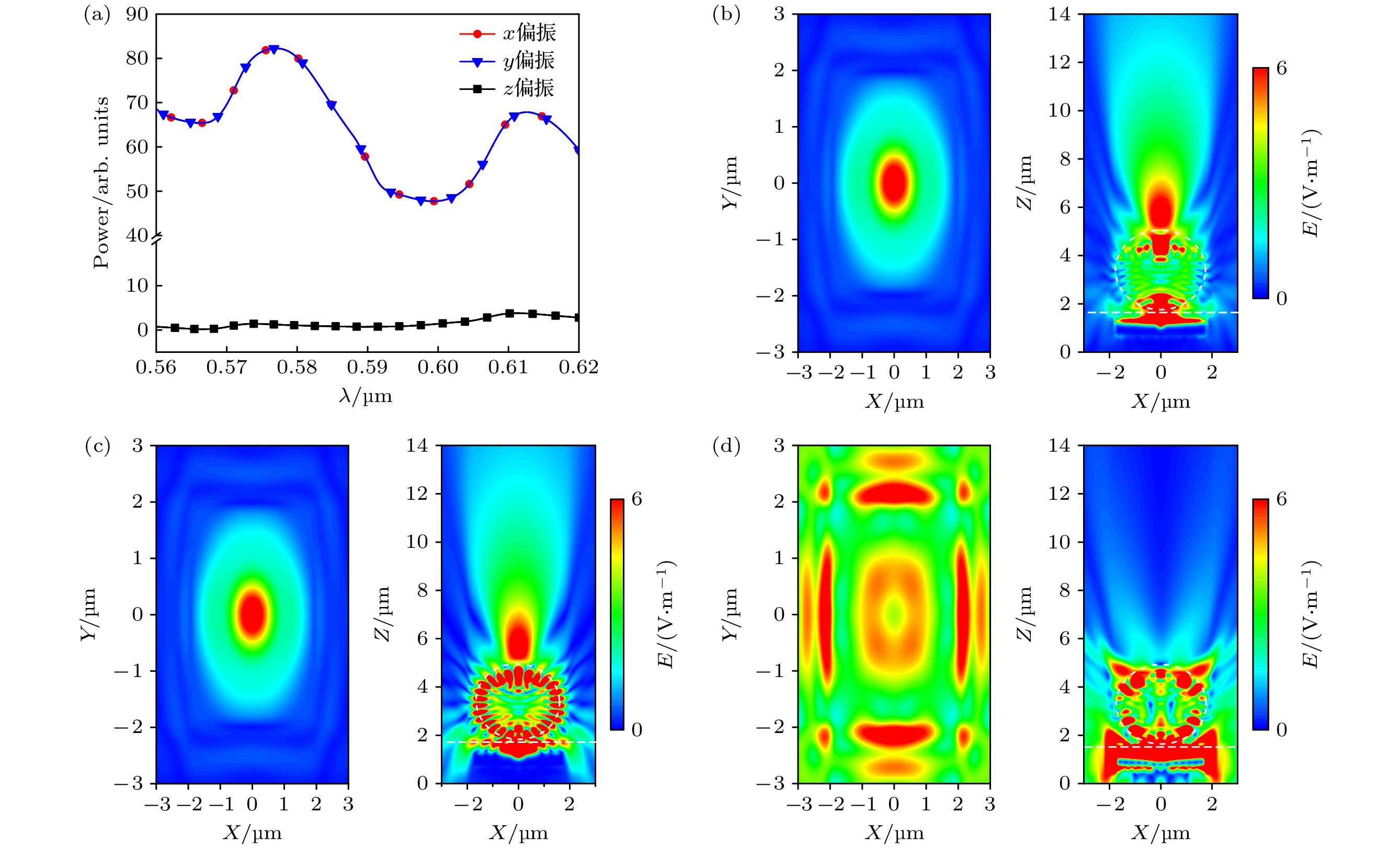
图 2 (a) 不同偏振态下偶极子光源的功率曲线; (b)—(d) 依次为x, y, z偏振态下的偶极子光源在中心波长590 nm处的俯视和横截面电场分布图
Figure 2. (a) Power curves of quantum dots in different polarization states; (b)–(d) top-view and cross-sectional electric field profiles of the dipole light source at the center wavelength of 590 nm under the x, y, z polarization states in turn, respectively.

图 3 量子点位于(0, 0, 0.78) μm处 (a) 3种结构的远场功率曲线图; (b)—(d) R = 2 μm, n = 1.5, 结构gp, ga和gs横截面处的电场分布图
Figure 3. Quantum dots are located at (0, 0, 0.78) μm: (a) Far-field power curves of the three structures; (b)–(d) plots of the electric field distribution at the cross-section of the gp, ga and gs structures at R = 2 μm, n = 1.5.

图 4 n = 1.5且量子点位于(0, 0, 0.78) μm处, 不同半径电介质微球的远场功率曲线
Figure 4. Far-field power curves of the dielectric microsphere with different radii for n = 1.5 and the quantum dots are located at (0, 0, 0.78) μm.
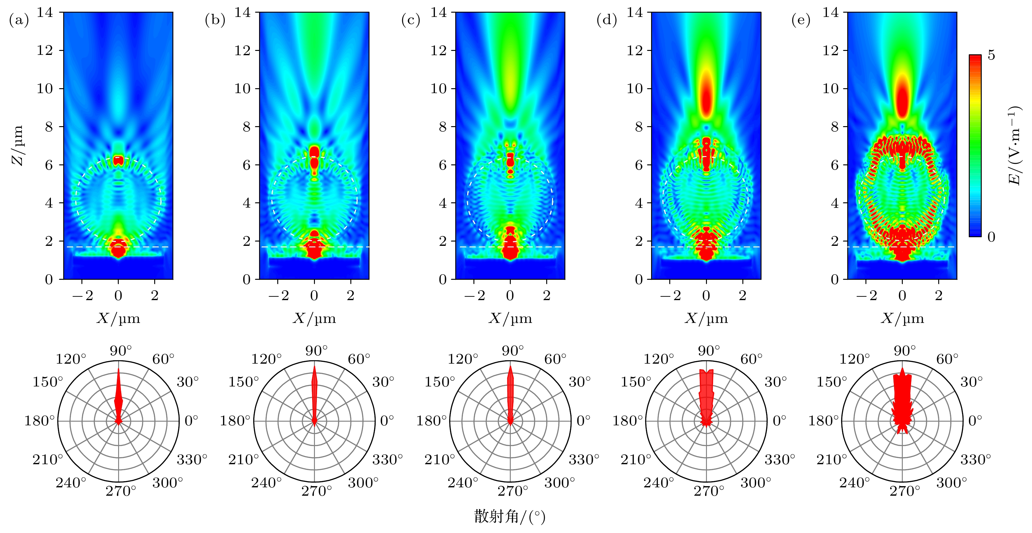
图 5 不同折射率电介质微球的电场强度和远场散射图 (a) n = 1.3; (b) n = 1.5; (c) n = 1.7; (d) n = 1.9; (e) n = 2.1
Figure 5. Eelectric field intensity and far-field scattering distributions of dielectric microsphere with different refractive indices: (a) n = 1.3; (b) n = 1.5; (c) n = 1.7; (d) n = 1.9; (e) n = 2.1.
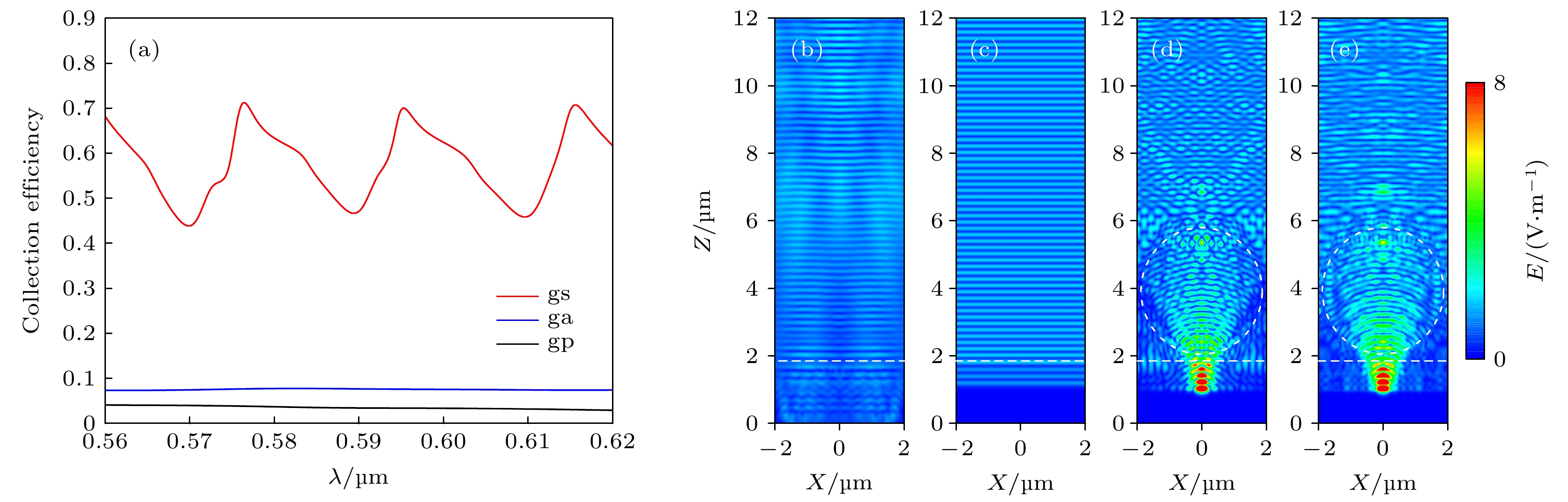
图 6 (a) R = 2 μm, n = 1.5时, 3个结构的远场收集效率; (b)—(e) 单色平面波长为405 nm处的激发电场图 (b) gp结构; (c) ga结构; (d), (e) gs结构的TE和TM偏振
Figure 6. (a) Far-field collection efficiencies of the three structures with R = 2 μm, n = 1.5; (b)–(e) excitation electric field maps at a wavelength of 405 nm in the monochromatic plane: (b) gp structure; (c) ga structure; (d), (e) the TE and TM polarizations of gs structure, respectively.
-
[1] Wang J, Sun C, Ji M, Wang B, Wang P, Zhou G, Dong B, Du W, Huang L, Wang H, Ren L 2021 Protein. Expr. Purif. 187 105952
Google Scholar
[2] Zhou M, Cao J, Akers W J 2016 Methods Mol. Biol. 1444 45
Google Scholar
[3] Zhou L, Zhou J, Lai W, Yang X, Meng J, Su L, Gu C, Jiang T, Pun E Y B, Shao L, Petti L, Sun X W, Jia Z, Li Q, Han J, Mormile P 2020 Nat. Commun. 11 1785
Google Scholar
[4] Itoh T 2012 Chem. Rev. 112 4541
Google Scholar
[5] Qian Z, Ma J, Shan X, Shao L, Zhou J, Chen J, Feng H 2013 RSC Advances 3 14571
Google Scholar
[6] Lu C Y, Browne D E, Yang T, Pan J W 2007 Phys. Rev. Lett. 99 250504
Google Scholar
[7] Fan L, Sun X, Xiong C, Schuck C, Tang H X 2013 Appl. Phys. Lett. 102 153507
Google Scholar
[8] Marcu L 2012 Ann. Biomed. Eng. 40 304
Google Scholar
[9] Wang Z, Zheng Y, Zhao D, Zhao Z, Liu L, Pliss A, Zhu F, Liu J, Qu J, Luan P 2017 J. Innov. Opt. Heal. Sci. 11 1830001
Google Scholar
[10] Ge F, Yang X 2017 J. Mater. Sci. 53 4840
Google Scholar
[11] Zhong K, Yu W, de Coene Y, Yamada A, Krylychkina O, Jooken S, Deschaume O, Bartic C, Clays K 2021 Biosens. Bioelectron. 194 113577
Google Scholar
[12] Cheng Q, Wang S, Liu N 2021 IEEE Sens. J. 21 17785
Google Scholar
[13] Li L, Wang W, Luk T S, Yang X, Gao J 2017 ACS Photonics 4 501
Google Scholar
[14] Luo S, Li Q, Yang Y, Chen X, Wang W, Qu Y, Qiu M 2017 Laser & Photonics Rev. 11 1600299
Google Scholar
[15] Karvinen P, Nuutinen T, Hyvarinen O, Vahimaa P 2008 Optics Express 16 16364
Google Scholar
[16] Muriano A, Thayil K N A, Salvador J P, Loza-Alvarez P, Soria S, Galve R, Marco M P 2012 Sensor. Actuat. B:Chem. 174 394
Google Scholar
[17] Lin J H, Liou H Y, Wang C D, Tseng C Y, Lee C T, Ting C C, Kan H C, Hsu C C 2015 ACS Photonics 2 530
Google Scholar
[18] Walia S, Shah C M, Gutruf P, Nili H, Chowdhury D R, Withayachumnankul W, Bhaskaran M, Sriram S 2015 Appl. Phys. Rev. 2 011303
Google Scholar
[19] Quaranta G, Basset G, Martin O J F, Gallinet B 2018 Laser & Photonics Rev. 12 1800017
Google Scholar
[20] Choudhury S D, Badugu R, Nowaczyk K, Ray K, Lakowicz J R 2013 J. Phys. Chem. Lett. 4 227
Google Scholar
[21] Yan Y, Zeng Y, Wu Y, Zhao Y, Ji L, Jiang Y, Li L 2014 Opt. Express. 22 23552
Google Scholar
[22] Golmakaniyoon S, Hernandez-Martinez P L, Demir H V, Sun X W 2017 Appl. Phys. Lett. 111 093302
Google Scholar
[23] Nyman M, Shevchenko A, Shavrin I, Ando Y, Lindfors K, Kaivola M 2019 APL Photonics 4 076101
Google Scholar
[24] Huang Y, Lin W, Chen K, Zhang W, Chen X, Zhang M Q 2014 Phys. Chem. Chem. Phys. 16 11584
Google Scholar
[25] Liu Y S, Lin H C, Xu H L 2018 IEEE Photonics J. 10 1
Google Scholar
[26] Hong F, Tang C, Xue Q, Zhao L, Shi H, Hu B, Zhang X 2019 Langmuir 35 14833
Google Scholar
[27] Chen Z, Taflove A, Backman V 2004 Opt. Express 12 1214
Google Scholar
[28] Liu C Y 2019 Crystals 9 198
Google Scholar
[29] Liu C Y, Lin F C 2016 Opt. Commun. 380 287
Google Scholar
[30] Mahariq I, Abdeljawad T, Karar A S, Alboon S A, Kurt H, Maslov A V 2020 Photonics 7 50
Google Scholar
[31] Sergeev A A, Sergeeva K A, Leonov A A, Voznesenskiy S S 2020 4th International Conference on Metamaterials and Nanophotonics (METANANO) Tbilisi, Georgia, 2020, Sep 14–18 pp261–263
[32] Zhang W, Lei H 2020 Nanoscale 12 6596
Google Scholar
[33] Zhou S, Zhou T 2020 Appl. Phys. Express 13 042010
Google Scholar
[34] Kong S C, Simpson J J, Backman V 2008 IEEE Microw. Wirel. Compon. Lett. 18 4
Google Scholar
[35] Sullivan D 2013 Electromagnetic Simulation Using the FDTD Method, Second Edition (Hoboken: IEEE Press) pp85–96
[36] Duan J, Song L, Zhan J 2010 Nano Res. 2 61
Google Scholar
[37] Johnson P B, Christy R W 1972 Phys. Rev. B 6 4370
Google Scholar
[38] Palik E D 1985 Handbook of Optical Constants of Solids First Edition (Orlando: Academic Press) pp286–287
[39] Das G M, Ringne A B, Dantham V R, Easwaran R K, Laha R 2017 Opt. Express 25 19822
Google Scholar
[40] Garrett C G B, Kaiser W, Bond W L 1961 Phys. Rev. 124 1807
Google Scholar
[41] Guo M, Ye Y H, Hou J, Du B 2015 Photonics Res. 3 339
Google Scholar
[42] Zhu H, Chen M, Zhou S, Wu L 2017 Macromolecules 50 660
Google Scholar

 DownLoad:
Catalog
Metrics
- Abstract views: 3009
- PDF Downloads: 99
- Cited By: 0
