


-
X-ray spectrum plays an important role in computed tomography (CT) beam hardening correction, dual spectral X-ray CT imaging, and radiation dose calculation. The commonly used method to estimate X-ray spectrum is to estimate the spectra indirectly by using the attenuation data of X-ray passing through the phantoms with different thickness. Since the problem is seriously ill-conditioned, how to choose a suitable mold, establish scanning models and construct solving methods to improve the robustness and accuracy of energy spectrum estimation is the focus of this paper. In this work, in the absence scattering, we present a method to estimate the distribution of the X-ray spectrum by using CT scanning data. In this method, the mutual verification relationship between spectral estimation and image reconstruction is considered. That is, when the spectral estimation is correct, the spectral information can be used to construct a correction algorithm to remove hardening artifacts, and the image without hardening artifacts can be obtained. When the reconstructed image has no hardening artifact, it can indirectly prove that the estimated spectrum is accurate. For single-material molds, when there is no hardening artifact, CT images are fragmentation constant, which can be described by image total variation (TV) minimum. In this method, the mutual corroboration relationship is used to construct an optimization model, and then the X-ray spectrum is estimated and CT images without hardening artifacts are reconstructed through alternate iterative solutions. The characteristic of this method is that it does not necessitate obtaining the cross-line length of the measured mold with different thickness in advance, and it does not require high production precision of the said mold either. When there is a small amount of scattering in CT scanning data, the proposed method can also better estimate the energy spectrum, except for the large deviation in the high-energy part. However, as the scattering ratio increases, the high-energy portion of the energy spectrum will increase, resulting in the estimated spectrum differing greatly from the actual spectrum. Therefore, in the actual experiment, we add collimors in front of the X-ray source and detector to reduce the influence of scattering on the energy spectrum estimation. The numerical result and experimental result show that the proposed method can accurately and robustly estimate the X-ray energy spectrum.
-
Keywords:
- X-ray spectrum estimation /
- computed tomography images /
- computed tomography scanning data /
- alternative iteration algorithm
[1] Jarry G, Demarco J J, Beifuss U, Cagnon C H, Mcnitt-Gray M F 2003 Phys. Med. Biol 48 2645
 Google Scholar
[2] Zhao Y S, Zhao X, Zhang P 2015 IEEE Trans. Med. Imaging 34 761
Google Scholar
[3] Elbakri, Idris A, Jeffrey A, Fessler 2002 IEEE Trans. Med. Imaging 21 89
Google Scholar
[4] 张慧滔, 张朋 2013 光学学报 33 8
Google Scholar
Zhang H T, Zhang P 2013 Acta Opt. Sin. 33 8
Google Scholar
[5] Francois P, Catala A, Scouarnec C 1993 Med. Phys. 20 1695
Google Scholar
[6] Tominaga, Shoji 1986 Nucl Instrum. Methods Phys. Res. 243 530
Google Scholar
[7] Armbruster B, Hamilton R J, Kuehl A K 2004 Phys. Med. Biol 49 5087
Google Scholar
[8] Leinweber C, Maier J, Kachelrie M 2017 Med. Phys. 44 6183
Google Scholar
[9] Duan X, Wang J, Yu L, Leng S, McCollough C H 2011 Med. Phys. 38 993
Google Scholar
[10] 罗婷, 李孟飞, 赵云松 2018 电子学报 46 8
Google Scholar
Luo T, Li M F, Zhao Y S 2018 Acta Electron. Sin. 46 8
Google Scholar
[11] Sidky E Y, Yu L, Pan X, Zou Y, Vannier M 2005 J. Appl. Phys. 97 623
Google Scholar
[12] Zhang L, Zhang G, Chen Z, Xing Y, Cheng J, Xiao Y 2007 IEEE Nuclear Science Symposium Conference Record Honolulu, HI, October 26—November 3, 2007 p3089
[13] 杨莹, 牟轩沁, 余厚军, 陈希, 张砚博, 汤少杰 2010 电子学报 38 7
Google Scholar
Yang Y, Mou X Q, Yu H J, Chen X, Zhang Y B, Tang S J 2010 Acta Electron. Sin. 38 7
Google Scholar
[14] Perkhounkov B, Stec J, Sidky E Y, Pan X C 2016 Spie Medical Imaging San Diego, America, February 27–March 3, 2016 p1315
[15] Zhao W, Niu K, Schafer S, Royalty K 2014 Phys. Med. Biol 60 339
Google Scholar
[16] Zhao W, Xing L, Zhang Q, Xie Q, Niu T 2017 J. Med. Imaging 4 023506
Google Scholar
[17] Tucker D M, Barnes G T, Chakraborty D P 1991 Med. Phys. 18 211
Google Scholar
[18] Hernandez A M, Boone J M 2014 Med. Phys. 41 042101
Google Scholar
[19] Herman G T, Meyer L B 1993 IEEE Trans. Med. Imaging 12 600
Google Scholar
[20] Goldstein T, Osher S 2009 SIAM J. Imaging Sci. 2 323
Google Scholar
[21] Zhang H M, Wang L Y, Yan B, Li L, Xi X Q, Lu L Z 2013 Chin. Phys. B 22 078701
Google Scholar
[22] 黄力宇, 朱守平, 匡涛 2015 医学断层图像重建仿真实验 (西安: 西安电子科技大学出版社) 第138—145页
Huang L Y, Zhu S P, Kuang T 2015 Simulation Experiment of Medical Image Reconstruction (Xi’an: Xidian University Press) pp138–145 (in Chinese)
[23] Sun M, Star-Lack J M 2010 Phys. Med. Biol. 55 6695
Google Scholar
-
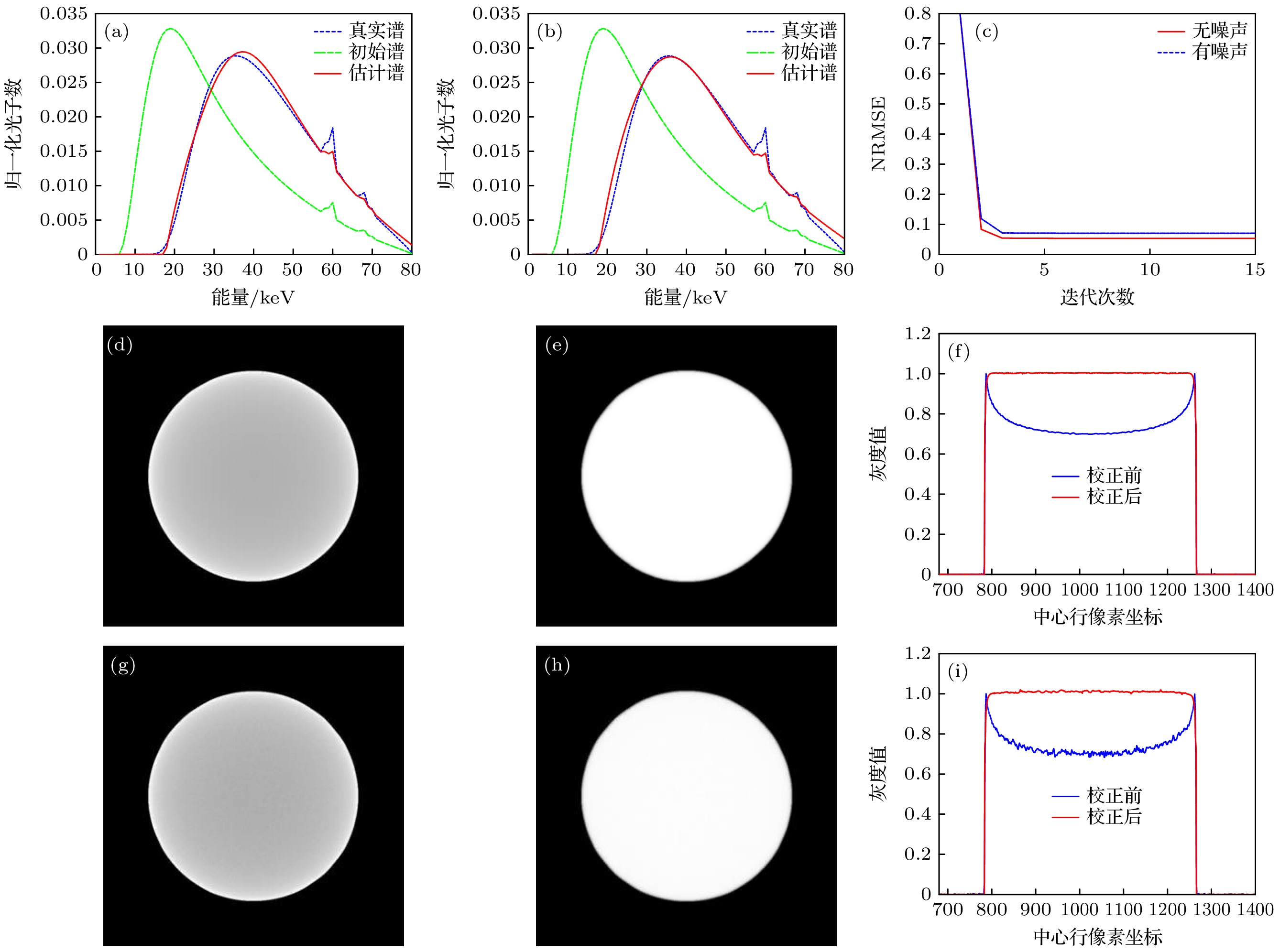
图 1 80 kV下X射线能谱估计及重建结果 (a) 无噪声条件下能谱估计结果; (b) 泊松噪声条件下能谱估计结果; (c) NRMSE随迭代次数的变化曲线; (d) 无噪声条件下ART重建结果; (e) 无噪声条件下本文方法重建图; (f) 图(d)和(e)中心行剖线对比图; (g) 有噪声条件下ART重建结果; (h) 有噪声条件下本文方法重建图; (i) 图(g)和(h)中心行剖线对比图
Figure 1. X-ray energy spectrum estimation and reconstruction results at 80 kV: (a) Results of X-ray spectrum estimation without noise; (b) X-ray spectrum estimation results under Poisson noise condition; (c) curve of NRMSE with the number of iterations; (d) ART reconstruction results in noise-free condition; (e) reconstruction image of the proposed method under noise-free condition; (f) Figs. (d) and (e) comparison of section lines in the center row; (g) ART reconstruction results in noisy conditions; (h) reconstruction image of the proposed method under noisy condition; (i) Figs. (g) and (h) comparison of section lines in the center row.

图 2 散射条件下的实验结果 (a) 散射占比为1%时的能谱估计图; (b) 散射占比为5%时的能谱估计图
Figure 2. Experimental results under scattering conditions: (a) Energy spectrum estimation for 1% scattering; (b) energy spectrum estimation for 5% scattering.
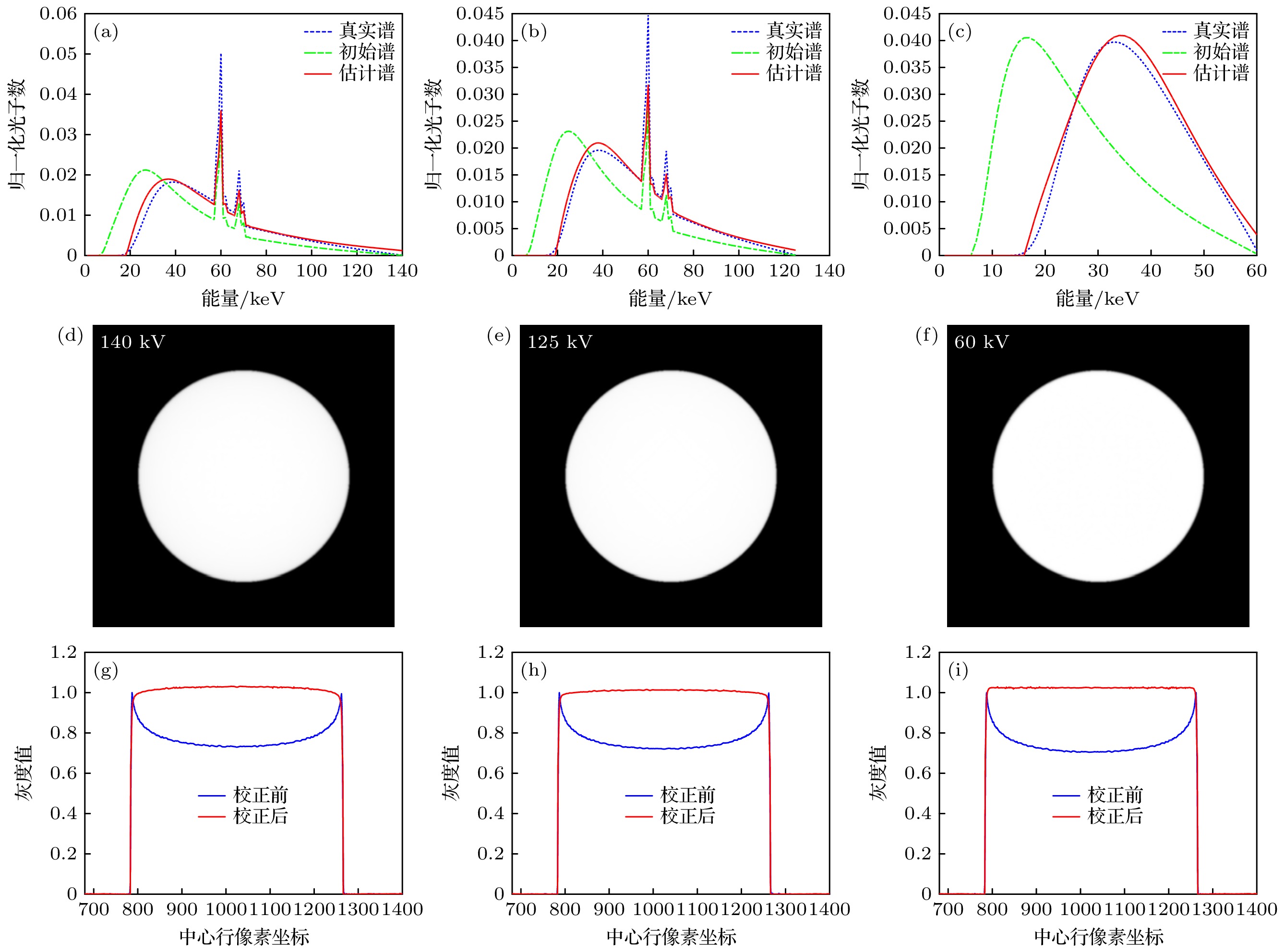
图 3 不同电压下X射线能谱估计及重建结果 (a) 140 kV下能谱估计结果; (b) 125 kV下能谱估计结果; (c) 60 kV下能谱估计结果; (d) 140 kV下本文方法重建图; (e) 125 kV下本文方法重建图; (f) 60 kV下本文方法重建图; (g) 140 kV下本文方法重建图和ART重建图中心行剖线对比图; (h) 125 kV下本文方法重建图和ART重建图中心行剖线对比图; (i) 60 kV下本文方法重建图和ART重建图中心行剖线对比图
Figure 3. X-ray energy spectrum estimation and reconstruction results under different voltages: (a) X-ray spectrum estimation at 140 kV; (b) X-ray spectrum estimation at 125 kV; (c) X-ray spectrum estimation at 60 kV; (d) reconstruction image of the proposed method at 140 kV; (e) reconstruction image of the proposed method at125 kV; (f) reconstruction image of the proposed method at 60 kV; (g) comparison of section lines in the center of the reconstructed image with the proposed method and the reconstructed image with ART at 140 kV; (h) comparison of section lines in the center of the reconstructed image with the proposed method and the reconstructed image with ART at 125 kV; (i) comparison of section lines in the center of the reconstructed image with the proposed method and the reconstructed image with ART at 60 kV.
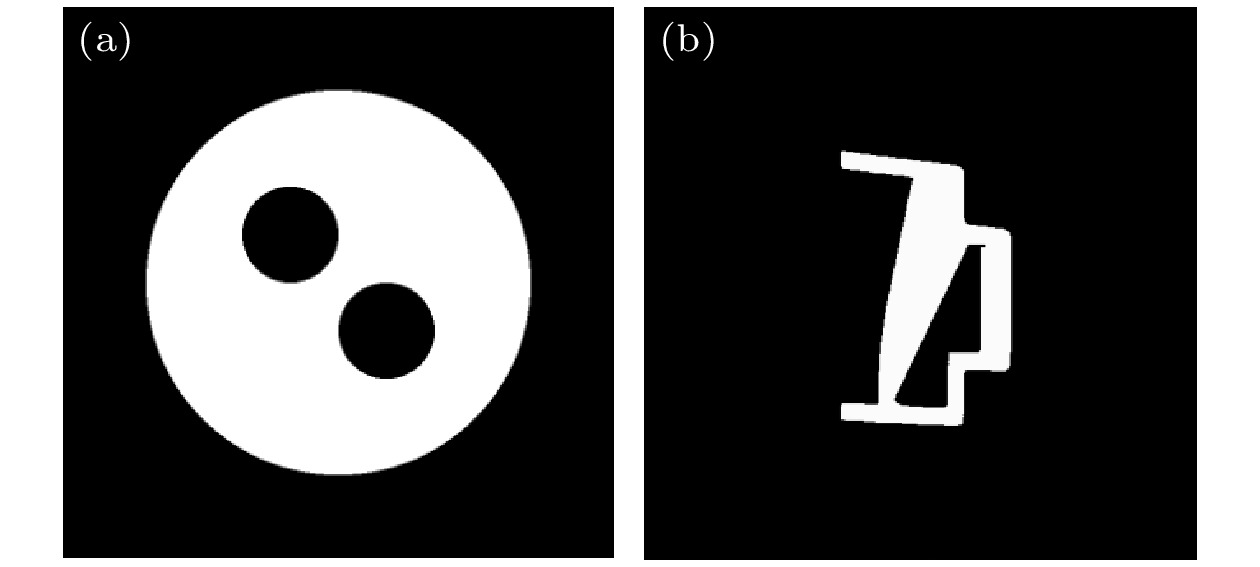
图 4 不规则模体图 (a) 模体a是在实验1的圆铝的基础上内部掏空了两个直径为10 mm的小圆; (b) 模体b是一个不规则的单材质铝材料模体
Figure 4. Irregular phantom diagram: (a) Phantom a was hollowed out with two small circles of 10 mm on the basis of the round aluminum in experiment 1; (b) phantom b is an irregular, single-material aluminum phantom.

图 5 模体a实验结果图 (a) 能谱估计结果; (b) 本文方法重建图; (c) 本文方法重建图和ART重建图中心行剖线对比图
Figure 5. Experimental results of phantom a: (a) Results of X-ray spectrum estimation; (b) reconstruction image of the proposed method; (c) comparison of section lines in the center of the reconstructed image with the proposed method and the reconstructed image with ART.
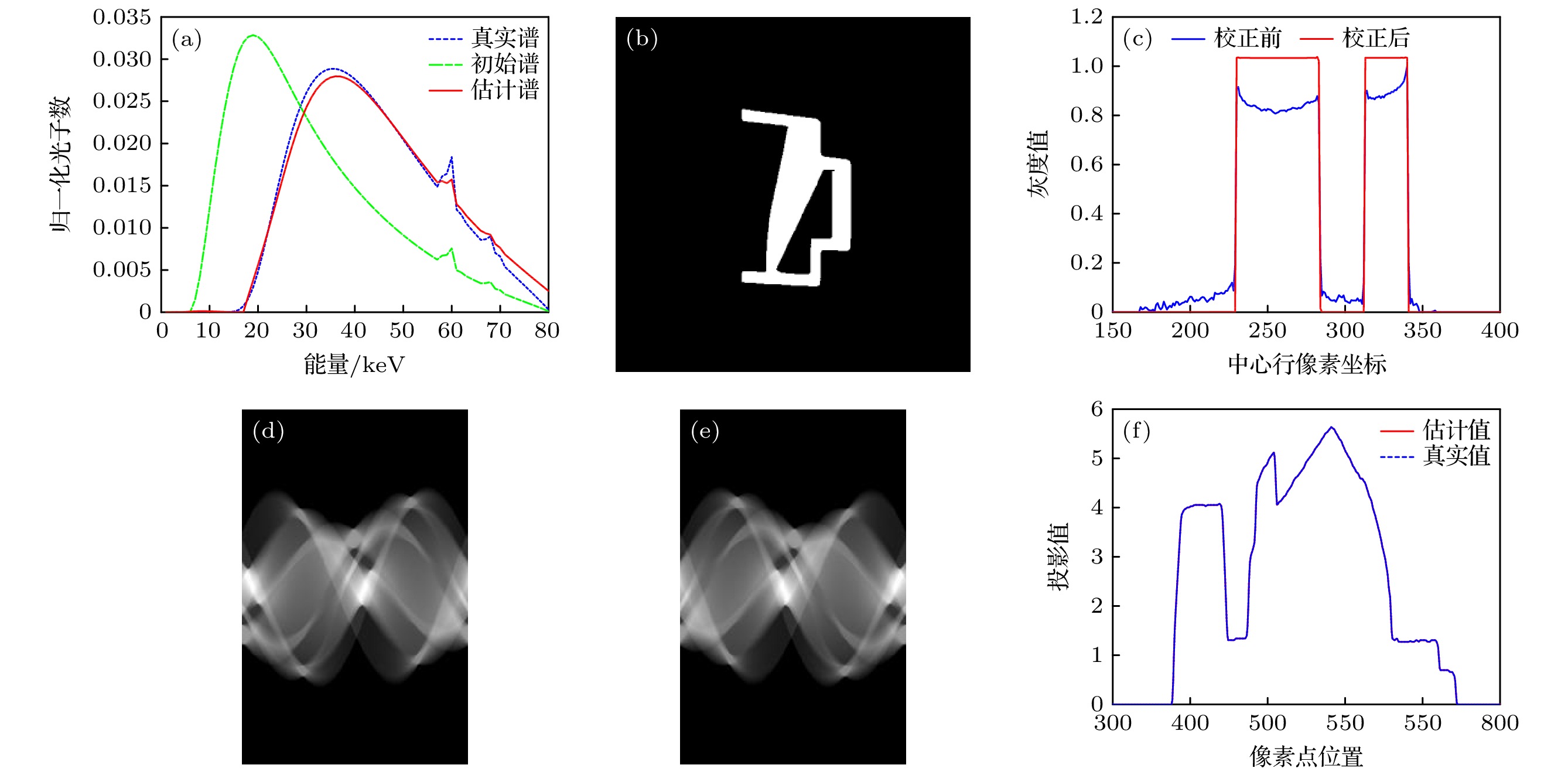
图 6 模体b实验结果图 (a) 能谱估计结果; (b) 本文方法重建图; (c) 本文方法重建图和ART重建图中心行剖线对比图; (d) 真实投影图; (e) 估计投影图; (f) 同一个扫描角度下真实投影数据和估计投影数据对比图
Figure 6. Experimental results of phantom b: (a) Results of X-ray spectrum estimation; (b) reconstruction image of the proposed method ; (c) comparison of section lines in the center of the reconstructed image with the proposed method and the reconstructed image with ART; (d) real projection diagram; (e) estimated projection diagram; (f) comparison of real and estimated projected data at one angle.
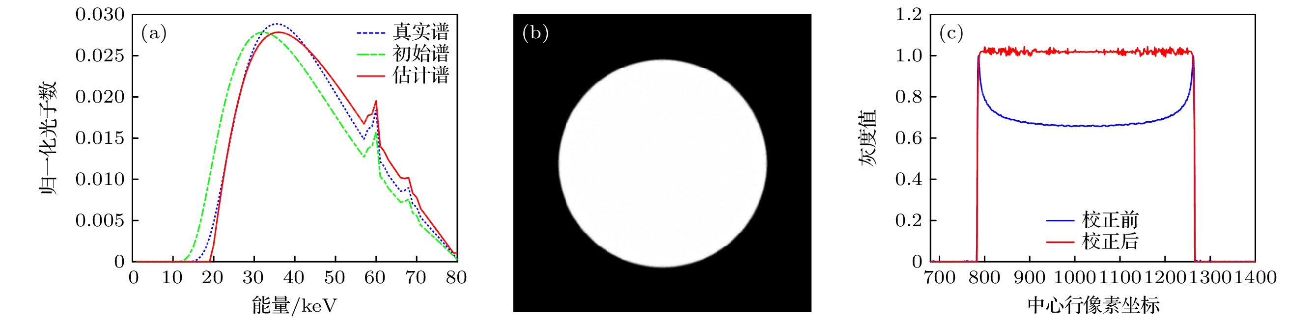
图 7 铜材质谱估计及重建结果图 (a) 能谱估计结果; (b) 本文方法重建图; (c) 本文方法重建图和ART重建图中心行剖线对比图
Figure 7. Spectrum estimation and reconstruction results of copper: (a) Results of X-ray spectrum estimation; (b) reconstruction image of the proposed method; (c) comparison of section lines in the center of the reconstructed image with the proposed method and the reconstructed image with ART.
图 8 实采CT系统示意图 (a) 实采CT扫描系统; (b) 模体图
Figure 8. Schematic diagram of real CT system: (a) Real CT scanning system; (b) phantom.
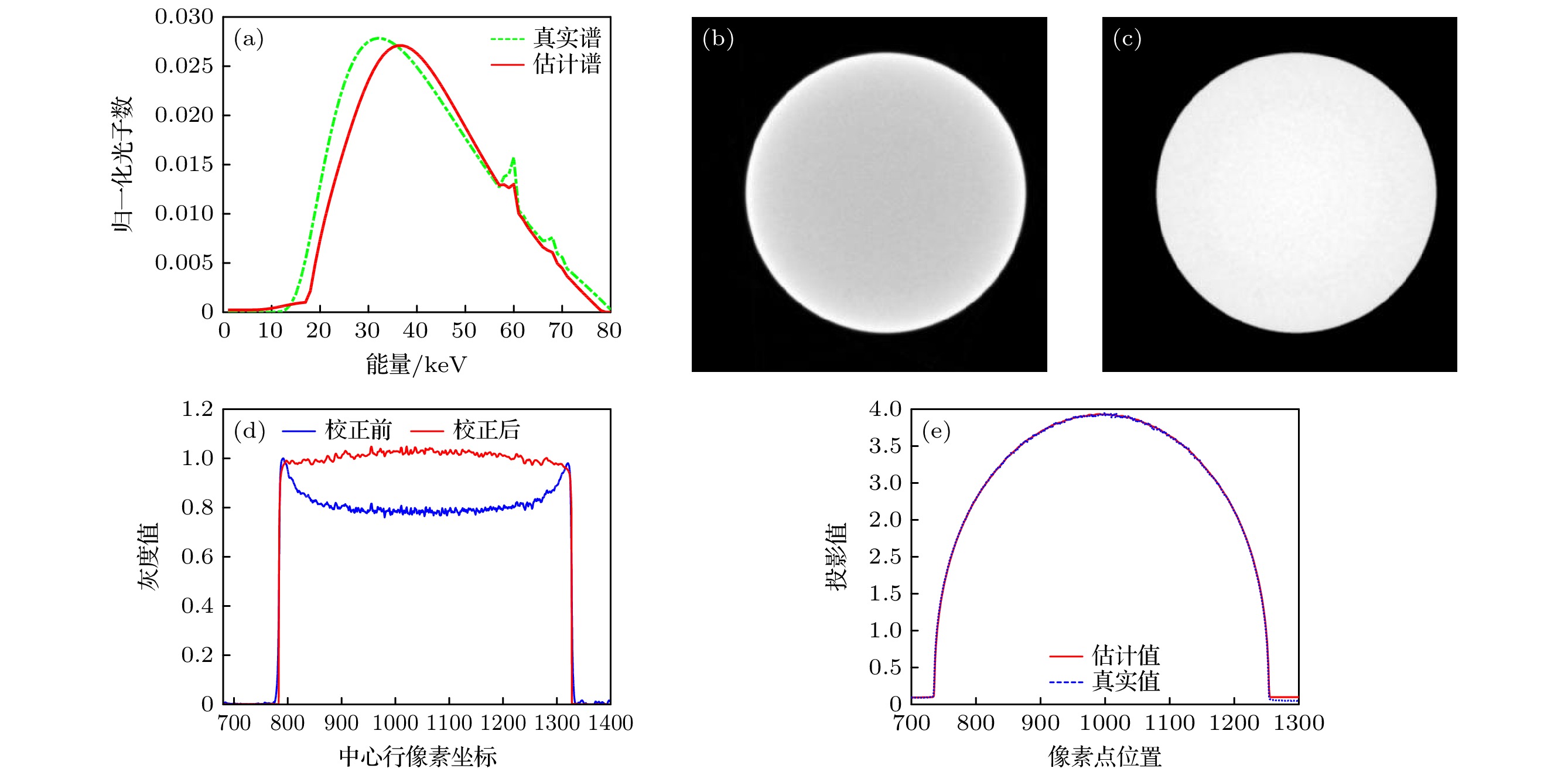
图 9 实际CT系统的实验结果 (a) 能谱估计图; (b) ART重建图; (c) 本文方法重建图; (d) 图(b)和(c)中心行的剖线对比图; (e) 一个角度下真实投影数据和估计投影数据的对比图
Figure 9. Experimental results of a real CT system: (a) X-ray spectrum estimation; (b) ART reconstruction image; (c) reconstruction image of the proposed method; (d) Figs. (b) and (c) comparison of section lines in the center row; (e) comparison of real and estimated projected data at one angle.
表 1 模拟CT扫描系统的实验参数
Table 1. Experimental parameters of simulated CT scanning system.
参数名称 射线源到探测器的距离/mm 1080 射线源到旋转中心的距离/mm 589.3525 扫描角度/(°) $ 360 $ 扫描角度个数 $ 720 $ 探测器单元大小/mm 0.278 探测器个数 $ 1136 $  DownLoad: CSV
表 2 实际CT扫描系统的实验参数
Table 2. Experimental parameters of real CT scanning system.
参数名称 射线源到探测器的距离/mm 634 射线源到旋转中心的距离/mm 239 扫描角度/(°) $ 360 $ 扫描角度个数 $ 720 $ 探测器单元大小/mm 0.2 探测器个数 $2048×2048$
DownLoad: CSV
-
[1] Jarry G, Demarco J J, Beifuss U, Cagnon C H, Mcnitt-Gray M F 2003 Phys. Med. Biol 48 2645
Google Scholar
[2] Zhao Y S, Zhao X, Zhang P 2015 IEEE Trans. Med. Imaging 34 761
Google Scholar
[3] Elbakri, Idris A, Jeffrey A, Fessler 2002 IEEE Trans. Med. Imaging 21 89
Google Scholar
[4] 张慧滔, 张朋 2013 光学学报 33 8
Google Scholar
Zhang H T, Zhang P 2013 Acta Opt. Sin. 33 8
Google Scholar
[5] Francois P, Catala A, Scouarnec C 1993 Med. Phys. 20 1695
Google Scholar
[6] Tominaga, Shoji 1986 Nucl Instrum. Methods Phys. Res. 243 530
Google Scholar
[7] Armbruster B, Hamilton R J, Kuehl A K 2004 Phys. Med. Biol 49 5087
Google Scholar
[8] Leinweber C, Maier J, Kachelrie M 2017 Med. Phys. 44 6183
Google Scholar
[9] Duan X, Wang J, Yu L, Leng S, McCollough C H 2011 Med. Phys. 38 993
Google Scholar
[10] 罗婷, 李孟飞, 赵云松 2018 电子学报 46 8
Google Scholar
Luo T, Li M F, Zhao Y S 2018 Acta Electron. Sin. 46 8
Google Scholar
[11] Sidky E Y, Yu L, Pan X, Zou Y, Vannier M 2005 J. Appl. Phys. 97 623
Google Scholar
[12] Zhang L, Zhang G, Chen Z, Xing Y, Cheng J, Xiao Y 2007 IEEE Nuclear Science Symposium Conference Record Honolulu, HI, October 26—November 3, 2007 p3089
[13] 杨莹, 牟轩沁, 余厚军, 陈希, 张砚博, 汤少杰 2010 电子学报 38 7
Google Scholar
Yang Y, Mou X Q, Yu H J, Chen X, Zhang Y B, Tang S J 2010 Acta Electron. Sin. 38 7
Google Scholar
[14] Perkhounkov B, Stec J, Sidky E Y, Pan X C 2016 Spie Medical Imaging San Diego, America, February 27–March 3, 2016 p1315
[15] Zhao W, Niu K, Schafer S, Royalty K 2014 Phys. Med. Biol 60 339
Google Scholar
[16] Zhao W, Xing L, Zhang Q, Xie Q, Niu T 2017 J. Med. Imaging 4 023506
Google Scholar
[17] Tucker D M, Barnes G T, Chakraborty D P 1991 Med. Phys. 18 211
Google Scholar
[18] Hernandez A M, Boone J M 2014 Med. Phys. 41 042101
Google Scholar
[19] Herman G T, Meyer L B 1993 IEEE Trans. Med. Imaging 12 600
Google Scholar
[20] Goldstein T, Osher S 2009 SIAM J. Imaging Sci. 2 323
Google Scholar
[21] Zhang H M, Wang L Y, Yan B, Li L, Xi X Q, Lu L Z 2013 Chin. Phys. B 22 078701
Google Scholar
[22] 黄力宇, 朱守平, 匡涛 2015 医学断层图像重建仿真实验 (西安: 西安电子科技大学出版社) 第138—145页
Huang L Y, Zhu S P, Kuang T 2015 Simulation Experiment of Medical Image Reconstruction (Xi’an: Xidian University Press) pp138–145 (in Chinese)
[23] Sun M, Star-Lack J M 2010 Phys. Med. Biol. 55 6695
Google Scholar

 DownLoad:
Catalog
Metrics
- Abstract views: 1976
- PDF Downloads: 64
- Cited By: 0
