


-
Microwave thermoacoustic imaging (MTAI) is an exciting imaging technique rooted from the underlying principle of exploiting the distinct electrical properties of biological tissues. By using short-pulsed microwaves as a stimulation source and their interaction with the human body, MTAI has paved the way for revolutionary advancements in medical imaging. When microwaves are absorbed by polar molecules and ions within the tissues, an ingenious thermoelastic effect gives rise to ultrasound waves. These ultrasound waves, brimming with invaluable pathological and physiological insights, propagate outward, carrying the essence of the composition and functionality of biological tissue. Through a meticulous collection of ultrasound signals from all directions surrounding the tissue, it becomes possible to reconstruct intricate internal structures and visualize the tissue's functional dynamics. The MTAI excels in non-invasiveness, capable of delving several centimeters beneath the surface with a microscopic resolution on the order of micrometers. The magic lies in converting microwave energy into ultrasound waves, entering into the hidden depths of tissues without causing harm. This groundbreaking imaging modality unlocks a realm of possibilities for acquiring profound insights into the intricate structures and functionality of deep-seated tissues. Furthermore, the inherent polarization characteristics of microwaves empower MTAI to capture additional dimensions of information, unraveling the intricate polarization properties and illuminating a richer understanding of the tissue's complexity. The great potential of MTAI extends far and wide within the medicine field. It has made remarkable achievements in non-invasive imaging of brain structures, screening breast tumors, visualizing human arthritis, and detecting liver fat content. These accomplishments have laid a solid foundation, firmly establishing MTAI as a trailblazing medical imaging technique. The present study offers a comprehensive and in-depth exploration of the physical principles underpinning MTAI, the sophisticated system devices involved, and the recent groundbreaking research breakthroughs. Moreover, it delves into the exciting prospects and challenges that lie ahead in the future development of MTAI. As the technology continues to progress by leaps and bounds, MTAI is ready to break down barriers, and usher in a new era of unmatched imaging quality and performance. This, in turn, will open the floodgates for transformative innovations and applications in medical diagnosis and treatment. The anticipation is palpable as MTAI strives to make substantial contributions to the ever-developing medical imaging field, bestowing upon humanity more accurate, reliable, and life-enhancing diagnostic capabilities.
-
Keywords:
- microwave-induced thermoacoustic imaging /
- electrical properties of biological tissues /
- microwave sources /
- technique of brain imaging
[1] Ketcham R A, Carlson W D 2001 Comput. Geosci. 27 381
 Google Scholar
[2] Bushong S C, Clarke G 2003 Magnetic Resonance Imaging: Physical and Biological Principles (Amsterdam: Elsevier Health Sciences) pp58–65
[3] Bushberg J T, Boone J M 2011 The Essential Physics of Medical Imaging (Philadelphia: Lippincott Williams & Wilkins) pp171–202
[4] Haribabu V, Girigoswami K, Sharmiladevi P, Girigoswami A 2020 ACS Biomater. Sci. Eng. 6 4377
Google Scholar
[5] Chan V, Perlas A 2011 Atlas of Ultrasound-guided Procedures in Interventional Pain Management (New York: Springer) p13
[6] Nguyen K C T, Le L H, Kaipatur N R, Zheng R, Lou E H, Major P W 2016 Ann. Biomed. Eng. 44 2874
Google Scholar
[7] Wang H, Liu N 2020 J. Med. Imaging Health Inf. 10 918
Google Scholar
[8] Xu M H, Ku G, Jin X, Wang L V, Fornage B D, Hunt K K 2005 The Sixth Conference on Biomedical Thermoacoustics, Optoacoustics, and Acousto-optics 5697 45
[9] Behari J 2019 Radio Frequency and Microwave Effects on Biological Tissues (New York: CRC Press) pp63–82
[10] Chen H, Tang X, Nie G, Wang Z, Hu J, Hu J, Qin H 2023 J. Innovative Opt. Health Sci. 16 2243002
Google Scholar
[11] Lin J C 2005 Advances in Electromagnetic Fields in Living Systems (Boston: Springer) p41
[12] Zhao S X, Wang H H, Li Y J, Nie L M, Zhang S X, Xing D, Qin H 2021 IEEE Trans. Biomed. Eng. 69 725
[13] Rahpeima R, Soltani M, Kashkooli F M 2020 Comput. Methods Programs Biomed. 196 105606
Google Scholar
[14] Liu Q, Liang X, Li T, Chao W, Qi W Z, Jin T, Gong Y, Jiang H B, Xi L 2023 IEEE Trans. Med. Imaging 42 2425
[15] Zhao Y, Shan T, Chi Z H, Jiang H B 2020 J. Xray Sci. Technol. 28 83
[16] Ren M Y, Cheng Z W, Wu L H, et al. 2023 IEEE Trans. Biomed. Eng. 70 175
Google Scholar
[17] Chi Z H, Huang L, Wu D, Long X J, Xu X L, Jiang H B 2022 Med. Phys. 49 84
Google Scholar
[18] Vander Vorst A, Rosen A, Kotsuka Y 2006 RF/microwave Interaction With Biological Tissues (Hoboken: John Wiley & Sons) pp30–38
[19] Schwan H P, Foster K R 1980 Proc. IEEE 68 104
Google Scholar
[20] Foster K R, Schwan H P 2019 CRC Handbook of Biological Effects of Electromagnetic Fields (Boca Raton: CRC press) pp27–76
[21] Fiedler T M, Ladd M E, Bitz A K 2018 Neuroimage 168 33
Google Scholar
[22] Williams J M 2001 arXiv: 0102007 [physics.gen-ph
[23] Bacon C, Guilliorit E, Hosten B, Chimenti D E 2001 J. Acoust. Soc. Am. 110 1398
Google Scholar
[24] Dagro A M, Wilkerson J W, Thomas T P, Kalinosky B T, Payne J A 2021 Sci. Adv. 7 eabd8405
Google Scholar
[25] Zhang X C, Xu J 2010 Introduction to THz Wave Photonics (Vol. 29) (New York: Springer) pp70–82
[26] Drain L 2019 Laser Ultrasonics: Techniques and Applications (New York: Routledge) pp305–322
[27] Paltauf G, Dyer P E 2003 Chem. Rev. 103 487
Google Scholar
[28] Harris C M, Piersol A G 2002 Harris’ Shock and Vibration Handbook (Vol. 5) (New York: McGraw-Hill) p21
[29] Drebushchak V 2020 J. Therm. Anal. Calorim. 142 1097
Google Scholar
[30] Gao F, Zheng Q, Zheng Y J 2014 Med. Phys. 41 053302
Google Scholar
[31] Luo W L, Ji Z, Yang S H, Xing D 2018 Phys. Rev. Appl. 10 1728
[32] Lou C G, Yang S H, Ji Z, Chen Q, Xing D 2012 Phys. Rev. Lett. 109 218101
Google Scholar
[33] Ji Z, Lou C G, Yang S H, Xing D 2012 Med. Phys. 39 6738
Google Scholar
[34] Yan J, Tao C J, Wu S Z 2005 IEEE Engineering in Medicine and Biology 27th Annual Conference Shanghai, China, 17–18 January 2006 p1521
[35] Lou C G, Nie L M, Xu D 2011 J. Appl. Phys. 110 083101
Google Scholar
[36] Ji Z, Ding W Z, Ye F H, Lou C G, Xing D 2015 Appl. Phys. Lett. 107 094104
Google Scholar
[37] Ji Z, Lou C G, Shi Y, Ding W Z, Yang S H, Xing D 2015 Appl. Phys. Lett. 107 839
[38] Wang X, Bauer D R, Vollin J L, Manzi D G, Witte R S, Xin H 2012 IEEE Antennas Wirel. Propag. Lett. 11 1634
Google Scholar
[39] Sharif-Khodaei Z, Aliabadi M 2014 Smart Mater. Struct. 23 075007
Google Scholar
[40] Berger C R, Demissie B, Heckenbach J, Willett P, Zhou S 2010 IEEE J. Sel. Top. Sign. Proces. 4 226
Google Scholar
[41] Li J, Wu R B 1998 IEEE Trans. Sign. Proces. 46 2231
Google Scholar
[42] Xu Q W, Zheng Z, Jiang H B 2021 Chin. Phys. B 30 024302
Google Scholar
[43] Zhang J L, Li C Z, Jiang W C, Wang Z C, Zhang L J, Wang X 2022 IEEE Trans. Antennas Propag. 70 6336
Google Scholar
[44] Li C Z, Xi Z J, Jin G F, Jiang W C, Wang B S, Cai X R, Wang X 2023 IEEE Trans. Biomed. Eng. 70 2350
Google Scholar
[45] Wang B S, Sun Y F, Wang Z C, Wang X 2020 IEEE Trans. Microwave Theory Tech. 68 377
Google Scholar
[46] Yu L, Antoni J, Wu H, Leclere Q, Jiang W 2019 Mech. Syst. Sig. Process. 134 106309
Google Scholar
[47] Song J, Shen T, Wang Q W 2022 IEEE J. Electromagn. RF Microwaves Med. Biol. 7 59
[48] Luo Z X, Li C Z, Liu D T, Wang B S, Zhang L J, Ma Y X, Xu K W, Wang X 2023 IEEE Trans. Microwave Theory Tech. 71 2652
Google Scholar
[49] Huang L, Rong J, Yao L, Qi W Z, Wu D, Xu J Y, Jiang H B 2013 Chin. Phys. Lett. 30 124301
Google Scholar
[50] Nie L M, Xing D, Zhou Q, Yang D W, Guo H 2008 Med. Phys. 35 4026
Google Scholar
[51] Huang L, Zheng Z, Chi Z H, Jiang H B 2021 Med. Phys. 48 4242
Google Scholar
[52] Ku G, Wang L V 2001 Med. Phys. 28 4
Google Scholar
[53] Liang X, Guo H, Liu Q, Wu C F, Gong Y B, Xi L 2020 Appl. Phys. Lett. 116 013702
Google Scholar
[54] Fu Y, Ji Z, Ding W Z, Ye F H, Lou C G 2014 Med. Phys. 41 110701
Google Scholar
[55] Ding W Z, Ji Z, Ye F H, Lou C G, Xing D 2015 IEEE Trans. Microwave Theory Tech. 63 3272
Google Scholar
[56] Volmer C, Weber J, Stephan R, Blau K, Hein M A 2008 IEEE Trans. Antennas Propag. 56 360
Google Scholar
[57] Nan H, Arbabian A 2017 IEEE Trans. Microwave Theory Tech. 65 2607
Google Scholar
[58] Ku G, Wang L V 2000 Med. Phys. 27 1195
Google Scholar
[59] Xu M H, Xu Y, Wang L V 2003 IEEE Trans. Biomed. Eng. 50 1086
Google Scholar
[60] Xu M H, Wang L V 2002 IEEE Trans. Med. Imaging 21 814
Google Scholar
[61] Zhao Z Q, Song J, Zhu X Z, Wang J G, Wu J N, Liu Y L, Nie Z P, Liu Q H 2012 Electromagn. Waves 134 323
[62] Cannata J M, Ritter T A, Chen W H, Silverman R H, Shung K K 2003 IEEE Trans. Ultrason. Ferroelectr. Freq. Control 50 1548
Google Scholar
[63] Sun X L, Yang X C, Zhu X Y, Liu H H 2017 IEEE Sens. J. 18 1373
[64] Li Z X, Chen D D, Fei C L, Li D, Feng W, Yang Y T 2021 IEEE Trans. Ultrason. Ferroelectr. Freq. Control 68 2202
Google Scholar
[65] Candès E J, Wakin M B 2008 IEEE Signal Process Mag. 25 21
Google Scholar
[66] Ye F H, Ji Z, Ding W Z, Lou C G, Yang S H, Xing D 2016 IEEE Trans. Med. Imaging 35 839
Google Scholar
[67] Chia S K, Speers C H, D’yachkova Y, Kang A, Malfair‐Taylor S, Barnett J, Coldman A, Gelmon K A, O’reilly S E, Olivotto I A 2007 Cancer 110 973
Google Scholar
[68] Youlden D R, Cramb S M, Dunn N A, Muller J M, Pyke C M, Baade P D 2012 Cancer Epidemiol. 36 237
Google Scholar
[69] Verkman A, Hara-Chikuma M, Papadopoulos M C 2008 J. Mol. Med. 86 523
Google Scholar
[70] Yu C H, Tang W, Wang Y H, Shen Q, Wang B, Cai C Q, Meng X J, Zou F 2016 Cancer Lett. 376 268
Google Scholar
[71] Baritaki S, Apostolakis S, Kanellou P, Dimanche‐Boitrel M T, Spandidos D A, Bonavida B 2007 Adv. Cancer Res. 98 149
[72] Li X, Davis S K, Hagness S C, Van der Weide D W, Van Veen B D 2004 IEEE Trans. Microwave Theory Tech. 52 1856
Google Scholar
[73] Celik A R, Kurt M B, Helhel S 2019 ACES 34 1549
[74] Kruger R A, Miller K D, Reynolds H E, Kiser Jr W L, Reinecke D R, Kruger G A 2000 Radiology 216 279
Google Scholar
[75] Wu L H, Cheng Z W, Ma Y Z, Li Y J, Ren M Y, Xing D, Qin H 2022 IEEE Trans. Med. Imaging 41 1080
Google Scholar
[76] Huang Y, Omar M, Tian W, Lopez-Schier H, Westmeyer G G, Chmyrov A, Sergiadis G, Ntziachristos V 2021 Sci. Adv. 7 eabd1505
Google Scholar
[77] Joines W T, Jirtle R L, Rafal M D, Schaefer D J 1980 Inte. J. Radiat. Oncol. Biol. Phys. 6 681
Google Scholar
[78] Zheng Z, Jiang Y C, Huang L, Zhao Y, Jiang H B 2020 J. X-Ray Sci. Technol. 28 137
[79] Zhao Y, Chi Z H, Huang L, Zheng Z, Yang J G, Jiang H B 2017 J. Innovative Opt. Health Sci. 10 1740001
Google Scholar
[80] Cunningham L S, Kelsey J L 1984 Am. J. Public Health 74 574
Google Scholar
[81] Tański W, Dudek K, Tomasiewicz A, Świątoniowska-Lonc N 2022 Int. J. Environ. Res. Public Health 19 3088
Google Scholar
[82] Gadeval A, Chaudhari S, Bollampally S P, et al. 2021 Drug Discovery Today 26 2315
Google Scholar
[83] Thornton G, Shrive N, Frank C 2001 J. Orthop. Res. 19 845
Google Scholar
[84] Buckwalter J A, Mow V C, Ratcliffe A 1994 JAAOS-J. Am. Acad. Orthopaedic Surgeons 2 192
Google Scholar
[85] Sultan K S, Mohammed B, Manoufali M, Abbosh A M 2021 IEEE Trans. Antennas Propag. 69 6824
Google Scholar
[86] Chi Z H, Zhao Y, Huang L, Zheng Z, Jiang H B 2016 Med. Phys. 43 6226
Google Scholar
[87] Chi Z H, Zhao Y, Yang J G, Li T T, Zhang G, Jiang H B 2019 IEEE Trans. Biomed. Eng. 66 1598
Google Scholar
[88] Gandhi M S, Kamalov G, Shahbaz A U, Bhattacharya S K, Ahokas R A, Sun Y, Gerling I C, Weber K T 2011 Heart Fail. Rev. 16 23
Google Scholar
[89] Weber K T, Sun Y, Bhattacharya S K, Ahokas R A, Gerling I C 2013 Nat. Rev. Cardiol. 10 15
Google Scholar
[90] Friedberg C K, Horn H 1939 J. Am. Med. Assoc. 112 1675
Google Scholar
[91] Smit M, Coetzee A, Lochner A 2020 J. Cardiothorac. Vasc. Anesth. 34 2501
Google Scholar
[92] Alpert J S 1989 Cardiology 76 85
Google Scholar
[93] Li Y J, Zhang S X, Wu L H, et al. 2022 Photonics Res. 10 1297
Google Scholar
-
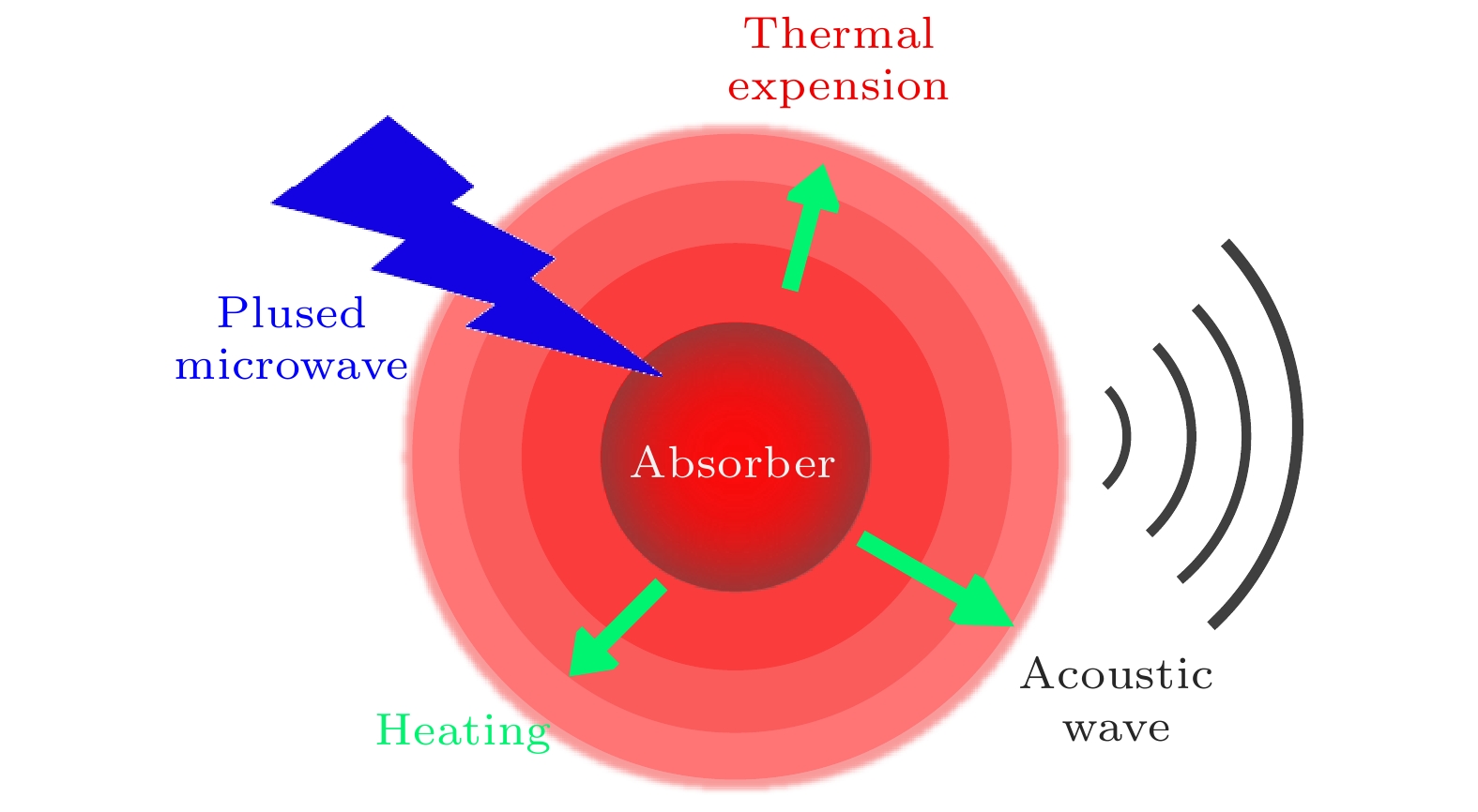
图 1 热声效应示意图
Figure 1. Schematic diagram of TA effect.
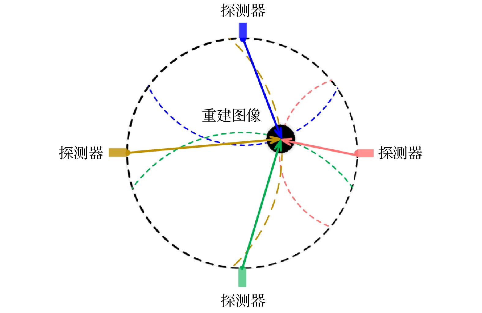
图 2 微波热声信号延时叠加算法示意图
Figure 2. Schematic diagram of the thermoacoustic signal delay superposition algorithm.
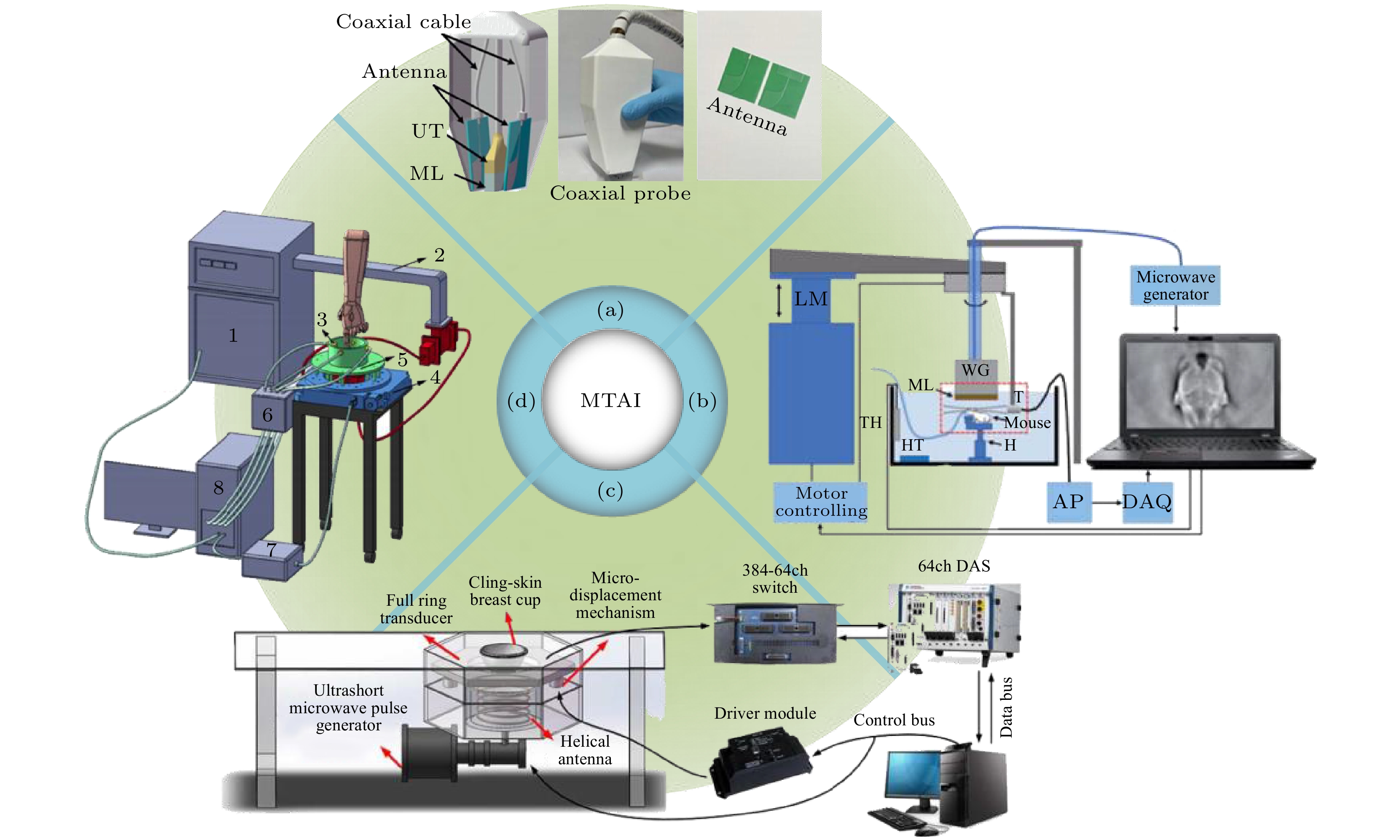
图 3 微波热声成像技术典型实现方案 (a)微波乳腺热声成像一体化探头装置图[16]; (b)微波热声脑成像装置图[15]; (c)微波乳腺热声成像装置[66]; (d)微波热声关节成像装置图[17]
Figure 3. Typical implementation scheme of microwave thermoacoustic imaging technology: (a) Microwave-induced breast thermoacoustic imaging integrated probe device[16]; (b) diagram of a Microwave-induced brain thermoacoustic imaging device[15]; (c) microwave-induced breast thermoacoustic imaging system[66]; (d) microwave-induced thermoacoustic joint imaging device[17].
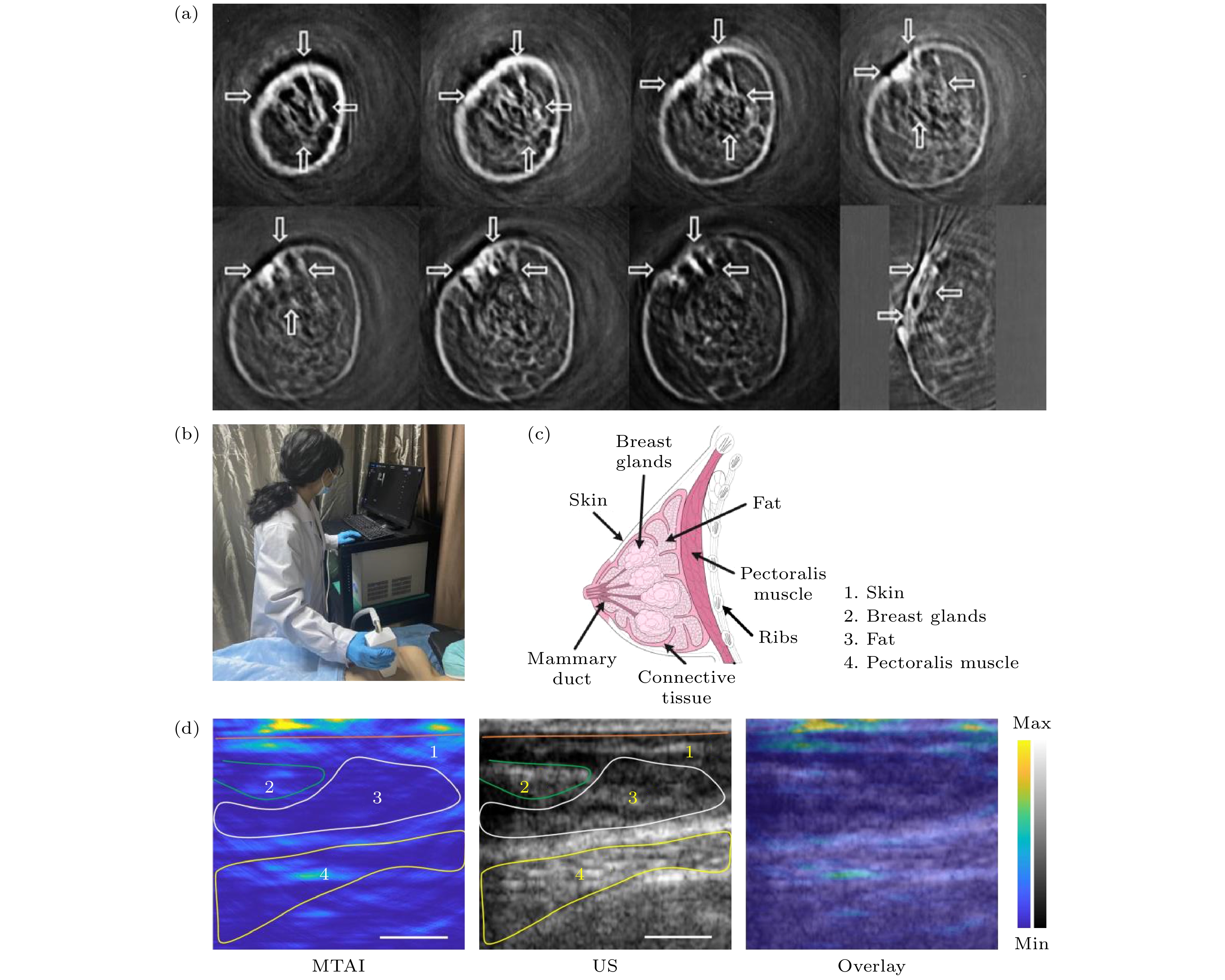
图 4 微波热声成像乳腺成像 (a) Kruger等[74]的乳腺成像图; (b) 微波热声乳腺成像实操图[16]; (c) 乳房的解剖结构示意图[16]; (d) 覃欢团队[16]乳腺成像图
Figure 4. MTAI breast imaging: (a) Kruger et al.[74] breast imaging; (b) microwave thermoacoustic breast imaging actual operation diagram[16]; (c) anatomical diagram of the breast[16]; (d) breast imaging of Professor Qin Huan’s team[16].
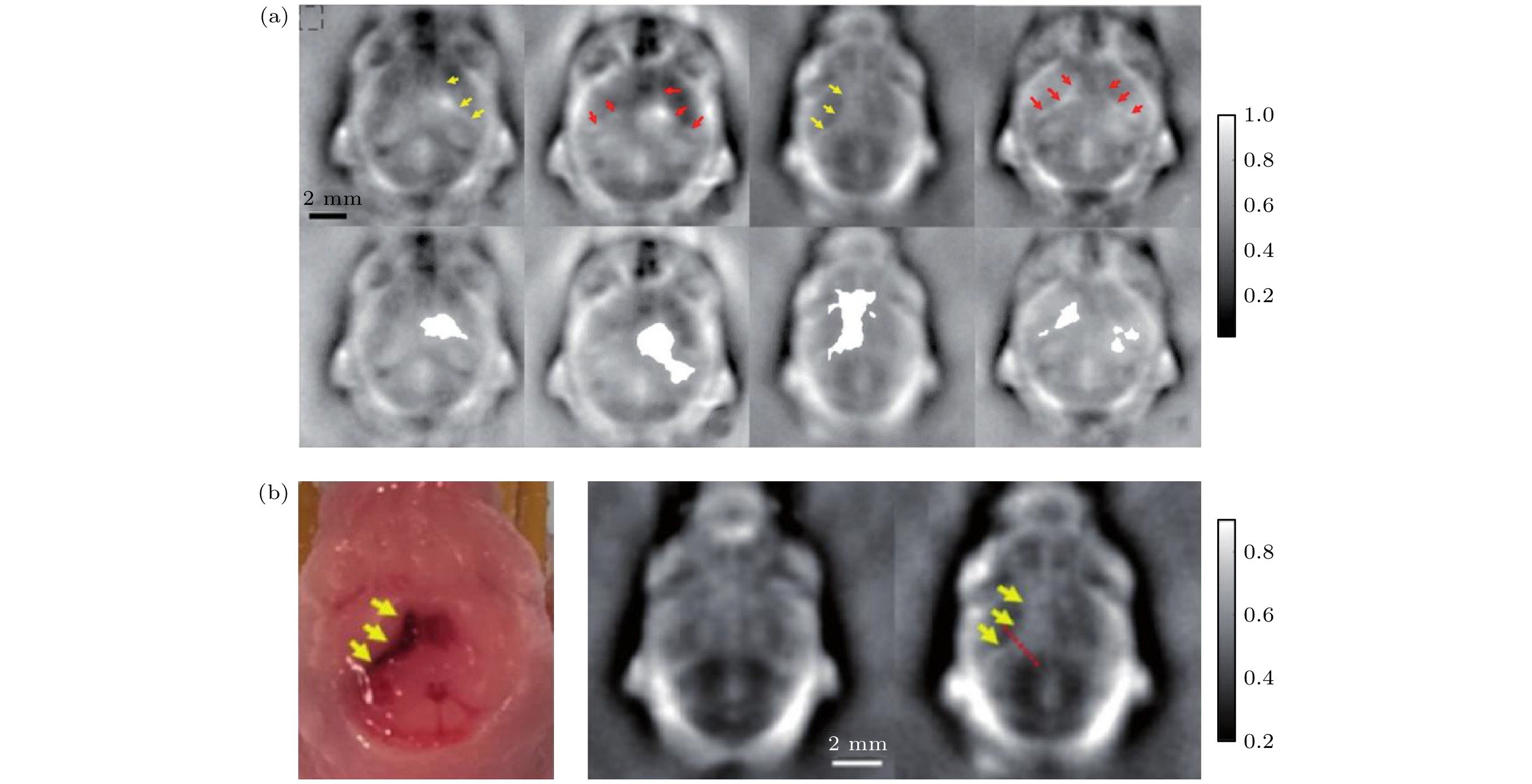
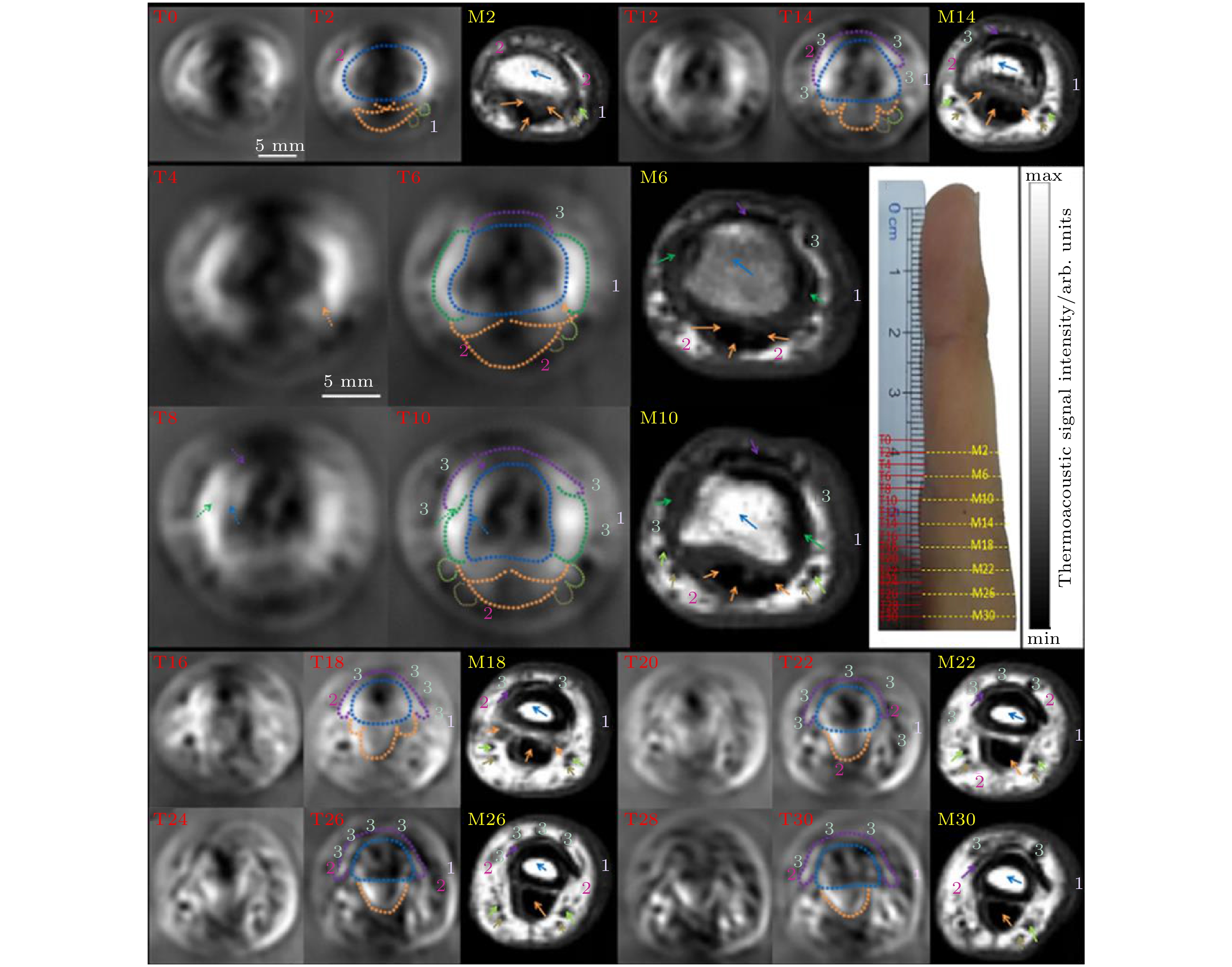
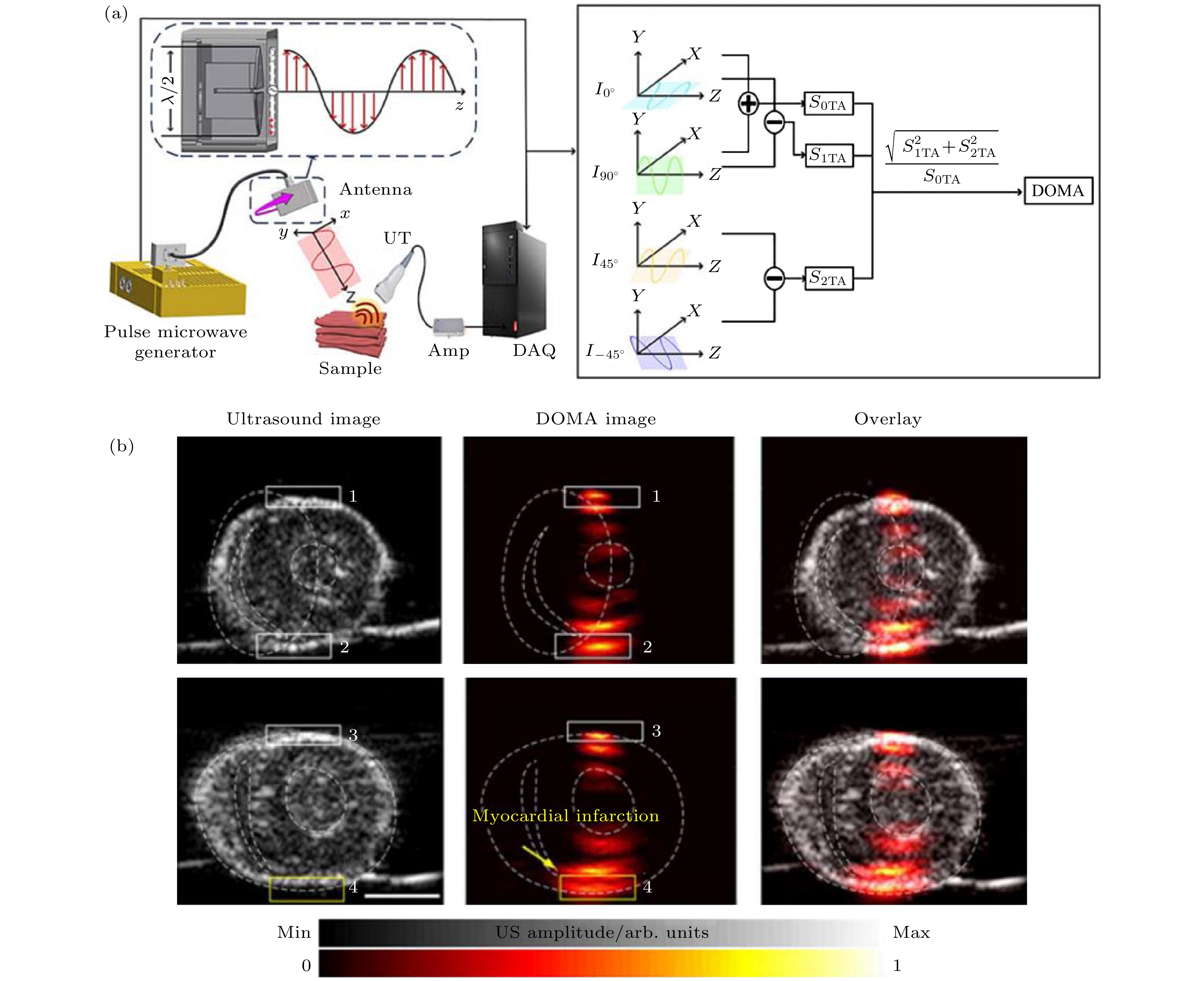
表 1 微波源各参数对成像影响
Table 1. Influence of various parameters of microwave source on imaging.
成像影响 微波源参数 分辨率 脉冲宽度、
脉冲波形信噪比 脉冲重复频率、
脉冲能量强度对比度 中心频率、
脉冲能量强度成像深度 中心频率、
脉冲能量强度 DownLoad: CSV
物质 电导率/(S·m–1) 脑灰质 2.2588 脑白质 1.5393 血液 3.0991 脑脊液 4.0592 小脑 2.5189 血管壁 1.8444 硬膜 2.0485 脊髓 1.3530
DownLoad: CSV
-
[1] Ketcham R A, Carlson W D 2001 Comput. Geosci. 27 381
Google Scholar
[2] Bushong S C, Clarke G 2003 Magnetic Resonance Imaging: Physical and Biological Principles (Amsterdam: Elsevier Health Sciences) pp58–65
[3] Bushberg J T, Boone J M 2011 The Essential Physics of Medical Imaging (Philadelphia: Lippincott Williams & Wilkins) pp171–202
[4] Haribabu V, Girigoswami K, Sharmiladevi P, Girigoswami A 2020 ACS Biomater. Sci. Eng. 6 4377
Google Scholar
[5] Chan V, Perlas A 2011 Atlas of Ultrasound-guided Procedures in Interventional Pain Management (New York: Springer) p13
[6] Nguyen K C T, Le L H, Kaipatur N R, Zheng R, Lou E H, Major P W 2016 Ann. Biomed. Eng. 44 2874
Google Scholar
[7] Wang H, Liu N 2020 J. Med. Imaging Health Inf. 10 918
Google Scholar
[8] Xu M H, Ku G, Jin X, Wang L V, Fornage B D, Hunt K K 2005 The Sixth Conference on Biomedical Thermoacoustics, Optoacoustics, and Acousto-optics 5697 45
[9] Behari J 2019 Radio Frequency and Microwave Effects on Biological Tissues (New York: CRC Press) pp63–82
[10] Chen H, Tang X, Nie G, Wang Z, Hu J, Hu J, Qin H 2023 J. Innovative Opt. Health Sci. 16 2243002
Google Scholar
[11] Lin J C 2005 Advances in Electromagnetic Fields in Living Systems (Boston: Springer) p41
[12] Zhao S X, Wang H H, Li Y J, Nie L M, Zhang S X, Xing D, Qin H 2021 IEEE Trans. Biomed. Eng. 69 725
[13] Rahpeima R, Soltani M, Kashkooli F M 2020 Comput. Methods Programs Biomed. 196 105606
Google Scholar
[14] Liu Q, Liang X, Li T, Chao W, Qi W Z, Jin T, Gong Y, Jiang H B, Xi L 2023 IEEE Trans. Med. Imaging 42 2425
[15] Zhao Y, Shan T, Chi Z H, Jiang H B 2020 J. Xray Sci. Technol. 28 83
[16] Ren M Y, Cheng Z W, Wu L H, et al. 2023 IEEE Trans. Biomed. Eng. 70 175
Google Scholar
[17] Chi Z H, Huang L, Wu D, Long X J, Xu X L, Jiang H B 2022 Med. Phys. 49 84
Google Scholar
[18] Vander Vorst A, Rosen A, Kotsuka Y 2006 RF/microwave Interaction With Biological Tissues (Hoboken: John Wiley & Sons) pp30–38
[19] Schwan H P, Foster K R 1980 Proc. IEEE 68 104
Google Scholar
[20] Foster K R, Schwan H P 2019 CRC Handbook of Biological Effects of Electromagnetic Fields (Boca Raton: CRC press) pp27–76
[21] Fiedler T M, Ladd M E, Bitz A K 2018 Neuroimage 168 33
Google Scholar
[22] Williams J M 2001 arXiv: 0102007 [physics.gen-ph
[23] Bacon C, Guilliorit E, Hosten B, Chimenti D E 2001 J. Acoust. Soc. Am. 110 1398
Google Scholar
[24] Dagro A M, Wilkerson J W, Thomas T P, Kalinosky B T, Payne J A 2021 Sci. Adv. 7 eabd8405
Google Scholar
[25] Zhang X C, Xu J 2010 Introduction to THz Wave Photonics (Vol. 29) (New York: Springer) pp70–82
[26] Drain L 2019 Laser Ultrasonics: Techniques and Applications (New York: Routledge) pp305–322
[27] Paltauf G, Dyer P E 2003 Chem. Rev. 103 487
Google Scholar
[28] Harris C M, Piersol A G 2002 Harris’ Shock and Vibration Handbook (Vol. 5) (New York: McGraw-Hill) p21
[29] Drebushchak V 2020 J. Therm. Anal. Calorim. 142 1097
Google Scholar
[30] Gao F, Zheng Q, Zheng Y J 2014 Med. Phys. 41 053302
Google Scholar
[31] Luo W L, Ji Z, Yang S H, Xing D 2018 Phys. Rev. Appl. 10 1728
[32] Lou C G, Yang S H, Ji Z, Chen Q, Xing D 2012 Phys. Rev. Lett. 109 218101
Google Scholar
[33] Ji Z, Lou C G, Yang S H, Xing D 2012 Med. Phys. 39 6738
Google Scholar
[34] Yan J, Tao C J, Wu S Z 2005 IEEE Engineering in Medicine and Biology 27th Annual Conference Shanghai, China, 17–18 January 2006 p1521
[35] Lou C G, Nie L M, Xu D 2011 J. Appl. Phys. 110 083101
Google Scholar
[36] Ji Z, Ding W Z, Ye F H, Lou C G, Xing D 2015 Appl. Phys. Lett. 107 094104
Google Scholar
[37] Ji Z, Lou C G, Shi Y, Ding W Z, Yang S H, Xing D 2015 Appl. Phys. Lett. 107 839
[38] Wang X, Bauer D R, Vollin J L, Manzi D G, Witte R S, Xin H 2012 IEEE Antennas Wirel. Propag. Lett. 11 1634
Google Scholar
[39] Sharif-Khodaei Z, Aliabadi M 2014 Smart Mater. Struct. 23 075007
Google Scholar
[40] Berger C R, Demissie B, Heckenbach J, Willett P, Zhou S 2010 IEEE J. Sel. Top. Sign. Proces. 4 226
Google Scholar
[41] Li J, Wu R B 1998 IEEE Trans. Sign. Proces. 46 2231
Google Scholar
[42] Xu Q W, Zheng Z, Jiang H B 2021 Chin. Phys. B 30 024302
Google Scholar
[43] Zhang J L, Li C Z, Jiang W C, Wang Z C, Zhang L J, Wang X 2022 IEEE Trans. Antennas Propag. 70 6336
Google Scholar
[44] Li C Z, Xi Z J, Jin G F, Jiang W C, Wang B S, Cai X R, Wang X 2023 IEEE Trans. Biomed. Eng. 70 2350
Google Scholar
[45] Wang B S, Sun Y F, Wang Z C, Wang X 2020 IEEE Trans. Microwave Theory Tech. 68 377
Google Scholar
[46] Yu L, Antoni J, Wu H, Leclere Q, Jiang W 2019 Mech. Syst. Sig. Process. 134 106309
Google Scholar
[47] Song J, Shen T, Wang Q W 2022 IEEE J. Electromagn. RF Microwaves Med. Biol. 7 59
[48] Luo Z X, Li C Z, Liu D T, Wang B S, Zhang L J, Ma Y X, Xu K W, Wang X 2023 IEEE Trans. Microwave Theory Tech. 71 2652
Google Scholar
[49] Huang L, Rong J, Yao L, Qi W Z, Wu D, Xu J Y, Jiang H B 2013 Chin. Phys. Lett. 30 124301
Google Scholar
[50] Nie L M, Xing D, Zhou Q, Yang D W, Guo H 2008 Med. Phys. 35 4026
Google Scholar
[51] Huang L, Zheng Z, Chi Z H, Jiang H B 2021 Med. Phys. 48 4242
Google Scholar
[52] Ku G, Wang L V 2001 Med. Phys. 28 4
Google Scholar
[53] Liang X, Guo H, Liu Q, Wu C F, Gong Y B, Xi L 2020 Appl. Phys. Lett. 116 013702
Google Scholar
[54] Fu Y, Ji Z, Ding W Z, Ye F H, Lou C G 2014 Med. Phys. 41 110701
Google Scholar
[55] Ding W Z, Ji Z, Ye F H, Lou C G, Xing D 2015 IEEE Trans. Microwave Theory Tech. 63 3272
Google Scholar
[56] Volmer C, Weber J, Stephan R, Blau K, Hein M A 2008 IEEE Trans. Antennas Propag. 56 360
Google Scholar
[57] Nan H, Arbabian A 2017 IEEE Trans. Microwave Theory Tech. 65 2607
Google Scholar
[58] Ku G, Wang L V 2000 Med. Phys. 27 1195
Google Scholar
[59] Xu M H, Xu Y, Wang L V 2003 IEEE Trans. Biomed. Eng. 50 1086
Google Scholar
[60] Xu M H, Wang L V 2002 IEEE Trans. Med. Imaging 21 814
Google Scholar
[61] Zhao Z Q, Song J, Zhu X Z, Wang J G, Wu J N, Liu Y L, Nie Z P, Liu Q H 2012 Electromagn. Waves 134 323
[62] Cannata J M, Ritter T A, Chen W H, Silverman R H, Shung K K 2003 IEEE Trans. Ultrason. Ferroelectr. Freq. Control 50 1548
Google Scholar
[63] Sun X L, Yang X C, Zhu X Y, Liu H H 2017 IEEE Sens. J. 18 1373
[64] Li Z X, Chen D D, Fei C L, Li D, Feng W, Yang Y T 2021 IEEE Trans. Ultrason. Ferroelectr. Freq. Control 68 2202
Google Scholar
[65] Candès E J, Wakin M B 2008 IEEE Signal Process Mag. 25 21
Google Scholar
[66] Ye F H, Ji Z, Ding W Z, Lou C G, Yang S H, Xing D 2016 IEEE Trans. Med. Imaging 35 839
Google Scholar
[67] Chia S K, Speers C H, D’yachkova Y, Kang A, Malfair‐Taylor S, Barnett J, Coldman A, Gelmon K A, O’reilly S E, Olivotto I A 2007 Cancer 110 973
Google Scholar
[68] Youlden D R, Cramb S M, Dunn N A, Muller J M, Pyke C M, Baade P D 2012 Cancer Epidemiol. 36 237
Google Scholar
[69] Verkman A, Hara-Chikuma M, Papadopoulos M C 2008 J. Mol. Med. 86 523
Google Scholar
[70] Yu C H, Tang W, Wang Y H, Shen Q, Wang B, Cai C Q, Meng X J, Zou F 2016 Cancer Lett. 376 268
Google Scholar
[71] Baritaki S, Apostolakis S, Kanellou P, Dimanche‐Boitrel M T, Spandidos D A, Bonavida B 2007 Adv. Cancer Res. 98 149
[72] Li X, Davis S K, Hagness S C, Van der Weide D W, Van Veen B D 2004 IEEE Trans. Microwave Theory Tech. 52 1856
Google Scholar
[73] Celik A R, Kurt M B, Helhel S 2019 ACES 34 1549
[74] Kruger R A, Miller K D, Reynolds H E, Kiser Jr W L, Reinecke D R, Kruger G A 2000 Radiology 216 279
Google Scholar
[75] Wu L H, Cheng Z W, Ma Y Z, Li Y J, Ren M Y, Xing D, Qin H 2022 IEEE Trans. Med. Imaging 41 1080
Google Scholar
[76] Huang Y, Omar M, Tian W, Lopez-Schier H, Westmeyer G G, Chmyrov A, Sergiadis G, Ntziachristos V 2021 Sci. Adv. 7 eabd1505
Google Scholar
[77] Joines W T, Jirtle R L, Rafal M D, Schaefer D J 1980 Inte. J. Radiat. Oncol. Biol. Phys. 6 681
Google Scholar
[78] Zheng Z, Jiang Y C, Huang L, Zhao Y, Jiang H B 2020 J. X-Ray Sci. Technol. 28 137
[79] Zhao Y, Chi Z H, Huang L, Zheng Z, Yang J G, Jiang H B 2017 J. Innovative Opt. Health Sci. 10 1740001
Google Scholar
[80] Cunningham L S, Kelsey J L 1984 Am. J. Public Health 74 574
Google Scholar
[81] Tański W, Dudek K, Tomasiewicz A, Świątoniowska-Lonc N 2022 Int. J. Environ. Res. Public Health 19 3088
Google Scholar
[82] Gadeval A, Chaudhari S, Bollampally S P, et al. 2021 Drug Discovery Today 26 2315
Google Scholar
[83] Thornton G, Shrive N, Frank C 2001 J. Orthop. Res. 19 845
Google Scholar
[84] Buckwalter J A, Mow V C, Ratcliffe A 1994 JAAOS-J. Am. Acad. Orthopaedic Surgeons 2 192
Google Scholar
[85] Sultan K S, Mohammed B, Manoufali M, Abbosh A M 2021 IEEE Trans. Antennas Propag. 69 6824
Google Scholar
[86] Chi Z H, Zhao Y, Huang L, Zheng Z, Jiang H B 2016 Med. Phys. 43 6226
Google Scholar
[87] Chi Z H, Zhao Y, Yang J G, Li T T, Zhang G, Jiang H B 2019 IEEE Trans. Biomed. Eng. 66 1598
Google Scholar
[88] Gandhi M S, Kamalov G, Shahbaz A U, Bhattacharya S K, Ahokas R A, Sun Y, Gerling I C, Weber K T 2011 Heart Fail. Rev. 16 23
Google Scholar
[89] Weber K T, Sun Y, Bhattacharya S K, Ahokas R A, Gerling I C 2013 Nat. Rev. Cardiol. 10 15
Google Scholar
[90] Friedberg C K, Horn H 1939 J. Am. Med. Assoc. 112 1675
Google Scholar
[91] Smit M, Coetzee A, Lochner A 2020 J. Cardiothorac. Vasc. Anesth. 34 2501
Google Scholar
[92] Alpert J S 1989 Cardiology 76 85
Google Scholar
[93] Li Y J, Zhang S X, Wu L H, et al. 2022 Photonics Res. 10 1297
Google Scholar

 DownLoad:
Catalog
Metrics
- Abstract views: 2793
- PDF Downloads: 84
- Cited By: 0
