


-
Photoacoustic (PA) and thermoacoustic (TA) imaging can share a set of data acquisition and data processing system, in addition to different excitation sources. In this paper, a reflection mode PA/TA dual modality imaging based on a hollow concave array is proposed. The PA/TA signals are excited through the holes in the hollow array by using optical fiber and dipole antenna, respectively. The excited light, microwave and received ultrasonic signals are coaxial, forming a PA/TA dual modality imaging mode. Through the compensation and calibration of the transducer crystal phase and amplitude of the hollow part of the array, a 3-mm-diameter plastic tube filled with 0.9 wt.% salt water, safflower oil, human arm, back of hand and instep are successfully imaged, separately. These experimental results show that the spatial resolution of the PA/TA dual modality imaging system is 0.33 mm, and this technology has a potential to provide the optical and microwave absorption distribution of tissues at the same time by using the same hollow concave array, which is helpful in accurately detecting tumor, diabetic foot and other diseases, and has a wide range of clinical application prospects.
-
Keywords:
- photoacoustic/thermoacoustic /
- dual modality imaging /
- hollow array /
- hand-held
[1] Shi J, Wong T T W, He Y, Li L, Wang L V 2019 Nat. Photonics 13 609
 Google Scholar
[2] Wong T T W, Zhang R, Zhang C, Hsu H C, Maslov K, Wang L, Shi J, Chen R, Shung K K, Zhou Q F, Wang L V 2017 Nat. Commun. 8 1386
Google Scholar
[3] Wang Y C, Liang G R, Liu F, Chen Q, Xi L 2020 IEEE. Trans. Biomed. Eng. 99 1
[4] Gottschalk S, Degtyaruk O, Mc Larney B, Rebling J, Hutter M A, Deán-Ben X L, Shoham S, Razansky D 2019 Nat. Biomed. Eng. 3 392
Google Scholar
[5] Merčep E, Herraiz J L, Deán-Ben X L, Razansky D 2019 Ligh-Sci. Appl. 8 1
Google Scholar
[6] Jiang H B 2015 Photoacoustic Tomography (Boca Raton, FL: CRC Press) pp1−50
[7] Ivankovic I, Merčep, Elena, Schmedt, C G, Deán-Ben X L, Razansky D 2019 Radiology 291 45
Google Scholar
[8] Attia ABE, Balasundaram G, Moothanchery M, Dinish US, Bi R, Ntziachristos V, Olivo M 2019 Photoacoustics 16 100144
Google Scholar
[9] Kruger RA, Kopecky KK, Aisen AM, Reinecke DR, Kruger GA, Kiser WL Jr 1999 Radiology 211 275
Google Scholar
[10] Wang X, Bauer D R, Witte R, Xin H 2012 IEEE. Trans. Biomed. Eng. 59 2782
Google Scholar
[11] Huang L, Yao L, Liu L, Rong J, Jiang H B 2012 Appl. Phys. Lett. 101 244106
Google Scholar
[12] Qin H, Cui Y S, Wu Z J, Chen Q, Xing D 2020 IEEE. Photonics. J. 99 1
[13] Huang L, Li T, Jiang H 2017 Med. Phys. 44 1494
Google Scholar
[14] Chi Z H, Zhao Y, Yang J G, Li TT, Jiang H B 2018 IEEE. Trans. Biomed. Eng. 66 1598
[15] Zheng Z, Huang L, Jiang HB 2018 Appl. Phys. Lett. 113 253702
Google Scholar
[16] Eckhart AT, Balmer RT, See WA, et al. 2011 IEEE Trans. Biomed. Eng. 58 2238
Google Scholar
[17] Patch S, Hull D, See W, Hanson G W 2016 IEEE Trans. Ultrason. Ferroelectr. Freq. Control 63 245
Google Scholar
[18] Ku G, Fornage B D, Jin X, Xu M H, Hunt K K, Wang L V 2005 Technol. Cancer Res. T. 4 559
Google Scholar
[19] Pramanik M, Ku G, Li C H, Wang L V 2008 Med. Phys. 35 2218
Google Scholar
[20] Ke H, Erpelding T N, Jankovic L, Liu C, Wang L V 2012 J. Biomed. Opt. 15 056010
[21] Merčep E, Deán-Ben X L, Razansky D 2017 IEEE. Trans. Med. Imaging. 36 2129
Google Scholar
[22] Reinecke D R, Kruger R A, Lam R B, Delrio S P 2010 Proc. SPIE Int. Soc. Opt. Eng. 7564 489
[23] Li M C, Liu C B, Gong X J, Zheng R Q, Bai Y Y, Xing M Y, Du X M, Liu X Y, Zeng J, Lin R Q, Zhou H C, Wang S J, Lu G M, Zhu W, Fang C H, Song L 2018 Biomed. Opt. Express 9 1408
Google Scholar
[24] American Laser Institute. American National Standards for the Safe Use of Lasers ANSIZ136.1. Orlando, FL: American Laser Institute, 2014
[25] Huang L, Ge S, Zheng Z, Jiang H B 2018 Med. Phys. 46 851
[26] IEEE standard for safety levels with respect to human exposure to radio frequency electromagnetic fields 3 kHz to 300 GHz, IEEE Standard C95.1; 1999
[27] Hoelen C G A, de Mul F F M 2001 Appl. Opt. 39 5872
[28] Jeon S, Park E Y, Choi W, Managuli R, Kim C 2019 Photoacoustics 15 100136
Google Scholar
[29] Zhang Y P, Li E, Zhang J, Yu C Y, Zheng H, Guo G F 2018 Rev. Sci. Instrum. 89 024701
Google Scholar
[30] Wang X, Huang L, Chi Z H, Jiang H B. 2021 Phy. Med. Biol. (Under Review)
[31] Choi W, Park E Y, Jeon S, Kim C 2018 Biomed. Eng. Lett. 8 1
Google Scholar
[32] Ji Z, Ding W Z, Ye F H, Lou C G 2016 Ultrason. Imaging 38 276
Google Scholar
[33] Chi Z H, Liang X, Wang X, Huang L, Jiang H B 2020 IEEE J. Electromagnet. RF Microwaves Med. Biol. 99 1
[34] Yang J G, Zhang G, Wu M, Shang Q Q, Huang L, Jiang H B 2019 J. Biophotonics 12 e201900004
-
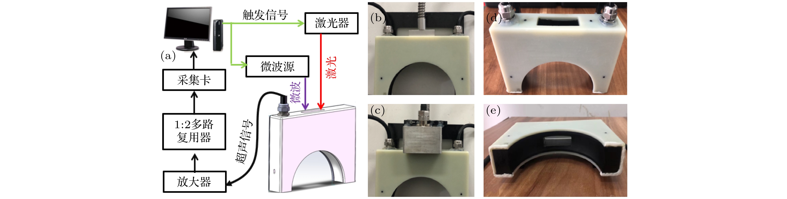
图 1 (a)为反射式光声/热声双模态成像系统框图; (b), (c)分别为反射式光声和热声成像探头接口实物图; (d), (e)分别为镂空探头俯视和侧视实物图
Figure 1. (a) Schematic of the photoacoustic (PA)/thermoacoustic (TA) dual modality imaging system; (b), (c) photograph of the PA and TA imaging system, respectively; (d), (e) Top view and side view of the hollow concave array, respectively.

图 2 镂空阵列探头校准结果图 (a) 第47和48晶元接收到的热声信号波形; (b) 第49晶元所接收热声信号校准前和校准后的波形图, 以及与第48晶元热声信号波形图; (c), (d) 分别为校准前和校准后的热声图像
Figure 2. The calibration results of hollow transducer array: (a) TA signal received by the 47 th and 48 th elements; (b) the TA signal before and after calibration of the 49 th element, and the TA signal of the 48 th element; (c), (d) are the TA images before and after calibration, respectively. TAM: Thermoacoustic Amplitude.

图 3 双模态成像性能验证实验 (a), (b) 分别为待成像物体示意图和实物图; (c), (d) 分别为热声图像和680 nm激发波长得到的光声图像; (e)融合后的热声/光声双模态图像
Figure 3. (a), (b) Schematic and photograph of the target, respectively; (c), (d) TA and PA images obtained at 680 nm, respectively; (e) the fused TA/PA image. PAM:Photoacoustic Amplitude

图 4 空间分辨率实验 (a) 两根直径66 μm铜丝的热声成像结果; (b) 沿(a)中红色虚线的热声图像一维轮廓分布
Figure 4. TAI of two copper wires for system spatial resolution evaluation: (a) Recovered TA image; (b) recovered microwave absorption profile along the red dashed line shown in (a). TAM: Thermoacoustic Amplitude.

图 5 正常人手臂双模态成像, 左侧为待成像平面示意图, A和B分别为自愿者1和2待成像手臂平面示意图; (a)−(d)和(e)−(h)依次为为自愿者1和2手臂的热声图像, 680, 720, 800 nm激发光声图像
Figure 5. The picture is the schematic of the opisthenar to be imaged, A and B are the detection plan of volunteers 1 and 2, respectively. (a)−(d) and (e)−(f) are TA image, 680 nm PA image, 720 nm PA image and 800 nm PA image of volunteers 1 and 2, respectively. TAM: Thermoacoustic Amplitude, PAM: Photoacoustic Amplitude.
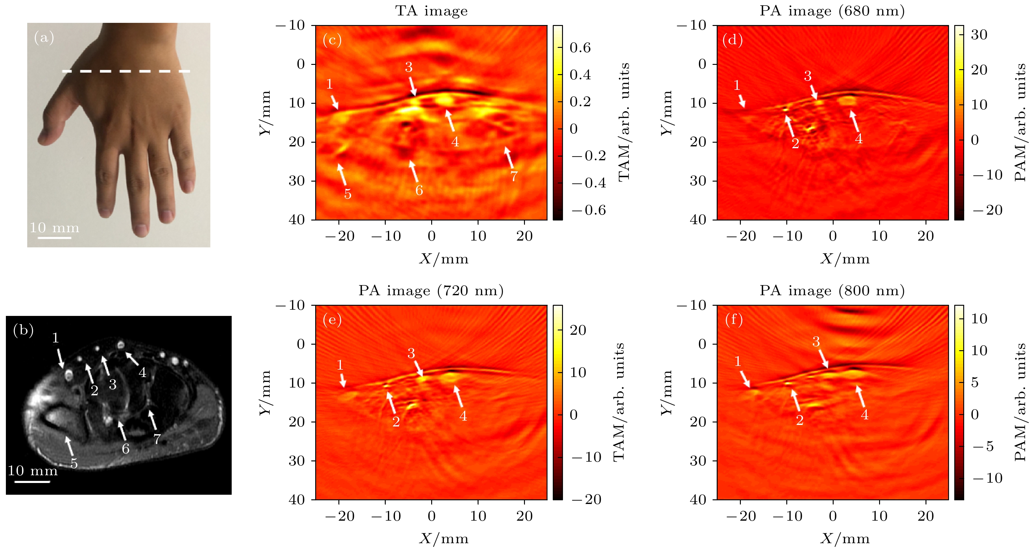
图 6 正常人手背双模态成像 (a) 待成像平面示意图; (b) 对应层面MRI图; (c)−(f) 依次为手背的热声图像, 680, 720和800 nm激发光声图像
Figure 6. (a) Schematic diagram of the plane to be imaged; (b) the corresponding MRI image; (c)−(f) are TA image, 680 nm PA image, 720 nm PA image and 800 nm image of hand, respectively. TAM: Thermoacoustic Amplitude, PAM: Photoacoustic Amplitude.

图 7 正常人脚背双模态成像 (a), (b) 待成像平面彩色多普勒超声图; (c)成像层面示意图; (d)−(g) 依次为脚背的热声图像, 680, 720和800 nm激发光声图像
Figure 7. (a), (b) The color Doppler ultrasound images; (c) the schematic of imaging plane; (d)−(g) TA image, 680 nm PA image, 720 nm PA image and 800 nm image of instep, respectively. TAM: Thermoacoustic Amplitude, PAM: Photoacoustic Amplitude.
-
[1] Shi J, Wong T T W, He Y, Li L, Wang L V 2019 Nat. Photonics 13 609
Google Scholar
[2] Wong T T W, Zhang R, Zhang C, Hsu H C, Maslov K, Wang L, Shi J, Chen R, Shung K K, Zhou Q F, Wang L V 2017 Nat. Commun. 8 1386
Google Scholar
[3] Wang Y C, Liang G R, Liu F, Chen Q, Xi L 2020 IEEE. Trans. Biomed. Eng. 99 1
[4] Gottschalk S, Degtyaruk O, Mc Larney B, Rebling J, Hutter M A, Deán-Ben X L, Shoham S, Razansky D 2019 Nat. Biomed. Eng. 3 392
Google Scholar
[5] Merčep E, Herraiz J L, Deán-Ben X L, Razansky D 2019 Ligh-Sci. Appl. 8 1
Google Scholar
[6] Jiang H B 2015 Photoacoustic Tomography (Boca Raton, FL: CRC Press) pp1−50
[7] Ivankovic I, Merčep, Elena, Schmedt, C G, Deán-Ben X L, Razansky D 2019 Radiology 291 45
Google Scholar
[8] Attia ABE, Balasundaram G, Moothanchery M, Dinish US, Bi R, Ntziachristos V, Olivo M 2019 Photoacoustics 16 100144
Google Scholar
[9] Kruger RA, Kopecky KK, Aisen AM, Reinecke DR, Kruger GA, Kiser WL Jr 1999 Radiology 211 275
Google Scholar
[10] Wang X, Bauer D R, Witte R, Xin H 2012 IEEE. Trans. Biomed. Eng. 59 2782
Google Scholar
[11] Huang L, Yao L, Liu L, Rong J, Jiang H B 2012 Appl. Phys. Lett. 101 244106
Google Scholar
[12] Qin H, Cui Y S, Wu Z J, Chen Q, Xing D 2020 IEEE. Photonics. J. 99 1
[13] Huang L, Li T, Jiang H 2017 Med. Phys. 44 1494
Google Scholar
[14] Chi Z H, Zhao Y, Yang J G, Li TT, Jiang H B 2018 IEEE. Trans. Biomed. Eng. 66 1598
[15] Zheng Z, Huang L, Jiang HB 2018 Appl. Phys. Lett. 113 253702
Google Scholar
[16] Eckhart AT, Balmer RT, See WA, et al. 2011 IEEE Trans. Biomed. Eng. 58 2238
Google Scholar
[17] Patch S, Hull D, See W, Hanson G W 2016 IEEE Trans. Ultrason. Ferroelectr. Freq. Control 63 245
Google Scholar
[18] Ku G, Fornage B D, Jin X, Xu M H, Hunt K K, Wang L V 2005 Technol. Cancer Res. T. 4 559
Google Scholar
[19] Pramanik M, Ku G, Li C H, Wang L V 2008 Med. Phys. 35 2218
Google Scholar
[20] Ke H, Erpelding T N, Jankovic L, Liu C, Wang L V 2012 J. Biomed. Opt. 15 056010
[21] Merčep E, Deán-Ben X L, Razansky D 2017 IEEE. Trans. Med. Imaging. 36 2129
Google Scholar
[22] Reinecke D R, Kruger R A, Lam R B, Delrio S P 2010 Proc. SPIE Int. Soc. Opt. Eng. 7564 489
[23] Li M C, Liu C B, Gong X J, Zheng R Q, Bai Y Y, Xing M Y, Du X M, Liu X Y, Zeng J, Lin R Q, Zhou H C, Wang S J, Lu G M, Zhu W, Fang C H, Song L 2018 Biomed. Opt. Express 9 1408
Google Scholar
[24] American Laser Institute. American National Standards for the Safe Use of Lasers ANSIZ136.1. Orlando, FL: American Laser Institute, 2014
[25] Huang L, Ge S, Zheng Z, Jiang H B 2018 Med. Phys. 46 851
[26] IEEE standard for safety levels with respect to human exposure to radio frequency electromagnetic fields 3 kHz to 300 GHz, IEEE Standard C95.1; 1999
[27] Hoelen C G A, de Mul F F M 2001 Appl. Opt. 39 5872
[28] Jeon S, Park E Y, Choi W, Managuli R, Kim C 2019 Photoacoustics 15 100136
Google Scholar
[29] Zhang Y P, Li E, Zhang J, Yu C Y, Zheng H, Guo G F 2018 Rev. Sci. Instrum. 89 024701
Google Scholar
[30] Wang X, Huang L, Chi Z H, Jiang H B. 2021 Phy. Med. Biol. (Under Review)
[31] Choi W, Park E Y, Jeon S, Kim C 2018 Biomed. Eng. Lett. 8 1
Google Scholar
[32] Ji Z, Ding W Z, Ye F H, Lou C G 2016 Ultrason. Imaging 38 276
Google Scholar
[33] Chi Z H, Liang X, Wang X, Huang L, Jiang H B 2020 IEEE J. Electromagnet. RF Microwaves Med. Biol. 99 1
[34] Yang J G, Zhang G, Wu M, Shang Q Q, Huang L, Jiang H B 2019 J. Biophotonics 12 e201900004

 DownLoad:
Catalog
Metrics
- Abstract views: 4370
- PDF Downloads: 96
- Cited By: 0
