


-
The fiber optic surface plasmon resonance (SPR) technologies can directly detect the change of the refractive index on the surface of the sensor, caused by the interaction of biochemical molecules. Fiber optic SPR technologies have advantages of small size, low cost, no labeling, high sensitivity, and are easy to realize the miniaturization, multi-parameter, real-time and in-situ detection. Two types of probe-type fiber optic SPR refractometers are constructed based on the novel two-dimensional nanomaterial, i.e., violet phosphorus (VP), the mature fabrication and characterization technologies. The fabrication processes of the fiber optic SPR refractometers are first introduced, and then the feasibility of the fabrication processes is verified via multiple characterization methods. In terms of the signal demodulation, the noise of the resonance spectrum is suppressed by the variational mode decomposition algorithm, and the resonance wavelength is interrogated and monitored in real time by the centroid method. The refractive index sensing performances of the near-field enhanced fiber optic SPR refractometers coated with different layers of VP are investigated. With the increase of the VP layer number, the resonance spectrum exhibits redshift and broadening and the sensitivity is enhanced. The refractive index sensing performance of the nearly guided wave fiber optic SPR refractometer is also investigated. In the low refractive index range of 1.33-1.34 corresponding to the refractive index of the low-concentration biological solution, the sensitivity and the figure of merit of the near-field enhanced fiber optic SPR refractometer with the sensing structure of fiber core/VP dielectric layer/Au layer/sample layer reach to 2335.64 nm/RIU and 24.15 RIU–1, respectively, which are 1.31 times and 1.25 times higher than the counterparts of the single Au layer fiber optic SPR refractometer, respectively. The sensitivity and the figure of merit of the nearly guided wave fiber optic SPR refractometer with the sensing structure of fiber core/Au layer/VP dielectric layer/sample layer can reach to 2802.06 nm/RIU and 22.53 RIU–1, respectively, which are 1.57 times and 1.16 times higher than the counterparts of the single Au layer fiber optic SPR refractometer. Finally, the near-field enhanced SPR and the nearly guided wave SPR are integrated into a single fiber probe to achieve the double-lane sensing. The fiber optic SPR refractometers developed in this study can realize the high-sensitivity, plug-and-play and double-lane detection of the combination of surface refractive index and volume refractive index. The probe-type refractometer also provides a new idea for detecting multi-type protein molecules and heavy metal ions in the biochemical field.
-
Keywords:
- two-dimensional violet phosphorus /
- surface plasmon resonance /
- fiber optic refractometry /
- sensitivity enhancement
[1] Liu Z W, Wu J N, Cai C, Yang B, Qi Z M 2022 Nat. Commun. 13 6475
 Google Scholar
[2] Ribeiro J A, Sales M G F, Pereira C M 2022 TrAC, Trends Anal. Chem. 157 116766
Google Scholar
[3] Tan J S, Chen Y Y, He J, Occhipinti L G, Wang Z H, Zhou X H 2023 J. Hazard. Mater. 455 131644
Google Scholar
[4] Cao S Q, Shao Y, Wang Y, Wu T S, Zhang L F, Huang Y J, Zhang F, Liao C R, He J, Wang Y P 2018 Opt. Express 26 3988
Google Scholar
[5] Jing J Y, Liu K, Jiang J F, Xu T H, Xiao L, Zhan X H, Liu T G 2023 Adv. Sci. 10 2207437
Google Scholar
[6] Dastmalchi B, Tassin P, Koschny T, Soukoulis C M 2016 Adv. Opt. Mater. 4 177
Google Scholar
[7] Mai Z G, Zhang J H, Chen Y Z, Wang J Q, Hong X M, Su Q N, Li X J 2019 Biosens. Bioelectron. 144 111621
Google Scholar
[8] Li X G, Gong P Q, Zhao Q M, Zhou X, Zhang Y A, Zhao Y 2022 Sens. Actuators, B 359 131596
Google Scholar
[9] Yasli A 2021 Plasmonics 16 1605
Google Scholar
[10] Shakya A K, Singh S 2022 Opt. Laser Technol. 153 108246
Google Scholar
[11] Liu R C, Yang W, Lu J J, Shafi M, Jiang M S, Jiang S Z 2023 Nanotechnology 34 095501
Google Scholar
[12] Hu S Q, Chen J Y, Liang J H, Luo J J, Shi W C, Yuan J M, Chen Y F, Chen L, Chen Z, Liu G S, Luo Y H 2022 ACS Appl. Mater. Interfaces 14 42412
Google Scholar
[13] Liu Z H, Zhang M, Zhang Y, Zhang Y X, Liu K Q, Zhang J Z, Yang J, Yuan L B 2019 Opt. Lett. 44 2907
Google Scholar
[14] Chiavaioli F, Gouveia C A J, Jorge P A S, Baldini F 2017 Biosensors-Basel 7 23
Google Scholar
[15] Jing J Y, Liu K, Jiang J F, Xu T H, Wang S, Ma J Y, Zhang Z, Zhang W L, Liu T G 2021 Photonics Res. 10 126
Google Scholar
[16] Shalabney A, Abdulhalim I 2011 Laser Photonics Rev. 5 571
Google Scholar
[17] Zhang L H, Huang H Y, Zhang B, Gu M Y, Zhao D, Zhao X W, Li L R, Zhou J, Wu K, Cheng Y H, Zhang J Y 2020 Angew. Chem. Int. Ed. 59 1074
Google Scholar
[18] Cicirello G, Wang M J, Sam Q P, Hart J L, Williams N L, Yin H B, Cha J J, Wang J J 2023 J. Am. Chem. Soc. 145 8218
Google Scholar
[19] Qiao J S, Kong X H, Hu Z X, Yang F, Ji W 2014 Nat. Commun. 5 4475
Google Scholar
[20] Jing J Y, Liu K, Jiang J F, Xu T H, Wang S, Liu T G 2023 Opto-Electron. Adv. 6 220072
Google Scholar
[21] Rehman H U, Cord-Landwehr S, Shapaval V, Dzurendova S, Kohler A, Moerschbacher B M, Zimmermann B 2023 Carbohydr. Polym. 302 120428
Google Scholar
[22] Patil R S, Sancaktar E 2021 Polymer 233 124181
Google Scholar
[23] Zhao Y, Tong R J, Xia F, Peng Y 2019 Biosens. Bioelectron. 142 111505
Google Scholar
[24] Guo J F, Xie R H, Wang Y X, Xiao L Z, Fu J W, Jin G W, Luo S H 2023 IEEE Trans. Geosci. Remote Sens. 61 5902014
Google Scholar
[25] Zhan S Y, Wang X P, Liu Y L 2011 Meas. Sci. Technol. 22 025201
Google Scholar
[26] Wang Q, Cong X W, Zhao W M, Ren Z H, Du N N, Yan X, Zhu A S, Qiu F M, Chen B H, Zhang K K 2022 IEEE Trans. Instrum. Meas. 71 7005808
Google Scholar
[27] Li Y L, Sun Q, Zu S, Shi X, Liu Y Q, Hu X Y, Ueno K, Gong Q H, Misawa H 2020 Phys. Rev. Lett. 124 163901
Google Scholar
[28] Chen Y Z, Ge Y Q, Huang W C, Li Z J, Wu L M, Zhang H, Li X J 2020 ACS Appl. Nano Mater. 3 303
Google Scholar
[29] Zhou Y A, Yan X 2022 IEEE Photonics J. 14 7151607
Google Scholar
[30] Chopra A, Mohanta G C, Das B, Bhatnagar R, Pal, S S 2021 IEEE Sens. J. 21 12153
Google Scholar
[31] Zakaria R, Zainuddin N M, Fahri M A S A, Thirunavakkarasu P M, Patel S K, Harun S W 2021 Opt. Fiber Technol. 61 102449
Google Scholar
[32] Zakaria R, Zainuddin N A M, Raya S A, Alwi S A K, Anwar T, Sarlan A, Ahmed K, Amiri I S 2020 Micromachines-Basel 11 77
Google Scholar
[33] Zhang H, Chen Y F, Feng X J, Xiong X, Hu S Q, Jiang Z P, Dong J L, Zhu W G, Qiu W T, Guan H Y, Lu H H, Yu J H, Zhong Y C, Zhang J, He M, Luo Y H, Chen Z 2018 IEEE J. Sel. Top. Quantum Electron. 25 7101909
Google Scholar
[34] 赵静, 王英, 王义平 2019 激光与光电子学进展 56 230601
Google Scholar
Zhao J, Wang Y, Wang Y P 2019 Laser Optoelectron. Progress 56 230601
Google Scholar
[35] Zainuddin N A M, Ariannejad M M, Arasu P T, Harun S W, Zakaria R 2019 Results Phys. 13 102255
Google Scholar
[36] Amiri I S, Alwi S A K, Raya S A, Zainuddin N A M, Rohizat N S, Rajan M S M, Zakaria R 2019 J. Opt. Commun. 44 53
Google Scholar
[37] Wang W J, Mai Z G, Chen Y Z, Wang J Q, Li L, Su Q N, Li X J, Hong X M 2017 Sci. Rep. 7 16904
Google Scholar
[38] Zhao J, Cao S Q, Liao C R, Wang Y, Wang G J, Xu X Z, Fu C L, Xu G W, Lian J R, Wang Y P 2016 Sens. Actuators, B 230 206
Google Scholar
[39] Shi S, Wang L B, Su R X, Liu B S, Huang R L, Qi W, He Z M 2015 Biosens. Bioelectron. 74 454
Google Scholar
[40] Cennamo N, Massarotti D, Conte L, Zeni L 2011 Sensors-Basel 11 11752
Google Scholar
[41] Jing J Y, Liu K, Jiang J F, Xu T H, Wang S, Ma J Y, Zhang Z, Zhang W L, Liu T G 2021 Nanomaterials-Basel 11 2137
Google Scholar
[42] Qu J H, Leirs K, Maes W, Imbrechts M, Callewaert N, Lagrou K, Geukens N, Lammertyn J, Spasic D 2022 ACS Sens. 7 477
Google Scholar
[43] Zhang Y N, Zhang L B, Han B, Gao P, Wu Q L, Zhang A Z 2018 Sens. Actuators, B 272 331
Google Scholar
-

图 1 (a)近场增强型和(b)近导波型光纤SPR激发结构示意图
Figure 1. Schematic diagram of the sensing structure of (a) the near-field enhanced fiber SPR and (b) the nearly guided wave fiber SPR.

图 2 双通道光纤SPR (a)有限元分析模型和(b)有限元损耗光谱. 模型参数: 纤芯半径rcore = 4.1 μm, 包层半径rcladding = 62.5 μm, 抛磨剩余厚度Dr = 66.6 μm, 金膜厚度50 nm, 紫磷厚度10 nm
Figure 2. (a) Finite element analysis model and (b) the loss spectrum of the double-lane fiber optic SPR. Model parameter: rcore = 4.1 μm, rcladding = 62.5 μm, Dr = 66.6 μm, the thickness of the Au layer and the VP layer is 50 nm and 10 nm, respectively.

图 3 (a)光纤预处理流程示意图; (b)光纤端面研磨; (c)光纤端面金反射镜溅射
Figure 3. (a) Schematic diagram of fiber preprocessing process; (b) the grinding of the end face of the optical fiber; (c) the sputtering of the gold mirror on the fiber end face.

图 A1 光纤端面研磨各阶段扫描电子显微镜图
Figure A1. Scanning electron microscopy of the end face of the optical fiber at each step.
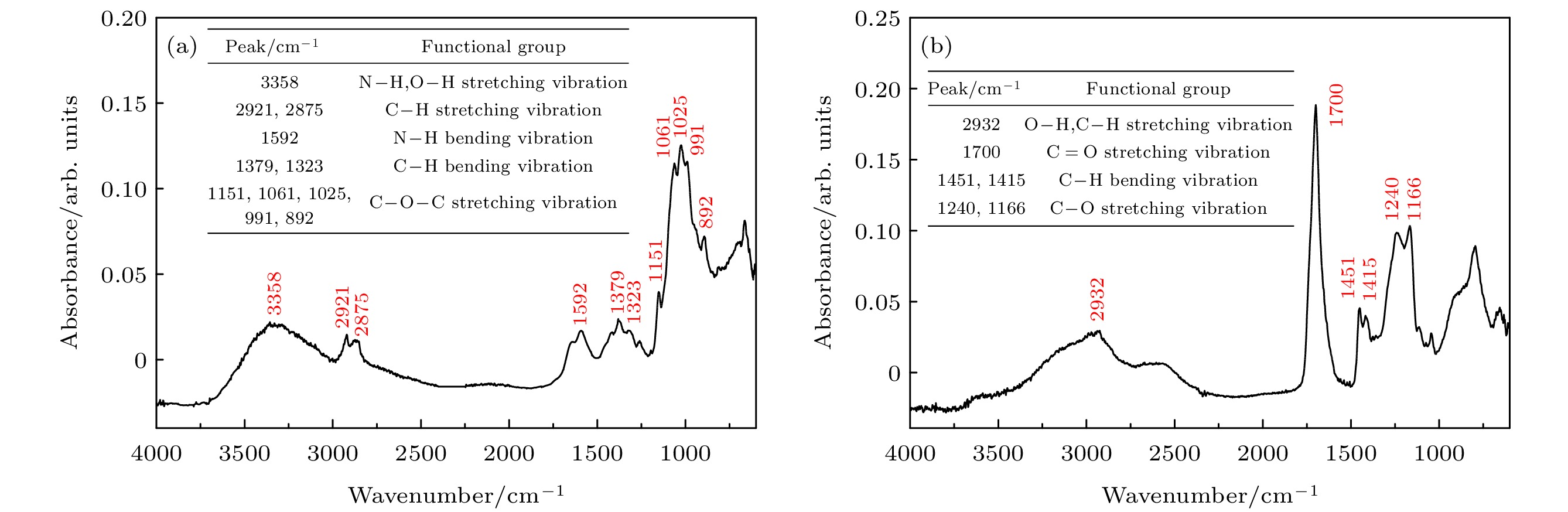
图 A2 (a)壳聚糖和(b)聚丙烯酸的傅里叶红外光谱图
Figure A2. Fourier transform infrared spectroscopy of (a) the CTS and (b) the PAA.

图 A3 壳聚糖(a)和聚丙烯酸(b)的Zeta电位
Figure A3. Zeta potential of (a) the CTS and (b) the PAA.

图 A4 紫磷的(a)拉曼光谱、(b)扫描电子显微镜能谱图和(c) X射线衍射光谱
Figure A4. (a) Raman spectrum, (b) the SEM energy spectrum and (c) the X-ray diffraction spectrum of the VP.

图 4 (a)近场增强型光纤SPR折射率计制备流程; (b)近场增强光纤SPR折射率计传感结构示意图; (c)紫磷层层自组装; (d)传感区域金层溅射
Figure 4. (a) Fabrication process of the near-field enhanced fiber SPR refractometer; (b) schematic diagram of sensing structure of the near-field enhanced fiber SPR refractometer; (c) the self-assembly of the VP layer; (d) the sputtering of the Au layer on the sensing area.

图 5 (a)近导波型光纤SPR折射率计制备流程; (b)近导波光纤SPR折射率计传感结构示意图; (c)传感区域金层溅射; (d)紫磷层层自组装
Figure 5. (a) Fabrication process of the nearly guided wave fiber SPR refractometer; (b) schematic diagram of sensing structure of the nearly guided wave fiber SPR refractometer; (c) the sputtering of the Au layer on the sensing area; (d) the self-assembly of the VP layer.

图 6 光纤SPR折射率计信号解调系统示意图
Figure 6. Schematic diagram of the signal demodulation system for the fiber SPR refractometer.

图 7 (a) 共振光谱信号噪声抑制; (b) 共振波长在线实时监测
Figure 7. (a) Noise suppression for the resonance spectra; (b) the online real-time monitoring of the resonance wavelength.

图 8 增覆(a) 1层、(b) 2层和(c) 3层紫磷电介质层的近场增强型光纤SPR折射率计共振光谱; (d)增覆不同紫磷电介质层的光纤SPR折射率计平均灵敏度, 插图为三种光纤SPR折射率计共振波长与折射率点二次拟合曲线
Figure 8. Resonance spectra of the near-field enhanced fiber SPR refractometer with (a) one-layer, (b) two-layer and (c) three-layer VP dielectric layers; (d) average sensitivity of the above three types of fiber SPR refractometers. Inset: binomial fitting curves of resonance wavelengths and refractive index points of three types of fiber SPR refractometers.

图 9 增覆2层紫磷电介质层的近导波型光纤SPR折射率计(a)共振光谱和(b)平均灵敏度, 插图: 近导波型光纤 SPR 折射率计共振波长与折射率点二次拟合曲线
Figure 9. (a) Resonance spectra and (b) the average sensitivity of the nearly guided wave fiber SPR refractometer coated with two-layer VP dielectric layer. Inset: the binomial fitting curve of resonance wavelengths and refractive index points.
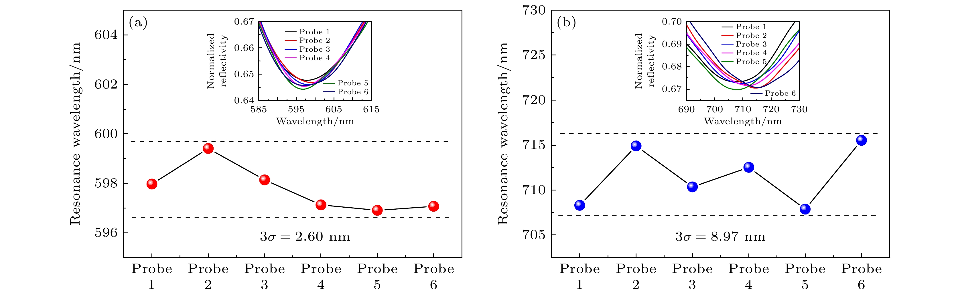
图 10 (a)近场增强型和(b)近导波型光纤SPR折射率计重复性测试
Figure 10. Repeatability of (a) the near-field enhanced fiber SPR refractometer and (b) the nearly guided wave fiber SPR refractometer

图 11 双通道光纤SPR折射率计(a)示意图和(b)实物图
Figure 11. (a) Schematic diagram and (b) realistic image of the double-lane optical fiber SPR refractometer.
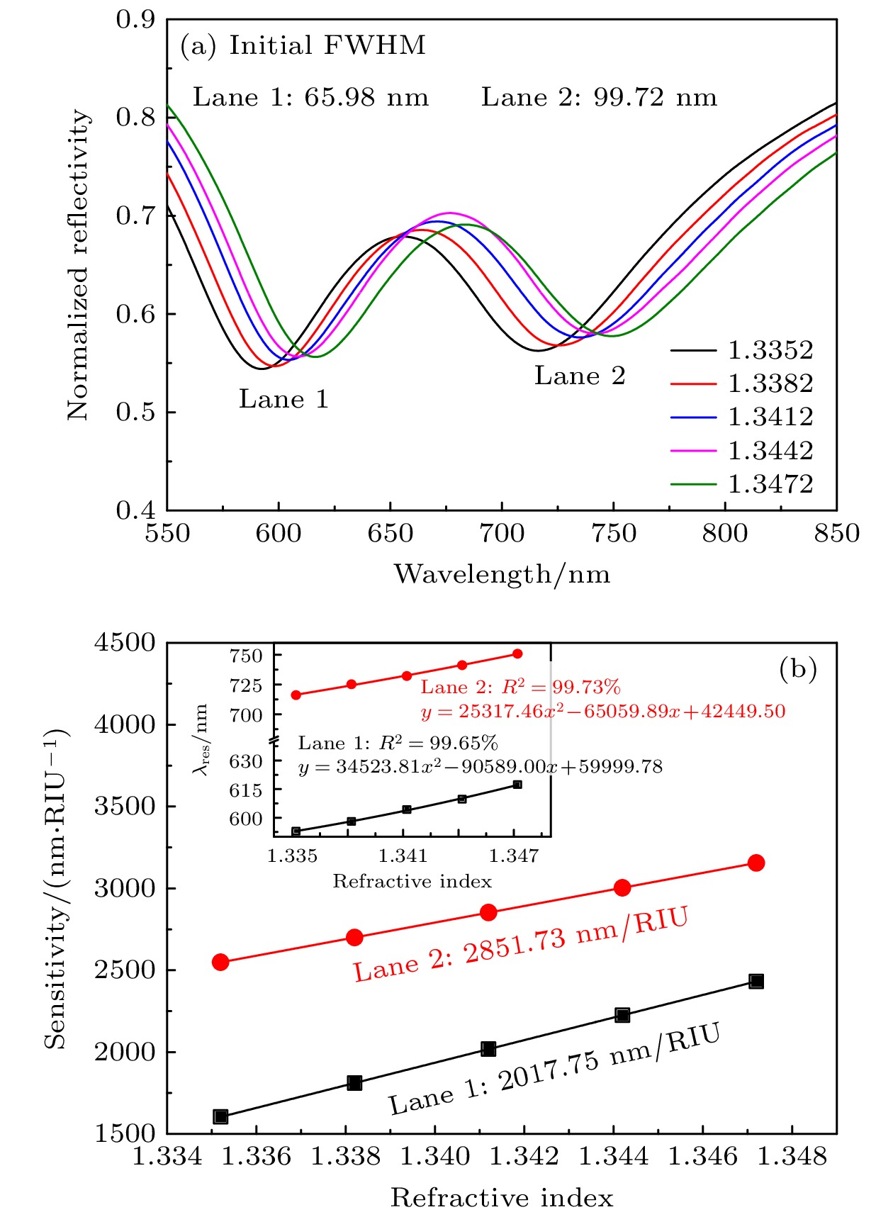
图 12 双通道光纤SPR折射率计(a)共振光谱与(b)平均灵敏度. 插图为双通道光纤SPR折射率计共振波长与折射率点二次拟合曲线
Figure 12. (a) Resonance spectra and (b) the average sensitivity of the double-lane optical fiber SPR refractometer. Inset: the binomial fitting curve of resonance wavelengths and refractive index points.

图 A5 (a)近场增强和(b)近导波型光纤SPR折射率计传感区截面扫描电子显微镜图(105倍)
Figure A5. Scanning electron microscopy images of cross sections of sensing areas of (a) the near-field enhanced and (b) the nearly guided wave fiber refractometers.
表 1 折射率传感特性对比
Table 1. Comparison of refractive index sensing characteristics.
传感结构 灵敏度/(nm·RIU–1) 半峰全宽/nm 品质因数/(RIU–1) 光纤/金/待测物 1787.93 92.43 19.34 光纤/一层紫磷/金/待测物 1927.61 79.82 24.15 光纤/两层紫磷/金/待测物 2140.53 93.22 22.96 光纤/三层紫磷/金/待测物 2335.64 116.94 24.15 光纤/金/两层紫磷/待测物 2802.06 124.39 22.53  DownLoad: CSV
表 2 本文光纤SPR折射率计光谱特性与已报道光纤SPR传感器光谱特性对比
Table 2. Comparison between the study in this work and reported works.
传感结构 折射率
测量范围灵敏度
计算方法灵敏度/
(nm·RIU–1)半峰全宽/
nm品质因数
(RIU–1)年份 参考
文献多模-单模-多模光纤/金/
Ti3C2Tx/待测物1.3343—1.3658 线性拟合 2180.2 — — 2022 [28] 侧抛单模光纤/金/Ti3C2Tx/待测物 1.32—1.34 线性拟合 3143 206 15.26 2022 [29] 锥形多模光纤/铬/金/待测物 1.337—1.359 线性拟合 2266 — — 2021 [30] 侧抛单模光纤/氟化镁/银/待测物 1.33—1.34 波长差与折射率差的比值 2812.50 35.95 78.23 2021 [31] 侧抛单模光纤/铜/待测物 1.3330—1.3573 波长差与折射率差的比值 425 2.5 — 2020 [32] 侧抛多模光纤/氟化镁/银/待测物 1.333—1.360 线性拟合 1603 47.80 33.54 2019 [33] 侧抛单模光纤/银/氧化石墨烯/待测物 1.32—1.34 波长差与折射率差的比值 2252.50 60.50 37.22 2019 [34] 侧抛单模光纤/银/待测物 1.333—1.345 波长差与折射率差的比值 2166.67 25 — 2019 [35] 侧抛单模光纤/银/氧化石墨烯/待测物 1.30—1.34 波长差与折射率差的比值 833.33 10 — 2019 [36] 多模光纤/金/待测物 1.3345—1.3592 线性拟合 2659.64 — — 2017 [37] 侧抛单模光纤/银/待测物 1.320—1.340 多项式
拟合1798.0 58.60 30.68 2016 [38] 多模光纤/化学镀金/待测物 1.333—1.359 线性拟合 2054 108.2 19 2015 [39] 多模光纤/光刻胶/金/待测物 1.332—1.352 波长差与折射率差的比值 2422 181 — 2011 [40] 多模光纤/三层紫磷/金/待测物 1.3335—1.3435 二次拟合 2335.64 116.94 24.15 本文工作 多模光纤/金/两层紫磷/待测物 1.3352—1.3472 二次拟合 2802.06 124.39 22.53
DownLoad: CSV
-
[1] Liu Z W, Wu J N, Cai C, Yang B, Qi Z M 2022 Nat. Commun. 13 6475
Google Scholar
[2] Ribeiro J A, Sales M G F, Pereira C M 2022 TrAC, Trends Anal. Chem. 157 116766
Google Scholar
[3] Tan J S, Chen Y Y, He J, Occhipinti L G, Wang Z H, Zhou X H 2023 J. Hazard. Mater. 455 131644
Google Scholar
[4] Cao S Q, Shao Y, Wang Y, Wu T S, Zhang L F, Huang Y J, Zhang F, Liao C R, He J, Wang Y P 2018 Opt. Express 26 3988
Google Scholar
[5] Jing J Y, Liu K, Jiang J F, Xu T H, Xiao L, Zhan X H, Liu T G 2023 Adv. Sci. 10 2207437
Google Scholar
[6] Dastmalchi B, Tassin P, Koschny T, Soukoulis C M 2016 Adv. Opt. Mater. 4 177
Google Scholar
[7] Mai Z G, Zhang J H, Chen Y Z, Wang J Q, Hong X M, Su Q N, Li X J 2019 Biosens. Bioelectron. 144 111621
Google Scholar
[8] Li X G, Gong P Q, Zhao Q M, Zhou X, Zhang Y A, Zhao Y 2022 Sens. Actuators, B 359 131596
Google Scholar
[9] Yasli A 2021 Plasmonics 16 1605
Google Scholar
[10] Shakya A K, Singh S 2022 Opt. Laser Technol. 153 108246
Google Scholar
[11] Liu R C, Yang W, Lu J J, Shafi M, Jiang M S, Jiang S Z 2023 Nanotechnology 34 095501
Google Scholar
[12] Hu S Q, Chen J Y, Liang J H, Luo J J, Shi W C, Yuan J M, Chen Y F, Chen L, Chen Z, Liu G S, Luo Y H 2022 ACS Appl. Mater. Interfaces 14 42412
Google Scholar
[13] Liu Z H, Zhang M, Zhang Y, Zhang Y X, Liu K Q, Zhang J Z, Yang J, Yuan L B 2019 Opt. Lett. 44 2907
Google Scholar
[14] Chiavaioli F, Gouveia C A J, Jorge P A S, Baldini F 2017 Biosensors-Basel 7 23
Google Scholar
[15] Jing J Y, Liu K, Jiang J F, Xu T H, Wang S, Ma J Y, Zhang Z, Zhang W L, Liu T G 2021 Photonics Res. 10 126
Google Scholar
[16] Shalabney A, Abdulhalim I 2011 Laser Photonics Rev. 5 571
Google Scholar
[17] Zhang L H, Huang H Y, Zhang B, Gu M Y, Zhao D, Zhao X W, Li L R, Zhou J, Wu K, Cheng Y H, Zhang J Y 2020 Angew. Chem. Int. Ed. 59 1074
Google Scholar
[18] Cicirello G, Wang M J, Sam Q P, Hart J L, Williams N L, Yin H B, Cha J J, Wang J J 2023 J. Am. Chem. Soc. 145 8218
Google Scholar
[19] Qiao J S, Kong X H, Hu Z X, Yang F, Ji W 2014 Nat. Commun. 5 4475
Google Scholar
[20] Jing J Y, Liu K, Jiang J F, Xu T H, Wang S, Liu T G 2023 Opto-Electron. Adv. 6 220072
Google Scholar
[21] Rehman H U, Cord-Landwehr S, Shapaval V, Dzurendova S, Kohler A, Moerschbacher B M, Zimmermann B 2023 Carbohydr. Polym. 302 120428
Google Scholar
[22] Patil R S, Sancaktar E 2021 Polymer 233 124181
Google Scholar
[23] Zhao Y, Tong R J, Xia F, Peng Y 2019 Biosens. Bioelectron. 142 111505
Google Scholar
[24] Guo J F, Xie R H, Wang Y X, Xiao L Z, Fu J W, Jin G W, Luo S H 2023 IEEE Trans. Geosci. Remote Sens. 61 5902014
Google Scholar
[25] Zhan S Y, Wang X P, Liu Y L 2011 Meas. Sci. Technol. 22 025201
Google Scholar
[26] Wang Q, Cong X W, Zhao W M, Ren Z H, Du N N, Yan X, Zhu A S, Qiu F M, Chen B H, Zhang K K 2022 IEEE Trans. Instrum. Meas. 71 7005808
Google Scholar
[27] Li Y L, Sun Q, Zu S, Shi X, Liu Y Q, Hu X Y, Ueno K, Gong Q H, Misawa H 2020 Phys. Rev. Lett. 124 163901
Google Scholar
[28] Chen Y Z, Ge Y Q, Huang W C, Li Z J, Wu L M, Zhang H, Li X J 2020 ACS Appl. Nano Mater. 3 303
Google Scholar
[29] Zhou Y A, Yan X 2022 IEEE Photonics J. 14 7151607
Google Scholar
[30] Chopra A, Mohanta G C, Das B, Bhatnagar R, Pal, S S 2021 IEEE Sens. J. 21 12153
Google Scholar
[31] Zakaria R, Zainuddin N M, Fahri M A S A, Thirunavakkarasu P M, Patel S K, Harun S W 2021 Opt. Fiber Technol. 61 102449
Google Scholar
[32] Zakaria R, Zainuddin N A M, Raya S A, Alwi S A K, Anwar T, Sarlan A, Ahmed K, Amiri I S 2020 Micromachines-Basel 11 77
Google Scholar
[33] Zhang H, Chen Y F, Feng X J, Xiong X, Hu S Q, Jiang Z P, Dong J L, Zhu W G, Qiu W T, Guan H Y, Lu H H, Yu J H, Zhong Y C, Zhang J, He M, Luo Y H, Chen Z 2018 IEEE J. Sel. Top. Quantum Electron. 25 7101909
Google Scholar
[34] 赵静, 王英, 王义平 2019 激光与光电子学进展 56 230601
Google Scholar
Zhao J, Wang Y, Wang Y P 2019 Laser Optoelectron. Progress 56 230601
Google Scholar
[35] Zainuddin N A M, Ariannejad M M, Arasu P T, Harun S W, Zakaria R 2019 Results Phys. 13 102255
Google Scholar
[36] Amiri I S, Alwi S A K, Raya S A, Zainuddin N A M, Rohizat N S, Rajan M S M, Zakaria R 2019 J. Opt. Commun. 44 53
Google Scholar
[37] Wang W J, Mai Z G, Chen Y Z, Wang J Q, Li L, Su Q N, Li X J, Hong X M 2017 Sci. Rep. 7 16904
Google Scholar
[38] Zhao J, Cao S Q, Liao C R, Wang Y, Wang G J, Xu X Z, Fu C L, Xu G W, Lian J R, Wang Y P 2016 Sens. Actuators, B 230 206
Google Scholar
[39] Shi S, Wang L B, Su R X, Liu B S, Huang R L, Qi W, He Z M 2015 Biosens. Bioelectron. 74 454
Google Scholar
[40] Cennamo N, Massarotti D, Conte L, Zeni L 2011 Sensors-Basel 11 11752
Google Scholar
[41] Jing J Y, Liu K, Jiang J F, Xu T H, Wang S, Ma J Y, Zhang Z, Zhang W L, Liu T G 2021 Nanomaterials-Basel 11 2137
Google Scholar
[42] Qu J H, Leirs K, Maes W, Imbrechts M, Callewaert N, Lagrou K, Geukens N, Lammertyn J, Spasic D 2022 ACS Sens. 7 477
Google Scholar
[43] Zhang Y N, Zhang L B, Han B, Gao P, Wu Q L, Zhang A Z 2018 Sens. Actuators, B 272 331
Google Scholar

 DownLoad:
Catalog
Metrics
- Abstract views: 1600
- PDF Downloads: 53
- Cited By: 0
