


-
基于多芯少模光纤结构特性, 提出了一种具有开放式感知通道的多芯少模光纤表面等离子体共振生物传感器. 建立了多芯少模光纤表面等离子体共振生物传感器的模型, 利用有限元方法分析了纤芯气孔间距、膜层厚度、膜层材料以及不同传输模式对传感器性能的影响, 并讨论了传感器多通道感知性能. 仿真分析发现, 纤芯气孔间距决定了倏逝波的耦合强度, 材料特性和模式共同影响了表面等离子体共振峰的位置和灵敏度. 经过计算可知: 当单个凹槽传感通道上沉积100 nm铟锡氧化物薄膜, 分析物折射率范围为1.33—1.39时, LP11ax模式对应的平均光谱灵敏度为12048 nm/RIU(其中RIU为折射率单位, 即refractive index unit), 最高灵敏度为20824.66 nm/RIU, 最大折射率分辨率可达4.8 × 10⁻⁶ RIU; 当光纤外围凹槽镀上不同厚度的金膜、银膜和铟锡氧化物膜时, 既可以单独探测生物物质, 也可以联合检测同一生物物质, 实现了传感通道的控制灵活性和测试物质的多样性.Based on the structural characteristics of the few-mode multicore fiber (FM-MCF), a multi-channel FM-MCF surface plasmon resonance (SPR) biosensor with open air-hole is presented. Due to the air-hole distribution of the FM-MCF, the six outer air-holes naturally become open air-holes, i.e. groove sensing channels, fabricated by chemical etching. Then, compared with D-shape structure, tapered structure of fiber and air-hole of photonic crystal fiber (PCF), the open groove structure is easy to accommodate the liquid analyte. In order to obtain better sensing performance, a sensing model of the presented FM-MCF SPR biosensor with sensitive dielectric layer is established and numerical simulations are performed using the finite element method. In the simulations, the effect of core-hole distance, coating thickness, sensing dielectrics, transmission modes in optical fiber on the sensing performance as well as the role of multi-channel are analyzed. The simulation results show that when the air-hole is tangent to the core (d = 0 μm), the FM-MCF SPR biosensor has the better performance because the core-hole distance d determines the leakage intensity of the evanescent wave. As the evanescent field excited by high-order mode (LP11ax mode) is stronger than that by fundamental mode (LP01x mode), the performance of biosensors for SPR excitation by using high-order mode is better than by using fundamental mode. Meanwhile when the coating thickness of gold, silver and indium tin oxides (ITOs) is 40 nm, 30 nm and 100 nm respectively, the FWHM of loss spectrum reaches a minimum value, which means that the presented biosensor has the better performance in this sense. For the case of different sensing dielectrics, it is observed that the resonance wavelength of gold and silver film are in the visible wavelength range, while the ITO is at near-infrared wavelength. Then it is useful for our biosensor to simultaneously detect many liquid analytes in one SPR transmittance spectrum. In addition, the calculation results also show that when one of the groove channels is coated with 100 nm ITO for the LP11ax mode, the FM-MCF SPR biosensor has a highest sensitivity of 20824.66 nm/RIU and refractive index (RI) resolution is 4.8 × 10⁻⁶ RIU with the surrounding RI changing from 1.33 to 1.39, in which the RI of bovine serum albumin (BSA) solution, human Immunoglobulin G and C-reactive protein can be detected. Moreover, when the outer groove channels of our biosensor are coated with gold, silver and ITO film with different thickness, many biological liquid analytes can be detected separately or the same biological liquid analyte can be detected jointly, which reveals that the control flexibility of the groove sensing channel and the diversity of the detection analytes .
-
Keywords:
- surface plasmon resonance /
- few-mode multicore fiber /
- multi-channel sensor /
- open air-hole
[1] Cooper M A 2002 Nat. Rev. Drug Discov. 1 515
 Google Scholar
[2] Borisov S M, Wolfbeis O S 2008 Chem. Rev. 108 423
Google Scholar
[3] 李加东, 程珺洁, 苗斌, 魏晓玮, 张志强, 黎海文, 吴东岷 2014 物理学报 63 070204
Google Scholar
Li J D, Cheng J J, Miao B, Wei X W, Zhang Z Q, Li H W, Wu D M 2014 Acta Phys. Sin. 63 070204
Google Scholar
[4] Shankaran D R, Gobi K V, Miura N 2007 Sensor Actuat. B-Chem. 121 158
Google Scholar
[5] Fan X D, White I M, Shopova S I, Zhu H Y, Suter J D, Sun Y Z 2008 Anal. Chim. Acta 620 8
Google Scholar
[6] Gupta B D, Verma R K 2009 J. Sens. 2009 1
[7] Homola J 2008 Chem. Rev. 108 462
Google Scholar
[8] Velázquez-González J S, Monzón-Hernández D, Moreno-Hernández D, Martínez-Piñón F, Hernández-Romano I 2017 Sensor Actuat. B-Chem. 242 912
Google Scholar
[9] 施伟华, 尤承杰, 吴静 2015 物理学报 64 224221
Google Scholar
Shi W H, You C J, Wu J 2015 Acta Phys. Sin. 64 224221
Google Scholar
[10] 冯李航, 曾捷, 梁大开, 张为公 2013 物理学报 62 124207
Google Scholar
Feng L H, Zeng J, Liang D K, Zhang W G 2013 Acta Phys. Sin. 62 124207
Google Scholar
[11] 张海志 2003 生物学通报 38 1
Google Scholar
Zhang H Z 2003 Bull. Biol. 38 1
Google Scholar
[12] Kassani S H, Khazaeinezhad R, Jung Y M, Kobelke J, Oh K 2015 IEEE Photonics J. 7 1
Google Scholar
[13] Liu C, Yang L, Lu X L, Liu Q, Wang F M, Lv J W, Sun T, Mu H W, Chu P K 2017 Opt. Express 25 14227
Google Scholar
[14] Yang Z, Xia L, Li C, Chen X, Liu D M 2019 Opt. Commun. 430 195
Google Scholar
[15] Wei Y, Su Y D, Liu C L, Zhang Y H, Nie X F, Liu Z H, Zhang Y, Peng F 2017 Opt. Express 25 21841
Google Scholar
[16] Li A, Wang Y F, Hu Q, Shieh W 2015 Opt. Express 23 1139
Google Scholar
[17] Sakaguchi J, Klaus W, Mendinueta J M D, Puttnam B J, Luís R S, Awaji Y, Wada N, Hayashi T, Nakanishi T, Watanabe T, Kokubun Y, Takahata T, Kobayashi T 2015 J. Lightwave Technol. 34 93
Google Scholar
[18] Xia C, Amezcua-Correa R, Bai N, Antonio-Lopez E, Arrioja D M, Schulzgen A, Richardson M, Liñares J, Montero C, Mateo E, Zhou X, Li G F 2012 IEEE Photonics Technol. Lett. 24 1914
Google Scholar
[19] Rakić A D, Djurišić A B, Elazar J M, Majewski M L 1998 Appl. Opt. 37 5271
Google Scholar
[20] Wang Y J, Dong J L, Luo Y H, Tang J Y, Lu H H, Yu J H, Guan H Y, Zhang J, Chen Z 2017 IEEE Photonics J. 9 1
Google Scholar
[21] Dong J L, Zhang Y X, Wang Y J, Yang F, Hu S Q, Chen Y F, Zhu W G, Qiu W T, Guan H Y, Lu H H, Yu J H, Zhong Y C, Zhang J, Luo Y H, Chen Z 2019 Opt. Express 27 11348
Google Scholar
[22] Zheng Y F, Lang T T, Cao B B, Jin J J, Dong R Q, Feng H 2018 Opt. Fiber Technol. 46 179
Google Scholar
[23] Wang W J, Mai Z G, Chen Y Z, Wang J Q, Li L, Su Q N, Li X J, Hong X M 2017 Sci. Rep. 7 1
Google Scholar
-
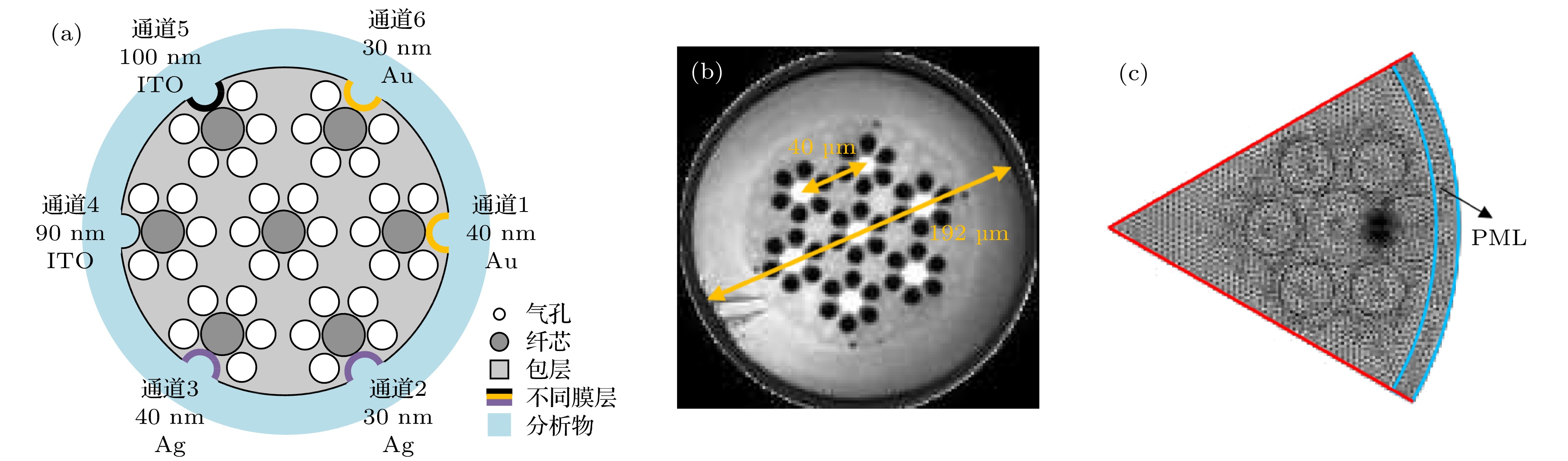
图 1 (a) FM-MCF SPR生物传感器的横截面; (b) FM-MCF横截面; (c) FEM网格划分
Fig. 1. (a) Cross section of the FM-MCF SPR biosensor; (b) cross section of the FM-MCF; (c) FEM mesh for calculation.
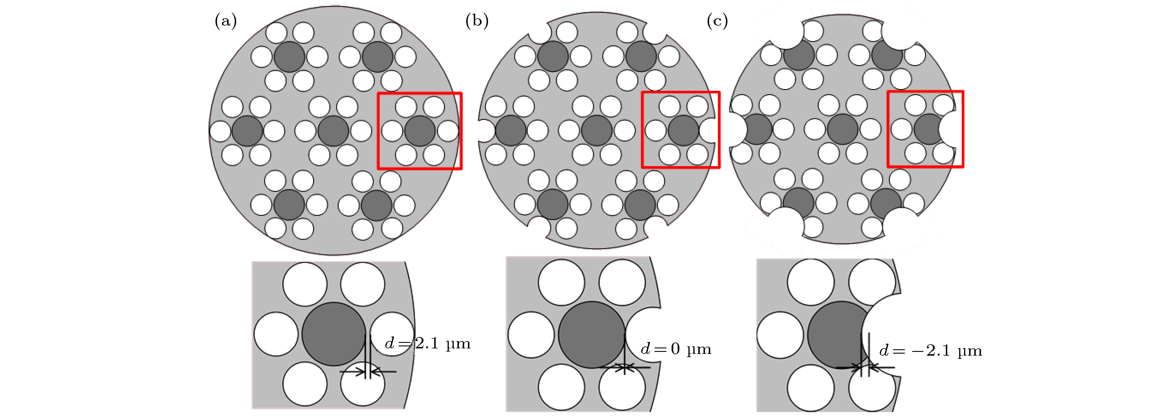
图 2 氢氟酸腐蚀FM-MCF过程中纤芯和气孔间距d的变化 (a)包层与最外侧气孔相切; (b)腐蚀气孔与纤芯恰好相切; (c)纤芯被腐蚀; 下方的插图是红色区域的放大示意图
Fig. 2. The core-hole distance d variation during using hydrofluoric acid to fabricate the groove channel: (a) Cladding is tangent to the outermost air-holes; (b) air-holes are tangent to the cores; (c) fiber cores are also etched. The inserts below are zoom-in of red region.

图 3 (a)当na = 1.33—1.36时不同d的芯模损耗光谱曲线; (b)不同RI下d对共振波长和灵敏度的影响; (c) d与传感器FWHM和FOM的关系
Fig. 3. (a) Loss spectra of the core mode with different d when na = 1.33—1.36; (b) the effect of d on both resonance wavelength and sensitivity with various RI; (c) relations between d and FWHM as well as FOM of the sensor.
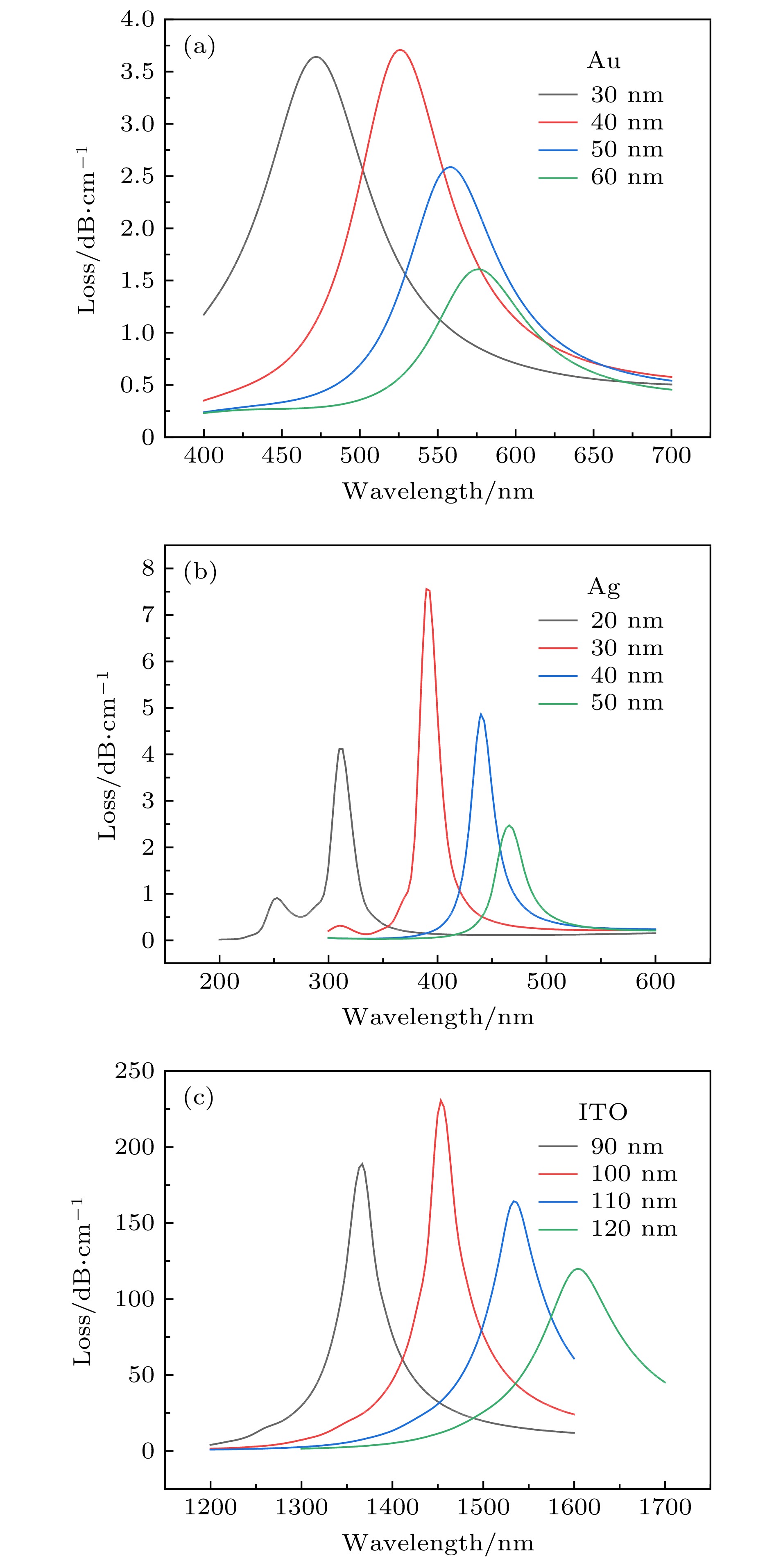
图 4 不同膜层厚度下的芯模损耗光谱 (a)金膜; (b)银膜; (c) ITO
Fig. 4. Loss spectra of the core mode for different coating thickness (t): (a) Au; (b) Ag; (c) ITO.

图 5 当na变化时传感器的共振光谱 (a)金膜厚度为40 nm; (b) ITO膜厚度为100 nm
Fig. 5. Loss spectra of the sensor with na increasing: (a) tAu = 40 nm; (b) tITO = 100 nm.

图 6 待测物RI与共振波长之间的关系 (a) 金膜厚度为40 nm; (b) ITO膜为100 nm
Fig. 6. Relations between na and resonance wavelength for different t: (a) tAu = 40 nm; (b) tITO = 100 nm.
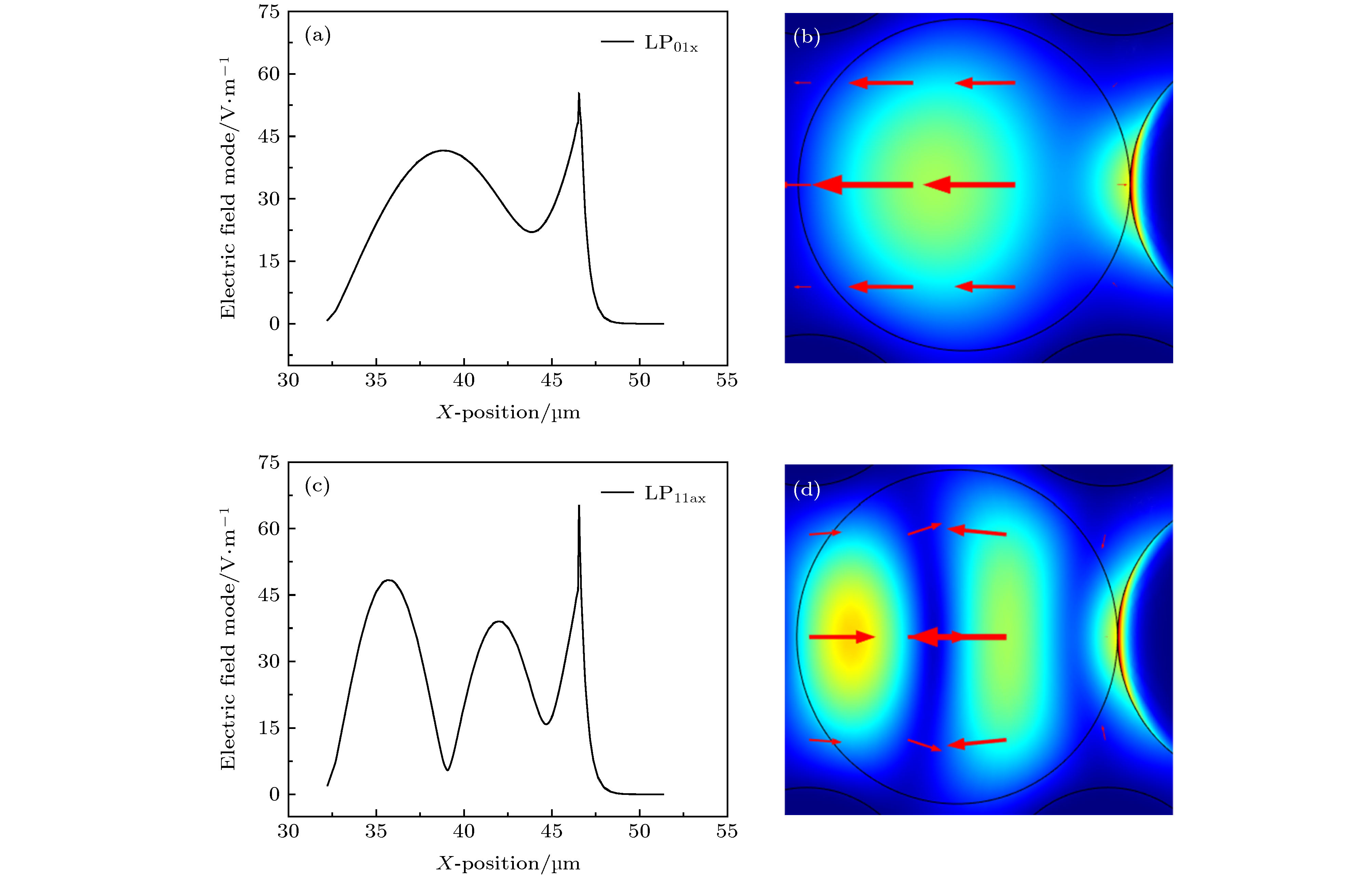
图 7 (a) LP01x模的电场分布; (b) LP01x模的光场分布; (c) LP11ax模的电场分布; (d) LP11ax模的光场分布
Fig. 7. (a) Electric field distributions of LP01x; (b) optical field distributions of LP01x; (c) electric field distributions of LP11ax; (d) optical field distributions of LP11ax.
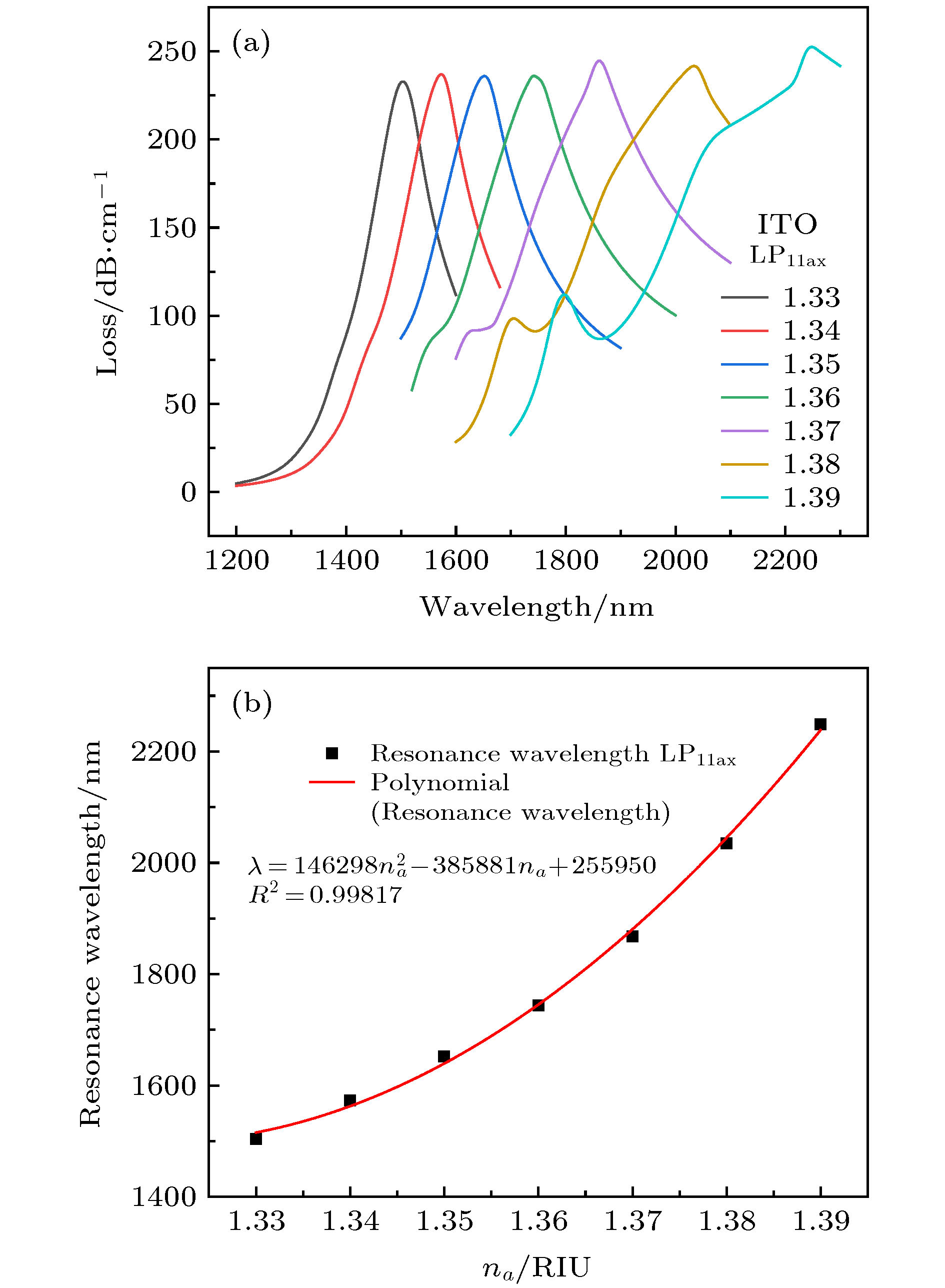
图 8 (a) LP11ax模激发的光谱损耗曲线; (b) LP11ax模激发时na与共振波长之间的关系
Fig. 8. (a) Loss spectra excited by LP11ax mode; (b) relations between na and resonance wavelength at LP11ax mode.
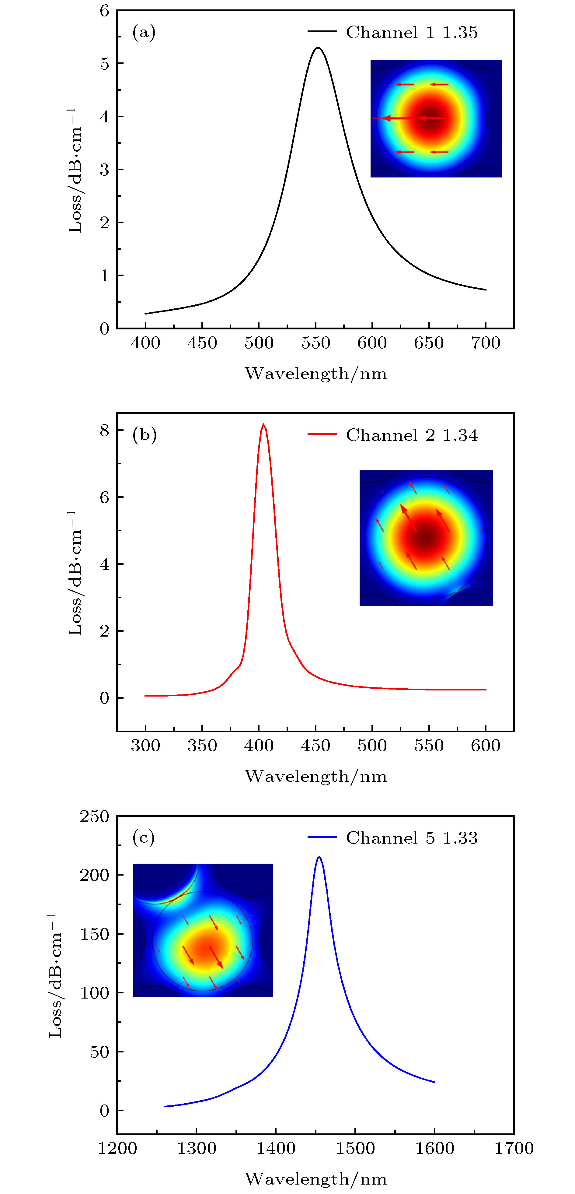
图 9 三种生物物质传感时光谱损耗 (a) 1通道na = 1.35, 金膜; (b) 2通道na = 1.34, 银膜; (c) 5通道na = 1.33, ITO膜; 插图是共振发生时通道中光场分布
Fig. 9. The loss spectra of three bio-substances sensing: (a) Channel 1 for Au coating with na = 1.35; (b) channel 2 for Ag coating with na = 1.34; (c) channel 5 for ITO coating with na = 1.33. Insets are optical field distributions for three channels.
-
[1] Cooper M A 2002 Nat. Rev. Drug Discov. 1 515
Google Scholar
[2] Borisov S M, Wolfbeis O S 2008 Chem. Rev. 108 423
Google Scholar
[3] 李加东, 程珺洁, 苗斌, 魏晓玮, 张志强, 黎海文, 吴东岷 2014 物理学报 63 070204
Google Scholar
Li J D, Cheng J J, Miao B, Wei X W, Zhang Z Q, Li H W, Wu D M 2014 Acta Phys. Sin. 63 070204
Google Scholar
[4] Shankaran D R, Gobi K V, Miura N 2007 Sensor Actuat. B-Chem. 121 158
Google Scholar
[5] Fan X D, White I M, Shopova S I, Zhu H Y, Suter J D, Sun Y Z 2008 Anal. Chim. Acta 620 8
Google Scholar
[6] Gupta B D, Verma R K 2009 J. Sens. 2009 1
[7] Homola J 2008 Chem. Rev. 108 462
Google Scholar
[8] Velázquez-González J S, Monzón-Hernández D, Moreno-Hernández D, Martínez-Piñón F, Hernández-Romano I 2017 Sensor Actuat. B-Chem. 242 912
Google Scholar
[9] 施伟华, 尤承杰, 吴静 2015 物理学报 64 224221
Google Scholar
Shi W H, You C J, Wu J 2015 Acta Phys. Sin. 64 224221
Google Scholar
[10] 冯李航, 曾捷, 梁大开, 张为公 2013 物理学报 62 124207
Google Scholar
Feng L H, Zeng J, Liang D K, Zhang W G 2013 Acta Phys. Sin. 62 124207
Google Scholar
[11] 张海志 2003 生物学通报 38 1
Google Scholar
Zhang H Z 2003 Bull. Biol. 38 1
Google Scholar
[12] Kassani S H, Khazaeinezhad R, Jung Y M, Kobelke J, Oh K 2015 IEEE Photonics J. 7 1
Google Scholar
[13] Liu C, Yang L, Lu X L, Liu Q, Wang F M, Lv J W, Sun T, Mu H W, Chu P K 2017 Opt. Express 25 14227
Google Scholar
[14] Yang Z, Xia L, Li C, Chen X, Liu D M 2019 Opt. Commun. 430 195
Google Scholar
[15] Wei Y, Su Y D, Liu C L, Zhang Y H, Nie X F, Liu Z H, Zhang Y, Peng F 2017 Opt. Express 25 21841
Google Scholar
[16] Li A, Wang Y F, Hu Q, Shieh W 2015 Opt. Express 23 1139
Google Scholar
[17] Sakaguchi J, Klaus W, Mendinueta J M D, Puttnam B J, Luís R S, Awaji Y, Wada N, Hayashi T, Nakanishi T, Watanabe T, Kokubun Y, Takahata T, Kobayashi T 2015 J. Lightwave Technol. 34 93
Google Scholar
[18] Xia C, Amezcua-Correa R, Bai N, Antonio-Lopez E, Arrioja D M, Schulzgen A, Richardson M, Liñares J, Montero C, Mateo E, Zhou X, Li G F 2012 IEEE Photonics Technol. Lett. 24 1914
Google Scholar
[19] Rakić A D, Djurišić A B, Elazar J M, Majewski M L 1998 Appl. Opt. 37 5271
Google Scholar
[20] Wang Y J, Dong J L, Luo Y H, Tang J Y, Lu H H, Yu J H, Guan H Y, Zhang J, Chen Z 2017 IEEE Photonics J. 9 1
Google Scholar
[21] Dong J L, Zhang Y X, Wang Y J, Yang F, Hu S Q, Chen Y F, Zhu W G, Qiu W T, Guan H Y, Lu H H, Yu J H, Zhong Y C, Zhang J, Luo Y H, Chen Z 2019 Opt. Express 27 11348
Google Scholar
[22] Zheng Y F, Lang T T, Cao B B, Jin J J, Dong R Q, Feng H 2018 Opt. Fiber Technol. 46 179
Google Scholar
[23] Wang W J, Mai Z G, Chen Y Z, Wang J Q, Li L, Su Q N, Li X J, Hong X M 2017 Sci. Rep. 7 1
Google Scholar

 下载:
下载:

计量
- 文章访问数: 11960
- PDF下载量: 193
- 被引次数: 0
