


-
近年来, 基于微泡非线性的造影超声技术得到了长足发展. 对比传统线性超声成像, 非线性造影超声在克服杂波滤除挑战的同时, 能进一步提高成像分辨率. 仿真可为超声成像新机制和技术研究提供有效工具, 是计算声学长期关注重点. 目前广泛采用的声场仿真工具主要基于有限元法、解析法、k空间伪谱法和时域有限差分法等实现. 有关组织非线性参数仿真已有较为成熟的解决方案. 然而, 因未考虑微泡非线性特点, 仍不适于微泡非线性造影超声仿真分析. 本文从微泡非线性出发, 结合经典k空间伪谱法求解组织的超声回波, 进而基于修正Rayleigh-Plesset方程数值计算微泡处的受迫振荡响应, 提出了一种非线性造影超声成像仿真方法. 随后, 结合平面波成像方法, 分别仿真了单个微泡和成簇微泡的B超图像, 并结合不同对比多脉冲序列成像策略(脉冲反转、幅度调制、幅相调制和阵元交替)和不同平面波发射角度验证了方法有效性. 相关技术有助于基于微泡非线性的造影超声技术发展.
-
关键词:
- 非线性超声仿真 /
- 超声微泡 /
- 非线性超声成像 /
- Rayleigh-Plesset方程
Contrast-enhanced ultrasound imaging (CEUS) based on the acoustic nonlinearity of ultrasonic microbubble has received great attention in recent years. Compared with conventional linear ultrasound imaging, nonlinear CEUS can further improve the imaging resolution while overcoming the challenge of clutter filtering. Simulation, acting as an effective tool for research on new mechanisms and technologies of ultrasound imaging, has been a long-term focus of computational acoustics. In the community of biomedical ultrasound, common sound field simulation tools are mainly based on finite element method (FEM), analytical method, k-space pseudospectral method and finite-difference time-domain method (FDTD), which are relatively mature solutions for simulating the nonlinear characteristics of tissue. However, it is still not trivial to simulate nonlinear CEUS by using the prevailing methods, as the nonlinearity of microbubble is often not considered. In this paper, we propose a simulation method of nonlinear CEUS imaging that successfully combines the microbubble nonlinearity and classic k-space pseudospectral method. Specifically, forced oscillation response of the microbubble is computed based on the modified Rayleigh-Plesset equation and such a nonlinear response is further dealt as an additional source for analyzing the nonlinear component propagation and CEUS imaging. To investigate the performance of the proposed method, B-mode images of single microbubble and clustered microbubbles are simulated based on plane wave imaging. The plane wave based CEUS imaging can thus be carried out with different compounding angles and different contrast pulse sequencing (CPS) strategies (pulse inversion, amplitude modulation, pulse inversion & amplitude modulation, and probe element alternation). Different soft-tissue and mechanical parameters of the microbubble can be adjusted by using the proposed nonlinear simulation strategy, thus providing efficient solution for CEUS simulation. Such a method can evaluate the performances of different CPS strategies, and further contribute to the CEUS development. -
Keywords:
- nonlinear ultrasound simulation /
- ultrasonic microbubble /
- nonlinear ultrasound imaging /
- Rayleigh-Plesset equation
[1] Stanziola A, Toulemonde M, Yildiz Y O, Eckersley R J, Tang M X 2016 IEEE Signal Process Mag. 33 111
 Google Scholar
[2] 郁钧瑾, 郭星奕, 隋怡辉, 宋剑平, 他得安, 梅永丰, 许凯亮 2022 物理学报 71 174302
Google Scholar
Yu J J, Guo X Y, Sui Y H, Song J P, Ta D A, Mei Y F, Xu K L 2022 Acta Phys. Sin. 71 174302
Google Scholar
[3] Guo X Y, Ta D A, Xu K L 2023 Ultrasonics 132 107009
Google Scholar
[4] 隋怡晖, 郭星奕, 郁钧瑾, Solovev A A, 他得安, 许凯亮 2022 物理学报 71 224301
Google Scholar
Sui Y H, Guo X Y, Yu J J, Solovev A A, Ta D A, Xu K L 2022 Acta Phys. Sin. 71 224301
Google Scholar
[5] Averkiou M A, Bruce M F, Powers J E, Sheeran P S, Burns P N 2020 Ultrasound Med. Biol. 46 498
Google Scholar
[6] Duck F A 2002 Ultrasound Med. Biol. 28 1
Google Scholar
[7] Brock-Fisher G A, Poland M D, Rafter P G 1996 US Patent 5577505
[8] Juin-Jet H, David H S 1999 US Patent 5951478
[9] Haider B, Chiao R Y 1999 IEEE International Ultrasonics Symposium (IUS) Tahoe, NV, USA, August 6, 2002 p1527
[10] Mor-Avi V, Caiani E G, Collins K A, Korcarz C E, Bednarz J E, Lang R M 2001 Circulation 104 352
Google Scholar
[11] Bouakaz A, Frigstad S, Ten-Cate F J, de-Jong N 2002 Ultrasound Med. Biol. 28 59
Google Scholar
[12] 刘贵栋, 沈毅, 王艳 2004 哈尔滨工业大学学报 36 599
Liu G D, Shen Y, Wang Y 2004 Journal of Harbin Institute of Technology 36 599
[13] 胡兵, 李佳, 应涛, 周永昌 2009 现代实用医学 21 299
Hu B, Li J, Ying T, Zhou Y C 2009 Modern Practical Medicine 21 299
[14] Couture O, Fink M, Tanter M 2012 IEEE Trans. Ultrason. Ferroelectr. Freq. Control 59 2676
Google Scholar
[15] Maresca D, Skachkov I, Renaud G, Jansen K, van Soest G, de-Jong N, van der-Steen A F 2014 Ultrasound Med. Biol. 40 1318
Google Scholar
[16] Muleki-Seya P, Xu K L, Tanter M, Couture O 2020 IEEE Trans. Ultrason. Ferroelectr. Freq. Control 67 598
Google Scholar
[17] Brown K G, Hoyt K 2021 IEEE Trans. Ultrason. Ferroelectr. Freq. Control 68 3347
Google Scholar
[18] Jenson J A 1996 Med. Biol. Eng. Comput. 34 351
Google Scholar
[19] Hallaj I M, Cleveland R O 1999 J. Acoust. Soc. Am. 105 7
Google Scholar
[20] Padilla F, Bossy E, Haiat G, Jenson F, Laugier P 2006 Ultrasonics 44 239
Google Scholar
[21] Treeby B E, Jaros J, Rendell A P, Cox B T 2012 J. Acoust. Soc. Am. 131 4324
Google Scholar
[22] 余锦华, 汪源源 2011 声学技术 30 33
Yu J H, Wang Y Y 2011 Technical Acoustics 30 33
[23] Martin E, Jaros J, Treeby B E 2020 IEEE Trans. Ultrason. Ferroelectr. Freq. Control 67 81
Google Scholar
[24] Leighton T 2012 The Acoustic Bubble (Massachusetts: Academic press) p1
[25] de-Jong N, Frinking P J A, Bouakaz A, Ten-Cate FJ 2000 Ultrasonics 38 87
Google Scholar
[26] Mezrich R 1995 Radiology 195 297
Google Scholar
[27] Treeby B E, Cox B T 2010 J. Biomed. Opt. 15 021314
Google Scholar
[28] de-Jong N, Hoff L, Skotland T, Bom N 1992 Ultrasonics 30 95
Google Scholar
[29] de-Jong N, Hoff L 1993 Ultrasonics 31 175
Google Scholar
[30] de-Jong N, Cornet R, Lancée C T 1994 Ultrasonics 32 447
Google Scholar
[31] Frinking P J A, de-Jong N, Céspedes E I 1999 J. Acoust. Soc. Am. 105 1989
Google Scholar
[32] de-Jong N, Bouakaz A, Frinking P J A 2002 Echocardiography 19 229
Google Scholar
[33] Plesset M S 1949 J. Appl. Mech. 16 277
Google Scholar
[34] Marmottant P, Meer S V D, Emmer M, Versluis M 2005 J. Acoust. Soc. Am. 118 3499
Google Scholar
[35] Tang M X, Eckersley R J 2006 IEEE Trans. Ultrason. Ferroelectr. Freq. Control 53 2406
Google Scholar
[36] Versluis M, Stride E, Lajoinie G, Dollet B, Segers T 2020 Ultrasound Med. Biol. 46 2117
Google Scholar
[37] Brown J, Christensen-Jeffries K, Harput S, Tang M X, Dunsby C, Eckersley R 2019 IEEE Trans. Ultrason. Ferroelectr. Freq. Control 66 676
Google Scholar
[38] Garcia D, Le-Tarnec L, Muth S, Montagnon E, Poree J, Cloutier G 2013 IEEE Trans. Ultrason. Ferroelectr. Freq. Control 60 1853
Google Scholar
-
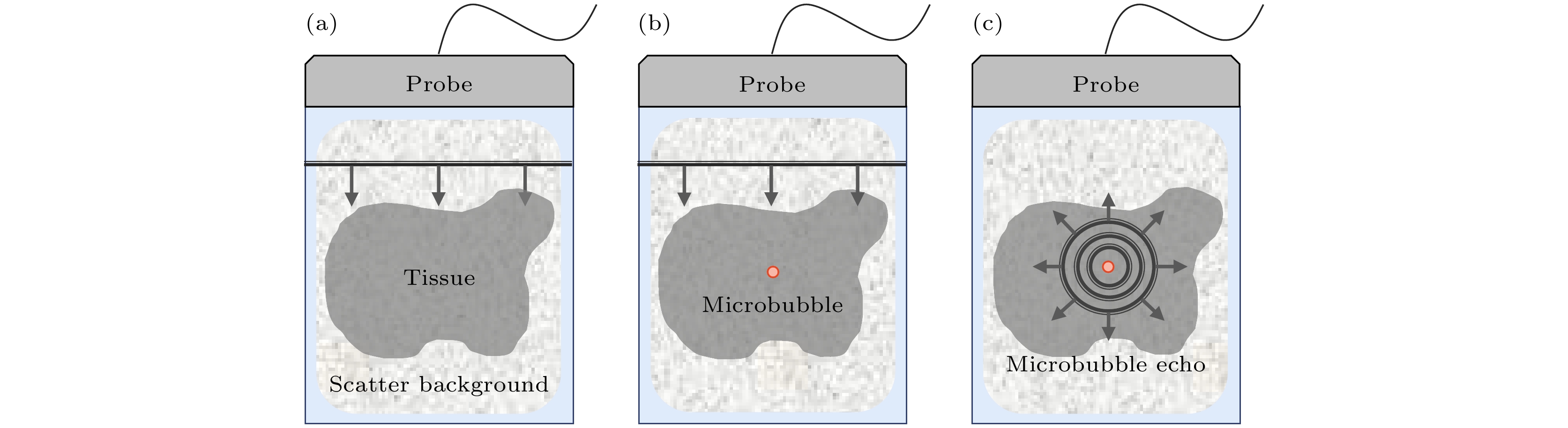
图 1 仿真方法部分步骤(发射多角度平面波, 图中仅示意) (a) 步骤1; (b) 步骤2; (c) 步骤3
Fig. 1. Schematic diagram of simulation method (transmitting multi-angle plane waves, the figure is only for illustration): (a) Step 1; (b) step 2; (c) step 3.

图 2 组织回波信号和微泡回波信号的频谱强度 (a) 时域; (b) 频域(f = 7.8)
Fig. 2. Spectra of tissue echo signal and microbubble echo signal: (a) Time domain; (b) frequency domain.

图 3 对比多脉冲序列成像策略处理后得到的微泡回波信号 (a) 时域; (b) 频域(f = 7.8)
Fig. 3. Microbubble echo signals after CPS strategies: (a) Time domain; (b) frequency domain.
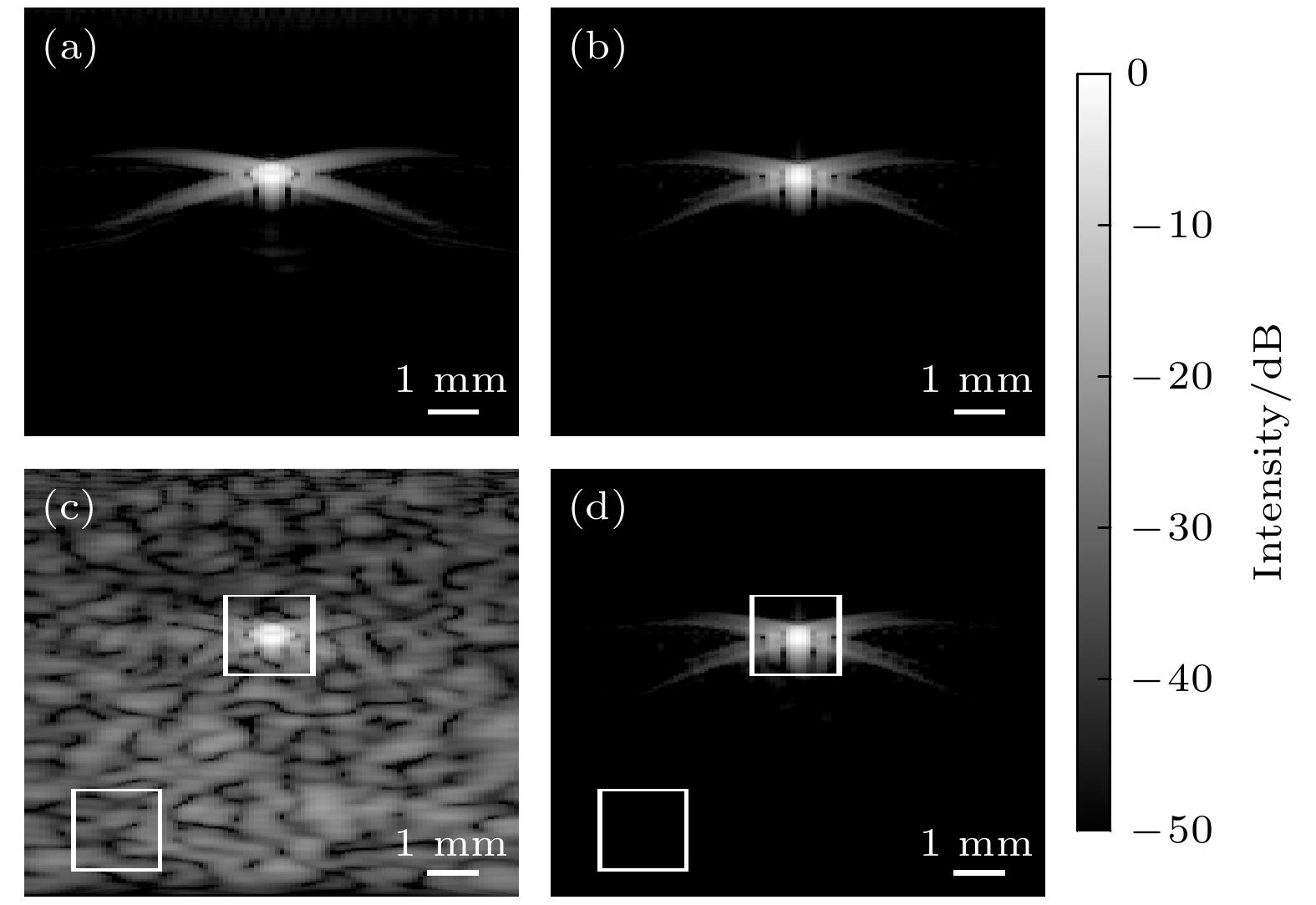
图 4 非线性超声仿真方法的微泡B超结果 (a) 无背景组织线性B超结果; (b) 无背景组织非线性B超结果; (c) 有背景组织线性B超结果; (d) 有背景组织非线性B超结果
Fig. 4. B-mode images of single microbubble under nonlinear ultrasound simulation method: (a) Linear B-mode result without tissue background; (b) nonlinear B-mode result without tissue background; (c) linear B-mode result with tissue background; (d) nonlinear B-mode result with tissue background.

图 5 非线性超声仿真方法的成簇微泡B超结果 (a) 有背景组织线性B超结果; (b) 有背景组织非线性B超结果
Fig. 5. B-mode images of clustered microbubbles under nonlinear ultrasound simulation method: (a) Linear B-mode result with tissue background; (b) nonlinear B-mode result with tissue background.
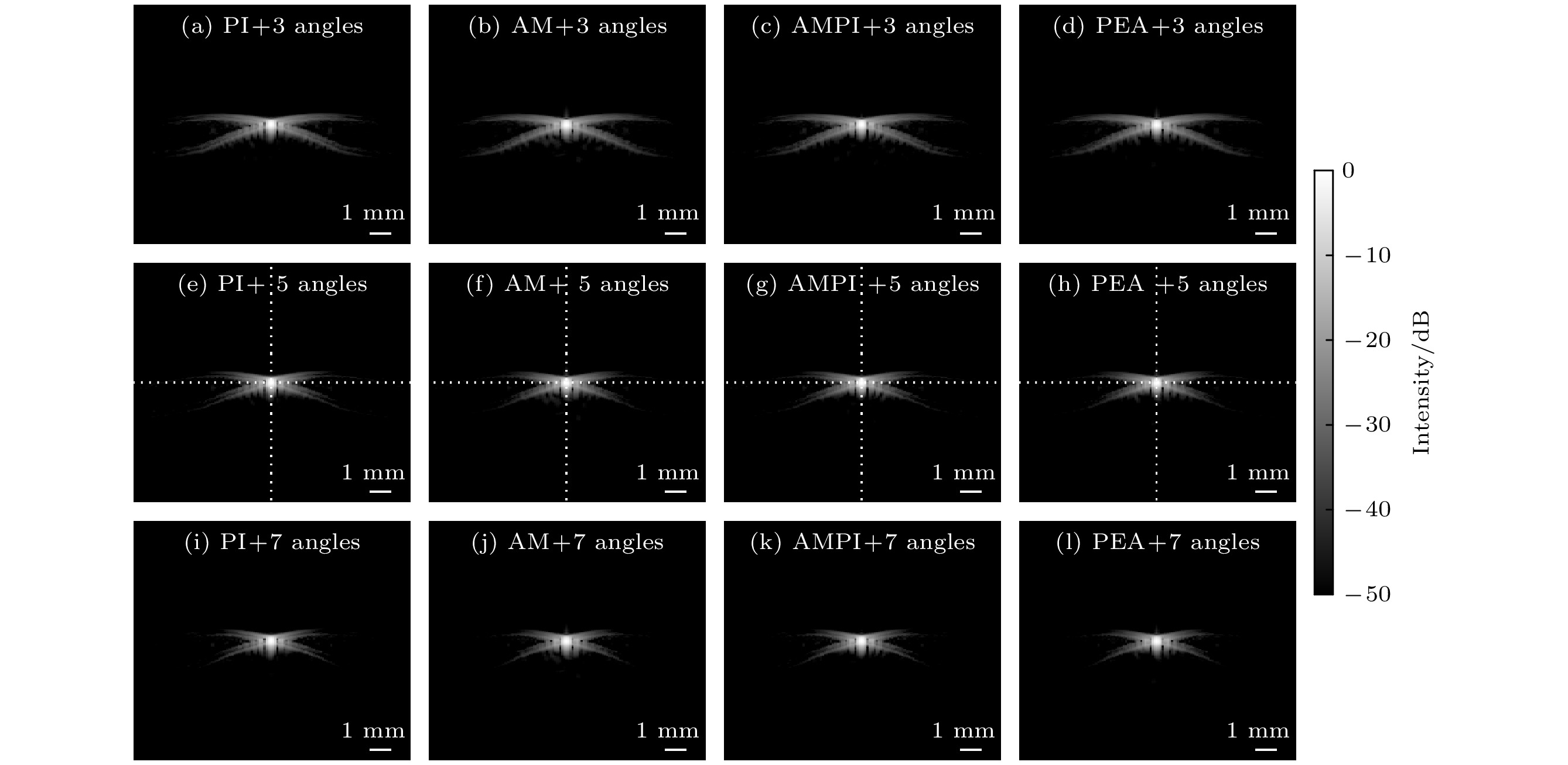
图 6 不同对比多脉冲序列成像策略下的B超结果 (a)—(d) 3个平面波发射角度; (e)—(h) 5个平面波发射角度; (i)—(l) 7个平面波发射角度
Fig. 6. B-mode images under different CPS strategies: (a)–(d) 3 emitting angles; (e)–(h) 5 emitting angles; (i)–(l) 7 emitting angles.
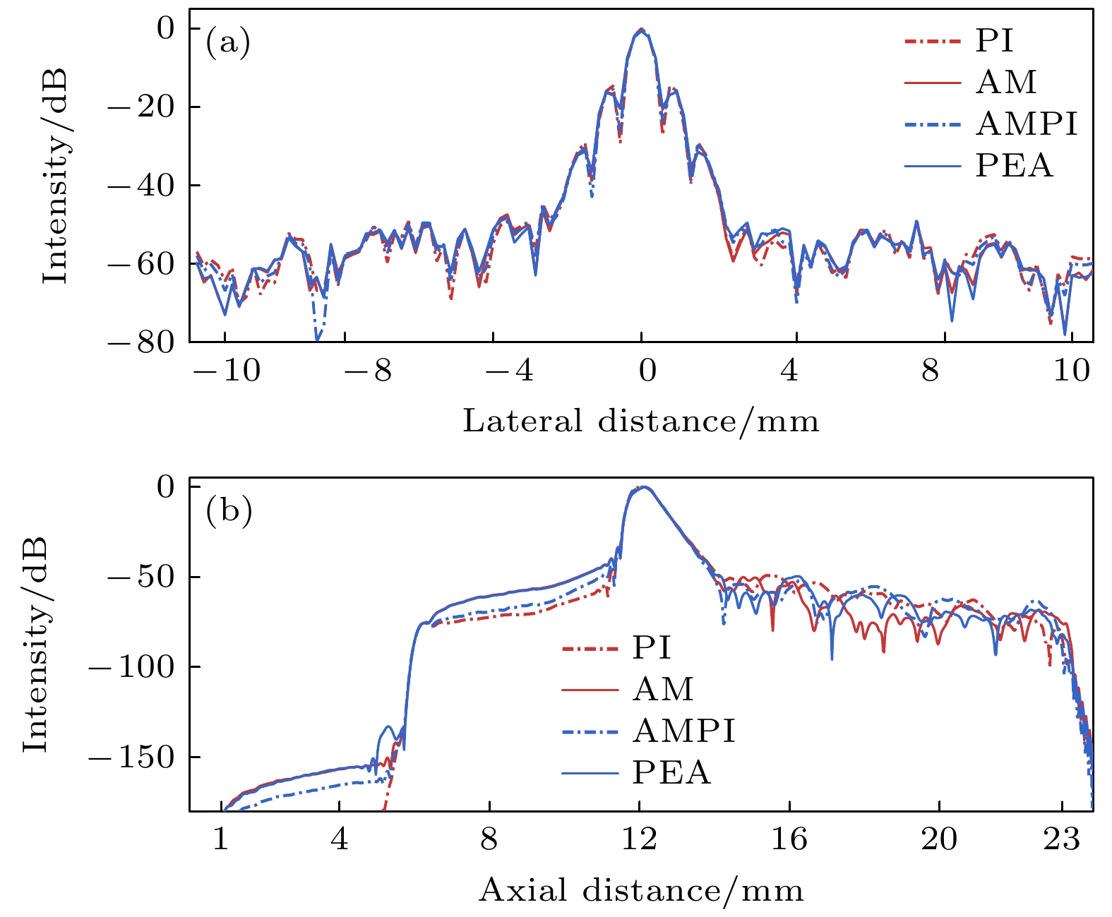
图 7 不同对比多脉冲序列成像策略下的点扩散函数 (a) 横向; (b) 纵向
Fig. 7. Point spread functions under different CPS strategies: (a) Lateral direction; (b) axial direction.
表 1 超声微泡仿真参数
Table 1. Simulation parameters of ultrasonic microbubble.
参数 数值 液体密度$ {\rho }_{0}/ $(${\rm{k} }{\rm{g} }{\cdot}{ {\rm{m} } }^{-3}$) $1055$ 表面张力$ {\sigma }_{0}/ $(${\rm{N} }{\cdot}{ {\rm{m} } }^{-1}$) $ 0.073 $ 液体黏度$ {\mu }_{0}/ $(${10}^{-3}\;{\rm{k} }{\rm{g} }{\cdot}{ {\rm{m} } }^{-1}{\cdot}{ {\rm{s} } }^{-1}$) $ 2 $ 液体声速$ {c}_{0}/ $(${\rm{m} }{\cdot}{ {\rm{s} } }^{-1}$) $1540$ 气体黏度指数 $ \nu $ $ 1.07 $ 壳黏度$ {\nu }_{{\rm{s}}}/ $(${10}^{-9}\;{\rm{k} }{\rm{g} }{\cdot}{ {\rm{s} } }^{-1}$) $ 6 $ 壳弹性模量$ \chi / $(${\rm{N} }{\cdot}{ {\rm{m} } }^{-1}$) $ 0.81 $ 环境压强$ {P}_{0}/ $(${\rm{N} }{\cdot}{ {\rm{m} } }^{-2}$) $101325$  下载: 导出CSV
表 2 不同策略下超声仿真成像的对比噪声比
Table 2. CNR of different strategies under ultrasound simulation.
成像方法 对比噪声比/dB 平面波超声成像 –0.65 PI 9.40 AM 9.90 AMPI 9.96 PEA 9.51
下载: 导出CSV
-
[1] Stanziola A, Toulemonde M, Yildiz Y O, Eckersley R J, Tang M X 2016 IEEE Signal Process Mag. 33 111
Google Scholar
[2] 郁钧瑾, 郭星奕, 隋怡辉, 宋剑平, 他得安, 梅永丰, 许凯亮 2022 物理学报 71 174302
Google Scholar
Yu J J, Guo X Y, Sui Y H, Song J P, Ta D A, Mei Y F, Xu K L 2022 Acta Phys. Sin. 71 174302
Google Scholar
[3] Guo X Y, Ta D A, Xu K L 2023 Ultrasonics 132 107009
Google Scholar
[4] 隋怡晖, 郭星奕, 郁钧瑾, Solovev A A, 他得安, 许凯亮 2022 物理学报 71 224301
Google Scholar
Sui Y H, Guo X Y, Yu J J, Solovev A A, Ta D A, Xu K L 2022 Acta Phys. Sin. 71 224301
Google Scholar
[5] Averkiou M A, Bruce M F, Powers J E, Sheeran P S, Burns P N 2020 Ultrasound Med. Biol. 46 498
Google Scholar
[6] Duck F A 2002 Ultrasound Med. Biol. 28 1
Google Scholar
[7] Brock-Fisher G A, Poland M D, Rafter P G 1996 US Patent 5577505
[8] Juin-Jet H, David H S 1999 US Patent 5951478
[9] Haider B, Chiao R Y 1999 IEEE International Ultrasonics Symposium (IUS) Tahoe, NV, USA, August 6, 2002 p1527
[10] Mor-Avi V, Caiani E G, Collins K A, Korcarz C E, Bednarz J E, Lang R M 2001 Circulation 104 352
Google Scholar
[11] Bouakaz A, Frigstad S, Ten-Cate F J, de-Jong N 2002 Ultrasound Med. Biol. 28 59
Google Scholar
[12] 刘贵栋, 沈毅, 王艳 2004 哈尔滨工业大学学报 36 599
Liu G D, Shen Y, Wang Y 2004 Journal of Harbin Institute of Technology 36 599
[13] 胡兵, 李佳, 应涛, 周永昌 2009 现代实用医学 21 299
Hu B, Li J, Ying T, Zhou Y C 2009 Modern Practical Medicine 21 299
[14] Couture O, Fink M, Tanter M 2012 IEEE Trans. Ultrason. Ferroelectr. Freq. Control 59 2676
Google Scholar
[15] Maresca D, Skachkov I, Renaud G, Jansen K, van Soest G, de-Jong N, van der-Steen A F 2014 Ultrasound Med. Biol. 40 1318
Google Scholar
[16] Muleki-Seya P, Xu K L, Tanter M, Couture O 2020 IEEE Trans. Ultrason. Ferroelectr. Freq. Control 67 598
Google Scholar
[17] Brown K G, Hoyt K 2021 IEEE Trans. Ultrason. Ferroelectr. Freq. Control 68 3347
Google Scholar
[18] Jenson J A 1996 Med. Biol. Eng. Comput. 34 351
Google Scholar
[19] Hallaj I M, Cleveland R O 1999 J. Acoust. Soc. Am. 105 7
Google Scholar
[20] Padilla F, Bossy E, Haiat G, Jenson F, Laugier P 2006 Ultrasonics 44 239
Google Scholar
[21] Treeby B E, Jaros J, Rendell A P, Cox B T 2012 J. Acoust. Soc. Am. 131 4324
Google Scholar
[22] 余锦华, 汪源源 2011 声学技术 30 33
Yu J H, Wang Y Y 2011 Technical Acoustics 30 33
[23] Martin E, Jaros J, Treeby B E 2020 IEEE Trans. Ultrason. Ferroelectr. Freq. Control 67 81
Google Scholar
[24] Leighton T 2012 The Acoustic Bubble (Massachusetts: Academic press) p1
[25] de-Jong N, Frinking P J A, Bouakaz A, Ten-Cate FJ 2000 Ultrasonics 38 87
Google Scholar
[26] Mezrich R 1995 Radiology 195 297
Google Scholar
[27] Treeby B E, Cox B T 2010 J. Biomed. Opt. 15 021314
Google Scholar
[28] de-Jong N, Hoff L, Skotland T, Bom N 1992 Ultrasonics 30 95
Google Scholar
[29] de-Jong N, Hoff L 1993 Ultrasonics 31 175
Google Scholar
[30] de-Jong N, Cornet R, Lancée C T 1994 Ultrasonics 32 447
Google Scholar
[31] Frinking P J A, de-Jong N, Céspedes E I 1999 J. Acoust. Soc. Am. 105 1989
Google Scholar
[32] de-Jong N, Bouakaz A, Frinking P J A 2002 Echocardiography 19 229
Google Scholar
[33] Plesset M S 1949 J. Appl. Mech. 16 277
Google Scholar
[34] Marmottant P, Meer S V D, Emmer M, Versluis M 2005 J. Acoust. Soc. Am. 118 3499
Google Scholar
[35] Tang M X, Eckersley R J 2006 IEEE Trans. Ultrason. Ferroelectr. Freq. Control 53 2406
Google Scholar
[36] Versluis M, Stride E, Lajoinie G, Dollet B, Segers T 2020 Ultrasound Med. Biol. 46 2117
Google Scholar
[37] Brown J, Christensen-Jeffries K, Harput S, Tang M X, Dunsby C, Eckersley R 2019 IEEE Trans. Ultrason. Ferroelectr. Freq. Control 66 676
Google Scholar
[38] Garcia D, Le-Tarnec L, Muth S, Montagnon E, Poree J, Cloutier G 2013 IEEE Trans. Ultrason. Ferroelectr. Freq. Control 60 1853
Google Scholar

 下载:
下载:
计量
- 文章访问数: 1747
- PDF下载量: 80
- 被引次数: 0
