


-
Magnetic resonance imaging is a medical imaging technique in which the phenomenon of nuclear magnetic resonance is used. This technique is widely used in clinical and scientific research at present. The diffusion of water molecules is isotropic in a homogeneous medium, while it is anisotropic in the structure of human tissue. Magnetic resonance diffusion tensor imaging (DTI) is for studying the microscopic structure inside body by using the water molecules’ diffusion effect which will reduce the signal intensity of magnetic resonance. Besides, it can quantitatively study the anisotropy of water molecules in three-dimensional space, and thus acquiring important pathological and physiological information without invading in vivo. In order to obtain the accurate result of the anisotropic diffusion of water molecules, according to a certain scheme, it is necessary to sequentially use diffusion sensitive gradient (DSG) magnetic fields in different diffusion orientations to measure the diffusion coefficient of water molecules for estimating the diffusion tensor. The precision of estimating diffusion tensor is affected by the applied DSG encoding scheme, and it is usually necessary to use a large number of linearly independent and evenly spatial distributions of DSG magnetic fields in order to make the tensor measurement result more accurate. Diffusion spectroscopy imaging technique and high angular resolution diffusion imaging (HARDI) technique are proposed for more complex fiber bundles crossing in human tissue, one of which, HARDI, has higher requirement for the number and the direction distribution uniformity of DSGs. In this paper, the basic principle of DTI and the DSG encoding schemes are reviewed, which includes completely random scheme, heuristic scheme, regular polyhedral scheme, numerically optimized scheme, etc. For the above various schemes their respective advantages and limitations are analyzed. At present, the Golden Ratio method is to be used in a new spherical DSG encoding scheme which meets the requirements for HARDI and can offer more accurate tensor estimation results in face of the corruption of data sets encountered in clinical practice.
[1] Le Bihan D, Mangin J F, Poupon C, Clark C A, Pappata S, Molko N, Chabriat H 2001 J. Magn. Reson. Imaging 13 534
 Google Scholar
[2] Basser P J, Mattiello J, LeBihan D 1994 Biophys. J. 66 259
Google Scholar
[3] Mattiello J, Basser P J, Lebihan D 1994 J. Magn. Reson., Ser. A 108 131
Google Scholar
[4] Alexander D C, Dyrby T B, Nilsson M, Zhang H 2019 NMR Biomed. 32 e3841
Google Scholar
[5] Novikov D S, Fieremans E, Jespersen S N, Kiselev V G 2019 NMR Biomed. 32 e3998
Google Scholar
[6] Tae W S, Ham B J, Pyun S B, Kang S H, Kim B J 2018 Clin. Neurol. 14 129
Google Scholar
[7] Okita G, Ohba T, Takamura T, Ebata S, Ueda R, Onishi H, Hori M 2018 Spine J. 8 268
Google Scholar
[8] Tuch D S, Weiskoff R M, Belliveau J W, Wedeen V J 1999 Proceedings of the 7 th Annual Meeting of ISMRM Philadelphia, USA, May 24−28, 1999 p321
[9] Torrey H C 1956 Phys. Rev. 104 563
Google Scholar
[10] Stejskal E O, Tanner J E 1965 J. Chem. Phys. 42 288
Google Scholar
[11] O’Donnell L J, Westin C F 2011 Neurosurg. Clin. N. Am. 22 185
Google Scholar
[12] Le Bihan D 1991 Magn. Reson. Q. 7 1
[13] Jones D K 2004 Magn. Reson. Med. 51 807
Google Scholar
[14] Hasan K M, Basser P J, Parker D L, Alexander A L 2001 Magn. Reson. 152 41
Google Scholar
[15] Westin C F, Maier S E, Mamata H, Nabavi A, Jolesz F A, Kikinis R 2002 Med. Image Anal. 6 93
Google Scholar
[16] Jonse D K, Knosche T R, Turner R 2013 NeuroImage 73 239
Google Scholar
[17] 张首誉, 包尚联, 亢孝俭 2013 物理学报 62 208703
Zhang S Y, Bao S L, Kang X J 2013 Acta Phys. Sin. 62 208703
[18] Basser P J 1995 NMR Biomed. 8 333
Google Scholar
[19] Basser P J, Mattiello J, Le Bihan D 1994 J. Magn. Reson. Ser. B 103 247
Google Scholar
[20] Jones D K, Horsfield M A, Simmons A 1999 Magn. Reson. Med. 42 515
Google Scholar
[21] Hasan K M, Parker D L, Alexander A L 2001 J. Magn. Reson. Imaging 13 769
Google Scholar
[22] Dubois J, Poupon C, Lethimonnier F, Le Bihan D 2006 Magn. Reson. Mater. Phys., Biol. Med. 19 134
Google Scholar
[23] Zhan L, Leow A.D, Jahanshad N, Chiang M C, Barysheva M, Lee A D, Toga A W, McMahon K L, Zubicaray G I, Wright M J, Thompson P M 2010 NeuroImage 47 1357
Google Scholar
[24] 高嵩, 朱艳春, 李硕, 包尚联 2014 物理学报 63 048704
Google Scholar
Gao S, Zhu Y C, Li S, Bao S L 2014 Acta Phys. Sin. 63 048704
Google Scholar
[25] Alderman D W, Sherwood M, Grant D 1990 J. Magn. Reson. 86 60
Google Scholar
[26] Basser P J, Pierpaoli C 1998 Magn. Reson. Med. 39 928
Google Scholar
[27] Conturo T E, McKinstry R C, Akbudak E, Robinson B H 1996 Magn. Reson. Med. 35 399
Google Scholar
[28] Shimony J S, McKinstry R C, Akbudak E, Aronovitz J A, Snyder A Z, Lori N F, Conturo T E 1999 Radiology 212 770
Google Scholar
[29] Nye J F 1985 Physical Properties of Crystals (Oxford: Clarendon Press) pp150−169
[30] Skare S, Nordell B 1999 Proceedings of the 7th Annual Meeting of ISMRM Philadelphia, USA, May 24−28, 1999 p322
[31] Kingsley P B 2006 Concepts Magn. Reson. A 28 123
Google Scholar
[32] Skare S, Hedehus M, Moseley M E, Li T Q 2000 J. Magn. Reson. 147 340
Google Scholar
[33] Batchelor P G, Atkinson D, Hill D L G, Calamante F, Connelly A 2003 Magn. Reson. Med. 49 1143
Google Scholar
[34] Papadakis N G, Xing D, Houston G C, Smith J M, Smith M I, James M F, Carpenter T A 1999 Magn. Reson. Imaging 17 881
Google Scholar
[35] Hasan K M, Parker D L, Alexander A L 2002 Image Anal. Stereol. 21 87
Google Scholar
[36] Akkerman E M 2003 Magn. Reson. Med. 49 599
Google Scholar
[37] Hasan K M, Narayana P A 2003 Magn. Reson. Med. 50 589
Google Scholar
[38] Tegmark M 1996 Astrophys. J. 470 L81
Google Scholar
[39] Sibley T Q 1998 The Geometric Viewpoint (New York: Addison-Wesley) pp41−45
[40] Stirnberg R, Stöcker T, Shah N J 2009 Proc. Intl. Soc. Mag. Reson. Med. 17 3574
[41] Wong S T S, Roos M S 1994 Magn. Reson. Med. 32 778
Google Scholar
[42] Jones D K, Leemans A 2011 Magnetic Resonance Neuroimaging (New York: Humana Press) pp127−144
[43] Cook P A, Symms M, Boulby P A, Alexander D C 2007 J. Magn. Reson. Imaging 25 1051
Google Scholar
[44] Alipoor M, Gu I Y 2015 IEEE 12th International Symposium on Biomedical Imaging New York, USA April 16−19, 2015 p959
[45] Dubois J, Cointepas Y, Poupon C, Lethimonnier F, Le Bihan D 2004 Proceedings of the 12th Annual Meeting of ISMRM Kyoto, Japan, May 15−21, 2004 p443
[46] Cook P A, Boulby P A, Symms M R, Alexander D C 2005 Proceedings of the 13th Annual Meeting of ISMRM Miami, USA, May 7−13, 2005 p1303
[47] Cook P A, Bai Y, Nedjati-Gilani S K K S, Seunarine K K, Hall M G, Parker G J, Alexander D C 2006 Proceedings 14th Scientific Meeting of the International Society for Magnetic Resonance in Medicine Seattle, Washington, USA, May 6−12, 2006 p2759
[48] Farquharson S, Tournier J D 2016 Diffusion Tensor Imaging (New York: Springer-Verlag) pp383−406
[49] Varentsova A, Zhang S, Arfanakis K 2014 NeuroImage 91 177
Google Scholar
[50] Sherbaf F G, Same K, Aarabi M H 2018 Acta Neurol. Belg. 118 573
Google Scholar
[51] -
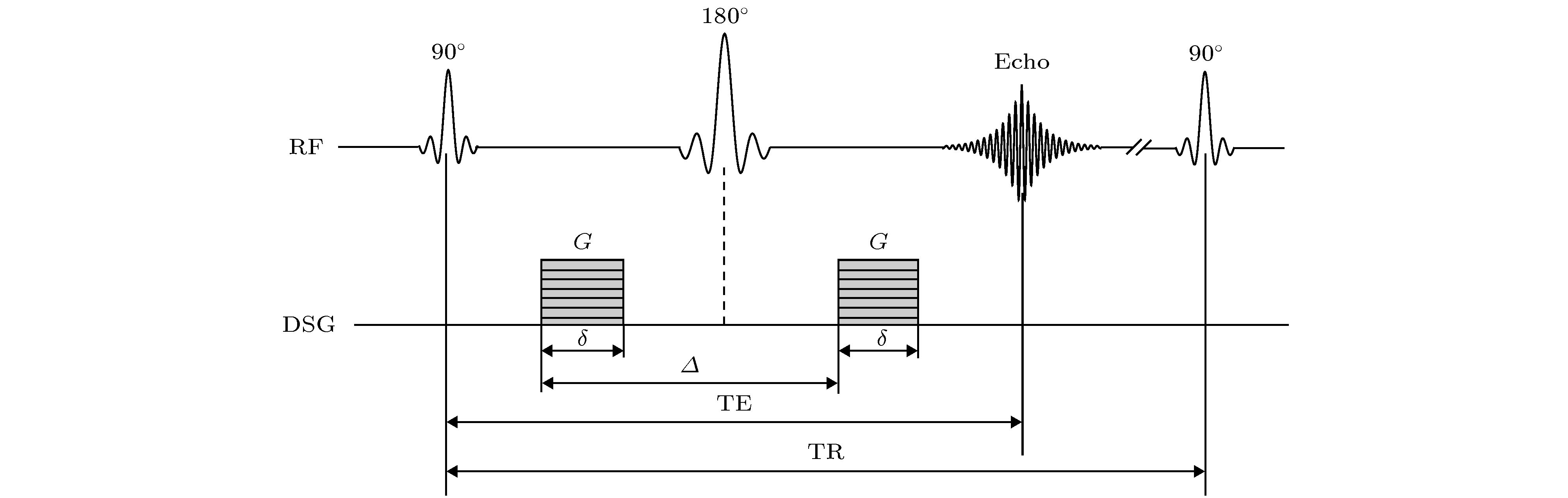
图 1 Stejskal-Tanner序列, 其中两个同等的DSG脉冲置于180°RF脉冲两侧, 强度为G, 宽度为δ, 时间间隔为Δ
Fig. 1. Stejskal-Tanner Scheme: Two diffusion sensitive gradients inserted before and after 180° RF refocusing pulse. G, amplitude; δ, duration of the DSG; Δ, time between the two sensitive gradient lobes.

图 2 随机分布60个方向DSG方向分布方案
Fig. 2. DSG encoding scheme with random distribution in 60 directions.

图 3 完全启发式13个方向DSG方向分布方案
Fig. 3. DSG encoding scheme with heuristic distribution in 13 directions.
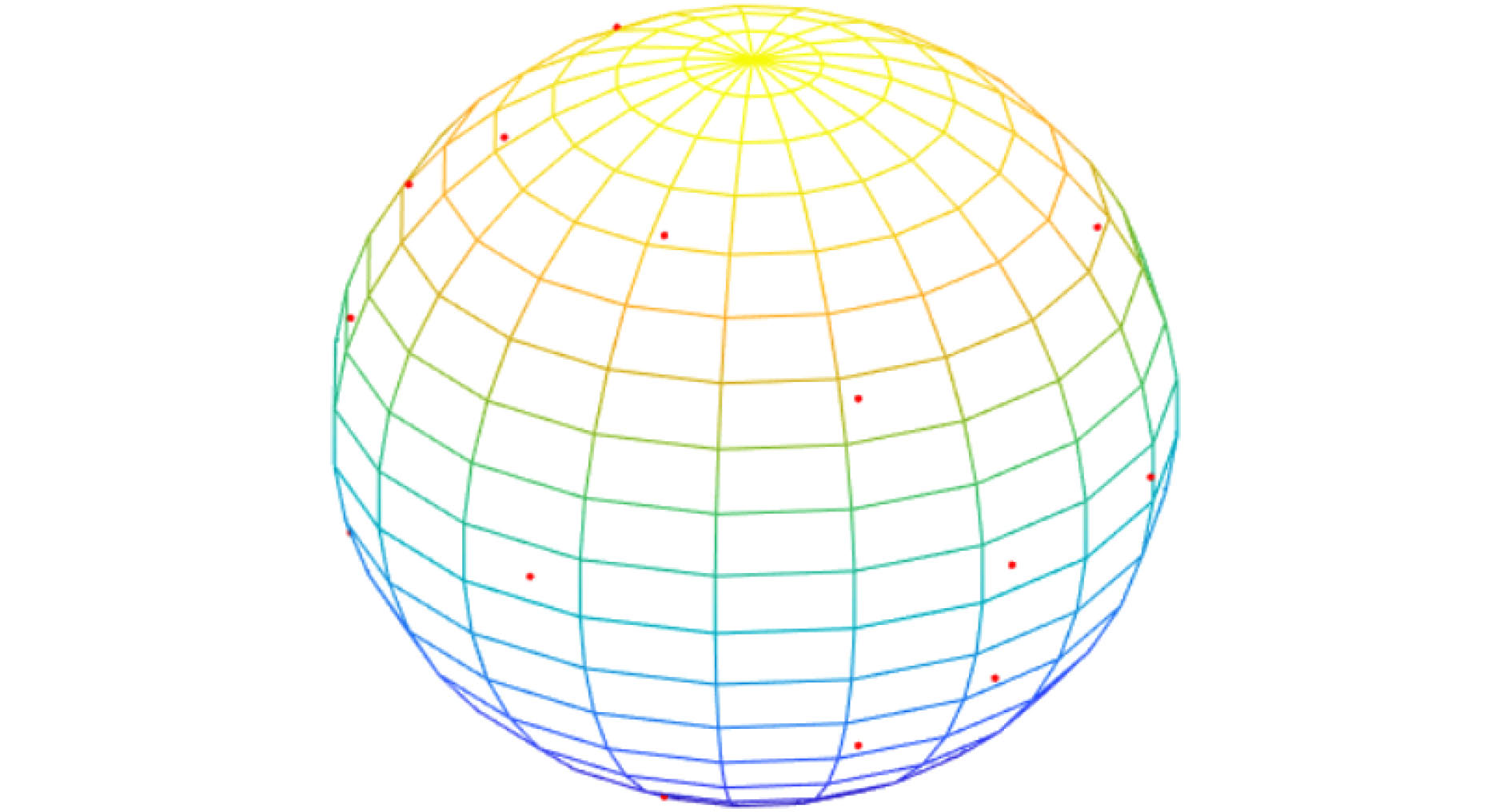
图 4 二十面体31个方向DSG方向分布方案
Fig. 4. DSG encoding scheme with icosahedron distribution in 31 directions.

图 5 DISCOBALL 30个方向DSG方向分布方案
Fig. 5. DSG encoding scheme with DISCOBALL distribution in 30 directions.
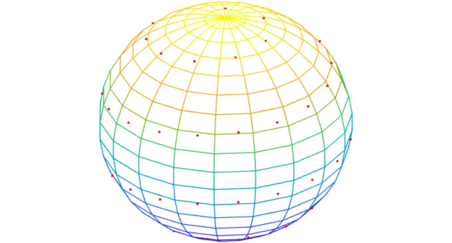
图 6 球面螺旋分布60个方向DSG方向分布方案
Fig. 6. DSG encoding scheme with spherical spiral distribution in 60 directions.
-
[1] Le Bihan D, Mangin J F, Poupon C, Clark C A, Pappata S, Molko N, Chabriat H 2001 J. Magn. Reson. Imaging 13 534
Google Scholar
[2] Basser P J, Mattiello J, LeBihan D 1994 Biophys. J. 66 259
Google Scholar
[3] Mattiello J, Basser P J, Lebihan D 1994 J. Magn. Reson., Ser. A 108 131
Google Scholar
[4] Alexander D C, Dyrby T B, Nilsson M, Zhang H 2019 NMR Biomed. 32 e3841
Google Scholar
[5] Novikov D S, Fieremans E, Jespersen S N, Kiselev V G 2019 NMR Biomed. 32 e3998
Google Scholar
[6] Tae W S, Ham B J, Pyun S B, Kang S H, Kim B J 2018 Clin. Neurol. 14 129
Google Scholar
[7] Okita G, Ohba T, Takamura T, Ebata S, Ueda R, Onishi H, Hori M 2018 Spine J. 8 268
Google Scholar
[8] Tuch D S, Weiskoff R M, Belliveau J W, Wedeen V J 1999 Proceedings of the 7 th Annual Meeting of ISMRM Philadelphia, USA, May 24−28, 1999 p321
[9] Torrey H C 1956 Phys. Rev. 104 563
Google Scholar
[10] Stejskal E O, Tanner J E 1965 J. Chem. Phys. 42 288
Google Scholar
[11] O’Donnell L J, Westin C F 2011 Neurosurg. Clin. N. Am. 22 185
Google Scholar
[12] Le Bihan D 1991 Magn. Reson. Q. 7 1
[13] Jones D K 2004 Magn. Reson. Med. 51 807
Google Scholar
[14] Hasan K M, Basser P J, Parker D L, Alexander A L 2001 Magn. Reson. 152 41
Google Scholar
[15] Westin C F, Maier S E, Mamata H, Nabavi A, Jolesz F A, Kikinis R 2002 Med. Image Anal. 6 93
Google Scholar
[16] Jonse D K, Knosche T R, Turner R 2013 NeuroImage 73 239
Google Scholar
[17] 张首誉, 包尚联, 亢孝俭 2013 物理学报 62 208703
Zhang S Y, Bao S L, Kang X J 2013 Acta Phys. Sin. 62 208703
[18] Basser P J 1995 NMR Biomed. 8 333
Google Scholar
[19] Basser P J, Mattiello J, Le Bihan D 1994 J. Magn. Reson. Ser. B 103 247
Google Scholar
[20] Jones D K, Horsfield M A, Simmons A 1999 Magn. Reson. Med. 42 515
Google Scholar
[21] Hasan K M, Parker D L, Alexander A L 2001 J. Magn. Reson. Imaging 13 769
Google Scholar
[22] Dubois J, Poupon C, Lethimonnier F, Le Bihan D 2006 Magn. Reson. Mater. Phys., Biol. Med. 19 134
Google Scholar
[23] Zhan L, Leow A.D, Jahanshad N, Chiang M C, Barysheva M, Lee A D, Toga A W, McMahon K L, Zubicaray G I, Wright M J, Thompson P M 2010 NeuroImage 47 1357
Google Scholar
[24] 高嵩, 朱艳春, 李硕, 包尚联 2014 物理学报 63 048704
Google Scholar
Gao S, Zhu Y C, Li S, Bao S L 2014 Acta Phys. Sin. 63 048704
Google Scholar
[25] Alderman D W, Sherwood M, Grant D 1990 J. Magn. Reson. 86 60
Google Scholar
[26] Basser P J, Pierpaoli C 1998 Magn. Reson. Med. 39 928
Google Scholar
[27] Conturo T E, McKinstry R C, Akbudak E, Robinson B H 1996 Magn. Reson. Med. 35 399
Google Scholar
[28] Shimony J S, McKinstry R C, Akbudak E, Aronovitz J A, Snyder A Z, Lori N F, Conturo T E 1999 Radiology 212 770
Google Scholar
[29] Nye J F 1985 Physical Properties of Crystals (Oxford: Clarendon Press) pp150−169
[30] Skare S, Nordell B 1999 Proceedings of the 7th Annual Meeting of ISMRM Philadelphia, USA, May 24−28, 1999 p322
[31] Kingsley P B 2006 Concepts Magn. Reson. A 28 123
Google Scholar
[32] Skare S, Hedehus M, Moseley M E, Li T Q 2000 J. Magn. Reson. 147 340
Google Scholar
[33] Batchelor P G, Atkinson D, Hill D L G, Calamante F, Connelly A 2003 Magn. Reson. Med. 49 1143
Google Scholar
[34] Papadakis N G, Xing D, Houston G C, Smith J M, Smith M I, James M F, Carpenter T A 1999 Magn. Reson. Imaging 17 881
Google Scholar
[35] Hasan K M, Parker D L, Alexander A L 2002 Image Anal. Stereol. 21 87
Google Scholar
[36] Akkerman E M 2003 Magn. Reson. Med. 49 599
Google Scholar
[37] Hasan K M, Narayana P A 2003 Magn. Reson. Med. 50 589
Google Scholar
[38] Tegmark M 1996 Astrophys. J. 470 L81
Google Scholar
[39] Sibley T Q 1998 The Geometric Viewpoint (New York: Addison-Wesley) pp41−45
[40] Stirnberg R, Stöcker T, Shah N J 2009 Proc. Intl. Soc. Mag. Reson. Med. 17 3574
[41] Wong S T S, Roos M S 1994 Magn. Reson. Med. 32 778
Google Scholar
[42] Jones D K, Leemans A 2011 Magnetic Resonance Neuroimaging (New York: Humana Press) pp127−144
[43] Cook P A, Symms M, Boulby P A, Alexander D C 2007 J. Magn. Reson. Imaging 25 1051
Google Scholar
[44] Alipoor M, Gu I Y 2015 IEEE 12th International Symposium on Biomedical Imaging New York, USA April 16−19, 2015 p959
[45] Dubois J, Cointepas Y, Poupon C, Lethimonnier F, Le Bihan D 2004 Proceedings of the 12th Annual Meeting of ISMRM Kyoto, Japan, May 15−21, 2004 p443
[46] Cook P A, Boulby P A, Symms M R, Alexander D C 2005 Proceedings of the 13th Annual Meeting of ISMRM Miami, USA, May 7−13, 2005 p1303
[47] Cook P A, Bai Y, Nedjati-Gilani S K K S, Seunarine K K, Hall M G, Parker G J, Alexander D C 2006 Proceedings 14th Scientific Meeting of the International Society for Magnetic Resonance in Medicine Seattle, Washington, USA, May 6−12, 2006 p2759
[48] Farquharson S, Tournier J D 2016 Diffusion Tensor Imaging (New York: Springer-Verlag) pp383−406
[49] Varentsova A, Zhang S, Arfanakis K 2014 NeuroImage 91 177
Google Scholar
[50] Sherbaf F G, Same K, Aarabi M H 2018 Acta Neurol. Belg. 118 573
Google Scholar
[51]

 下载:
下载:

计量
- 文章访问数: 15364
- PDF下载量: 101
- 被引次数: 0
