


-
By designing and fabricating a narrow-band Fabry-Perot multi-beam interference spectroscopic microcavity array, and integrating it with a visible light detector focal plane array, we demonstrate a small compact multispectral imaging detector. The micro-cavity filter array with 4×4 basic repeating units and a total of 2048×2048 pixels is obtained on a quartz substrate by the four-fractal combination lithograph-etching process. Then the micro miniatured multispectral imaging detector is formed by fitting with the detector chip. The depth and precision of the etching will determine the distribution and offset of the central wavelength of the narrowband spectral channel respectively. The results show that the etching rate of reactive ion is (3.6 ± 0.2) Å/s, and the process is stable and controllable. Due to the different etching depths, the basic repeating unit forms 16 different levels of steps, and the process achieves the design expectation well. The results are obtained as follows: the response spectrum peak of the microcavity array sample varies from 520 to 680 nm, the free spectrum range is 160 nm, the full width at the half-peak is less than 10 nm, the transmittance is about 70%, the relative half-width of the transmittance peak at 590 nm is 1.19%, and the waveform coefficient is 2.78. A 16-channel multispectral camera is constructed by using the optical micro-precision assembly device to realize the precise alignment and the fitting of the micro-cavity filter array and the image sensor. Xenon lamp and monochromator are used as tunable wavelength monochromatic cooperative light source to detect the effect of 16-channel snapshot multispectral imaging on a pixel scale. The results show that the multispectral imaging detector has 16 different narrow-band response spectra. The characteristic spectrum of the target can be clearly distinguished by spectral channel. When imaging the target with known spectral characteristics, for a certain frame of multi-spectral image, selecting a suitable spectral channel can eliminate the background in the field of view through image subtraction and improve the contrast of the target. In the dark room condition, we take the LED light source with center wavelength varying in a range between 528 and 589 nm as the target (the wavelength coincides with the working wavelength of the spectral channel), and effectively suppress the background through the spectral differential intensity subtraction, which can improve the accuracy and sensitivity of the target capture. The 16-channel snapshot multi-spectral imaging detector based on integrated Fabry-Perot microcavity array has the advantages of small size, high integration and strong environmental adaptability, and is expected to play a role in realizing the real-time detection of weak moving targets, auxiliary diagnosis of skin surface observation, and high dynamic range imaging of target observation under backlight conditions. -
Keywords:
- microcavity /
- snapshot /
- multi spectral imaging /
- detector
[1] Janssen P J C 1869 C. R. Acad. Sci. 68 93
[2] Fabry C, Pérot A 1897 Ann. Chim. Phys. 12 459
[3] Hagen N, Kudenov M W 2013 Opt. Eng. 52 090901
 Google Scholar
[4] Williams C, Gordon G S D, Wilkinson T D, Bohndiek S E 2019 ACS Photonics 6 3132
Google Scholar
[5] 周奎, 单政, 张倩, 王谢君, 周健, 邓宸伟, 虞益挺 2022 光学学报 42 080000
Google Scholar
Zhou K, Shan Z, Zhang Q, Wang X J, Zhou J, Deng C W, Yu Y T 2022 Acta Opt. Sin. 42 080000
Google Scholar
[6] Wang S W, Li M, Xia C S, Wang H Q, Chen X S, Lu W 2007 Appl. Phys. B 88 281
Google Scholar
[7] 王绪权, 黄松垒, 于月华, 叶捷敏, 邵秀梅, 方家熊 2018 光子学报 47 530001
Google Scholar
Wang X Q, Huang S L, Yu Y H, Ye J M, Shao X M, Fang J X 2018 Acta Photonica Sin. 47 530001
Google Scholar
[8] Xuan Z Y, Liu Q Q, Cui Z Z, Huang S L, Yang B, Li C L, Wang S W, Lu W 2022 Chin. Opt. Lett. 20 061302
Google Scholar
[9] Yin Z Q, Liu Q Q, Guan X Y, Xie M B, Lu W, Wang S W 2023 Opt. Lett. 48 3371
Google Scholar
[10] Xuan Z Y, Wang Z, Liu Q Q, Huang S L, Yang B, Yang L Y, Yin Z Q, Xie M B, Li C L, Yu J Y, Wang S W, Lu W 2022 Adv. Opt. Mater. 10 2200284
Google Scholar
[11] Liu Q Q, Xuan Z Y, Wang Z, Zhao X C, Yin Z Q, Li C L, Chen G, Wang S W, Lu W 2022 Opt. Lett. 47 2923
Google Scholar
[12] Liu Q Q, Li C L, Xie M B, Wang S W, Yang S M, Zuo Y, Zhao J, Xuan Z Y, Zhu Y Y, Li Z F, Wu Y Q, Lu W 2023 ACS Photonics 10 125
Google Scholar
[13] Li C L, Liu Q Q, Zhu Y Y, Zhao X C, Yang S M, Li Z F, Wang S W, Shen X C 2023 Nanoscale 15 11466
Google Scholar
[14] 肖树林, 胡长虹, 高路尧 2021 中国科技成果 23 26
Google Scholar
Xiao S L, Hu C H, Gao L Y 2021 China Achieve. Sci. Technol. 23 26
Google Scholar
[15] Kutteruf Mary R, Yetzbacher Michael K, Deprenger Michael J, Novak K M, Miller C A, Kanaev A, Henry D J, Lange D A, VonBerg D L, Rajan S R, Walls T J, Young D L 2014 Proc. SPIE 9076 90760M
Google Scholar
[16] Geelen B, Blanch C, Gonzalez P, Tack N, Lambrechts A 2015 Proc. SPIE 9374 937414
Google Scholar
[17] Zhang H, Cai Y, Chen Z L, Xie X L, Chang S L, Fang J Y 2020 Proc. SPIE 11567 115672P
Google Scholar
[18] Xiao G Y, Zhang H, Xie X L, Chang S L, Fang J Y 2021 Proc. SPIE 11763 117631Q
Google Scholar
[19] Thomas G 2021 Opt. Lett. 46 5926
Google Scholar
[20] 季家镕 2007 高等光学教程 (北京: 科学出版社) 第188页
Ji J R 2007 Advanced Optics Tutorial (Beijing: Science Press) p188
-

图 1 16通道快照式多光谱FP微腔阵列的制作工艺 (a) 底部分布式布拉格反射器(distributed Bragg reflector, DBR)和空腔层沉积在石英基底上; (b) 通过UV光刻和反应离子刻蚀组合工艺处理空腔层; (c) 顶部DBR沉积在处理过的空腔层上, 并形成具有4×4个不同FP腔厚度的微腔阵列
Figure 1. Fabrication process of a 16-channel snapshot type multispectral Fabry Perot microcavity array: (a) Bottom distributed Bragg reflector (DBR) and cavity layer are deposited on a quartz substrate; (b) cavity layer is processed by a combined process of UV photolithography and reactive ion etching; (c) top DBR is deposited on the treated cavity layer and formed with 4 × 4 microcavity arrays with different FP cavity thicknesses.

图 2 (a) 窄带膜系透射峰位置随FP腔厚度发生改变(∆h表示FP腔厚度的改变量); (b) 峰位改变量∆λ随厚度改变∆h的关系(红色直线为拟合结果)
Figure 2. (a) Transmission peak position of the narrowband film system changes with the thickness of the FP cavity (∆h represents the amount of change in the thickness of the FP cavity); (b) relationship between peak position change ∆λ and thickness change ∆h (Red line represents the fitting result).

图 3 (a) 依次使用A, B, C, D掩膜(以8×8阵列为例)对FP腔进行四步反应离子刻蚀的示意图; (b)—(e) 顺序进行的四步反应离子刻蚀的相对深度(颜色表示刻蚀区域, 数字表示台阶的相对高度)
Figure 3. (a) Using masks A, B, C, and D in sequence (with 8 × 8 array) schematic diagram of four step reactive-ion etching of FP cavity; (b)–(e) the relative depth of four steps of Reactive-ion etching in sequence (Color indicates the etching area, and number indicates the elevation of the steps).

图 4 (a)—(d)依次使用A, B, C, D掩膜进行刻蚀后, FP腔层(18×24微腔阵列区域)的光学显微图像
Figure 4. Optical microscopic image of FP cavity layer (18 × 24 microcavity array area) after etching with (a) A, (b) B, (c) C, and (d) D masks in sequence.
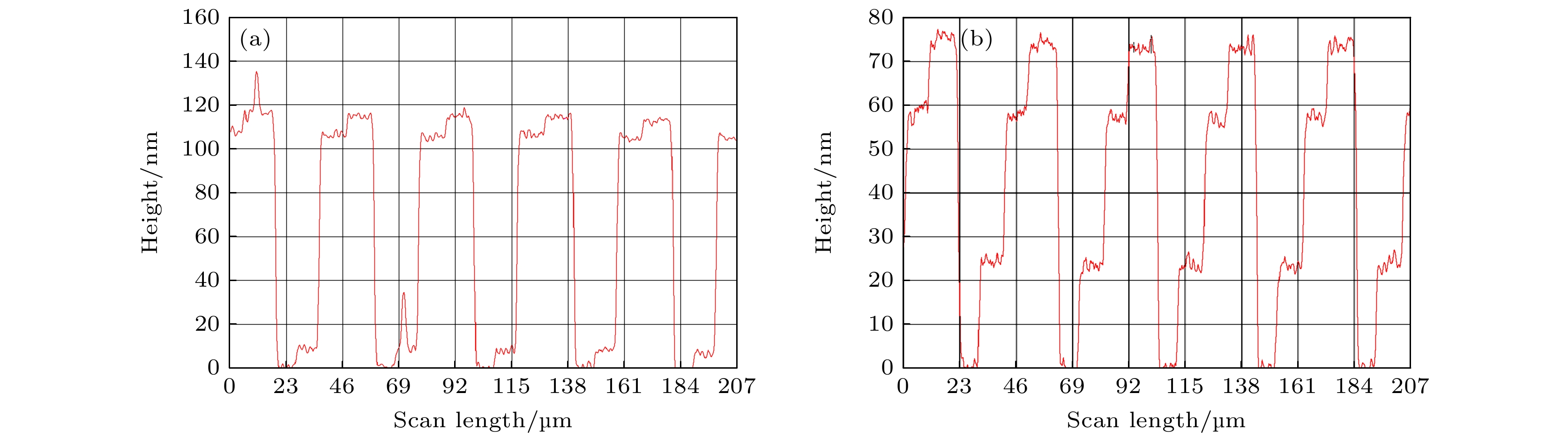
图 5 分别沿(a) A方向和(b) B方向扫描, 测量得到的样品台阶轮廓曲线
Figure 5. Measured step contour curves which are scanned along the (a) A and (b) B direction, respectively.
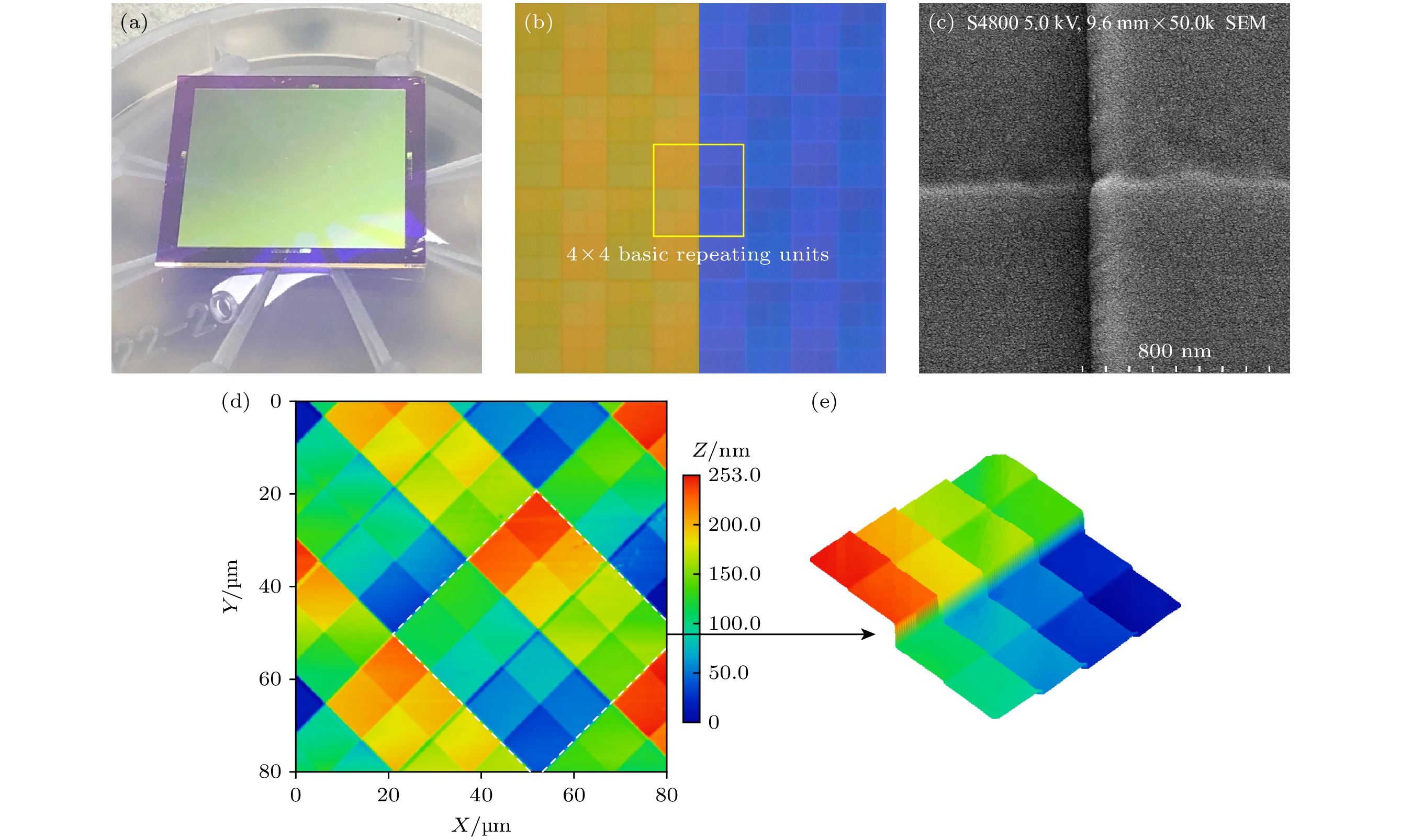
图 6 微腔阵列样品 (a) 实物照片; (b) 光学显微照片(左半部分为正片, 右半部分为负片); (c) 扫描电子显微图像; (d), (e) 原子力显微镜形貌图及局部放大的4×4重复单元的阶梯状三维形貌(横轴和纵轴是空间坐标; 颜色条表示AFM扫描的相对高度)
Figure 6. Microcavity array sample: (a) Physical photos; (b) optical micrographs (positive in the left half and negative in the right half); (c) scanning electron microscopy image of the Fabry Perot microcavity array sample; (d), (e) atomic force microscope morphology and step like three-dimensional morphology of locally enlarged 4×4 repeating units (Horizontal and vertical axes are spatial coordinates; the color bar indicates the relative height of the AFM scan).

图 7 微腔阵列样品(波长范围是520—680 nm)的(a)光谱通道中心波长设计值(虚线)与微区透射谱测量曲线(实线); (b)光谱通道中心波长相对设计值的偏离(以中心波长设计值的1%设置error bar); (c) 单个透射峰特性
Figure 7. (a) Design value of spectral channel center wavelength (dashed line) and measurement curve of micro region transmission spectrum (solid line) of the microcavity array sample (wavelength range 520–680 nm); (b) deviation of the center wavelength of the multispectral channel from the design value (set as error bar at 1% of the center wavelength design value); (c) single transmission peak characteristics.
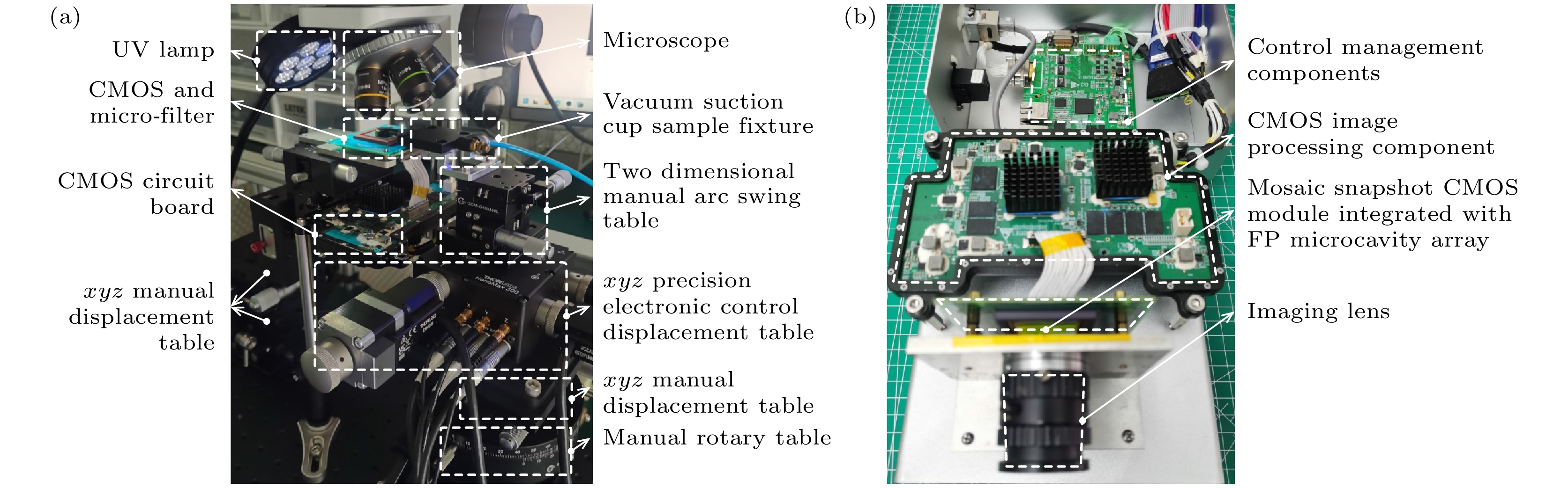
图 8 (a) 光学显微精密装配装置; (b) 多光谱相机
Figure 8. (a) Precision assembly device for optical microscopy; (b) multi spectral camera.
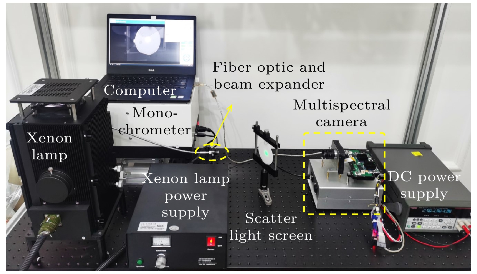
图 9 马赛克多通道光谱成像的检测光路
Figure 9. Detection optical path of mosaic multi-channel spectral imaging.

图 10 (a)—(p) 分别以553, 558, 567, 574, 582, 588, 593, 601, 608, 614, 627, 634, 643, 651, 663和546 nm单色光入射时, 某个4×4重复单元的像素级灰度响应结果
Figure 10. (a)–(p) Pixel level grayscale response results of a 4 × 4 repeating unit when incident with monochromatic light at 553, 558, 567, 574, 582, 588, 593, 601, 608, 614, 627, 634, 643, 651, 663, and 546 nm, respectively.

图 11 多光谱通道RGB颜色示意图
Figure 11. Schematic diagram of RGB color in multispectral channels.

图 12 (a), (b) 波长为529和540 nm的单色光入射引起的灰度响应
Figure 12. (a), (b) Grayscale response caused by monochromatic light incidence with wavelengths of 529 and 540 nm.

图 13 合作光源的多光谱观测结果
Figure 13. Multispectral observation results of cooperative light sources.
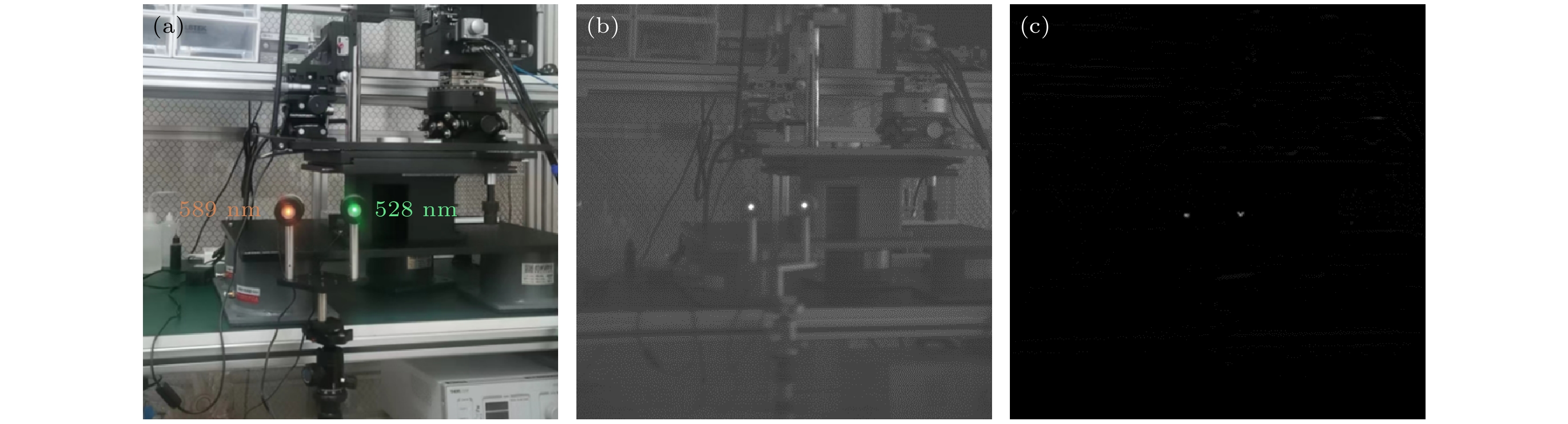
图 14 多光谱消背景观测结果 (a) 目标场景; (b) 多光谱图像; (c) 光谱差分结果
Figure 14. Multi spectral background cancellation observation results: (a) Target scenario; (b) multi spectral images; (c) spectral difference results.
表 1 沿A方向测得的高差
Table 1. Measured height difference along direction A.
Item Height difference/nm Etching time/s Etching rate/(Å·s–1) 1 2 3 4 5 6 7 8 Average ∆h1 9.1 9.5 9.2 10.3 9.2 8.9 9.7 9.4 9.41 28 3.36 Δh2 107.2 107.1 107.3 107.5 106.0 107.3 107.0 108.6 107.25 300 3.58 Δh3 113.5 113.0 116.2 123.6 112.0 115.0 113.3 119.3 115.74 328 3.53  DownLoad: CSV
表 2 沿B方向测得的高差
Table 2. Measured height difference along direction B.
Item Height difference/nm Etching time/s Etching rate/(Å·s–1) 1 2 3 4 5 6 7 8 Average ∆h1 24.7 23.5 25.0 24.3 24.5 24.3 26.0 24.4 24.59 70 3.51 Δh2 55.5 54.1 57.2 58.0 57.0 56.7 56.8 56.5 56.48 150 3.77 Δh3 76.5 76.5 72.0 73.5 72.6 78.3 77.3 78.1 75.60 220 3.44
DownLoad: CSV
-
[1] Janssen P J C 1869 C. R. Acad. Sci. 68 93
[2] Fabry C, Pérot A 1897 Ann. Chim. Phys. 12 459
[3] Hagen N, Kudenov M W 2013 Opt. Eng. 52 090901
Google Scholar
[4] Williams C, Gordon G S D, Wilkinson T D, Bohndiek S E 2019 ACS Photonics 6 3132
Google Scholar
[5] 周奎, 单政, 张倩, 王谢君, 周健, 邓宸伟, 虞益挺 2022 光学学报 42 080000
Google Scholar
Zhou K, Shan Z, Zhang Q, Wang X J, Zhou J, Deng C W, Yu Y T 2022 Acta Opt. Sin. 42 080000
Google Scholar
[6] Wang S W, Li M, Xia C S, Wang H Q, Chen X S, Lu W 2007 Appl. Phys. B 88 281
Google Scholar
[7] 王绪权, 黄松垒, 于月华, 叶捷敏, 邵秀梅, 方家熊 2018 光子学报 47 530001
Google Scholar
Wang X Q, Huang S L, Yu Y H, Ye J M, Shao X M, Fang J X 2018 Acta Photonica Sin. 47 530001
Google Scholar
[8] Xuan Z Y, Liu Q Q, Cui Z Z, Huang S L, Yang B, Li C L, Wang S W, Lu W 2022 Chin. Opt. Lett. 20 061302
Google Scholar
[9] Yin Z Q, Liu Q Q, Guan X Y, Xie M B, Lu W, Wang S W 2023 Opt. Lett. 48 3371
Google Scholar
[10] Xuan Z Y, Wang Z, Liu Q Q, Huang S L, Yang B, Yang L Y, Yin Z Q, Xie M B, Li C L, Yu J Y, Wang S W, Lu W 2022 Adv. Opt. Mater. 10 2200284
Google Scholar
[11] Liu Q Q, Xuan Z Y, Wang Z, Zhao X C, Yin Z Q, Li C L, Chen G, Wang S W, Lu W 2022 Opt. Lett. 47 2923
Google Scholar
[12] Liu Q Q, Li C L, Xie M B, Wang S W, Yang S M, Zuo Y, Zhao J, Xuan Z Y, Zhu Y Y, Li Z F, Wu Y Q, Lu W 2023 ACS Photonics 10 125
Google Scholar
[13] Li C L, Liu Q Q, Zhu Y Y, Zhao X C, Yang S M, Li Z F, Wang S W, Shen X C 2023 Nanoscale 15 11466
Google Scholar
[14] 肖树林, 胡长虹, 高路尧 2021 中国科技成果 23 26
Google Scholar
Xiao S L, Hu C H, Gao L Y 2021 China Achieve. Sci. Technol. 23 26
Google Scholar
[15] Kutteruf Mary R, Yetzbacher Michael K, Deprenger Michael J, Novak K M, Miller C A, Kanaev A, Henry D J, Lange D A, VonBerg D L, Rajan S R, Walls T J, Young D L 2014 Proc. SPIE 9076 90760M
Google Scholar
[16] Geelen B, Blanch C, Gonzalez P, Tack N, Lambrechts A 2015 Proc. SPIE 9374 937414
Google Scholar
[17] Zhang H, Cai Y, Chen Z L, Xie X L, Chang S L, Fang J Y 2020 Proc. SPIE 11567 115672P
Google Scholar
[18] Xiao G Y, Zhang H, Xie X L, Chang S L, Fang J Y 2021 Proc. SPIE 11763 117631Q
Google Scholar
[19] Thomas G 2021 Opt. Lett. 46 5926
Google Scholar
[20] 季家镕 2007 高等光学教程 (北京: 科学出版社) 第188页
Ji J R 2007 Advanced Optics Tutorial (Beijing: Science Press) p188

 DownLoad:
Catalog
Metrics
- Abstract views: 539
- PDF Downloads: 41
- Cited By: 0
