


-
多光子显微镜(multiphoton microscopy, MPM)已成为生物医学领域的重要研究工具. 目前, MPM的驱动激光基于钛蓝宝石激光器, 可提供720—950 nm的波长可调谐飞秒脉冲. 为覆盖1000—1350 nm的第二生物透射窗口, 通常需要引入复杂的光学参量振荡器. 而为增加成像深度, 位于1600—1750 nm 的第三生物透射窗口的光源同样也得到了广泛的关注. 然而, 迄今为止还没有能够同时覆盖三个透射窗口的超快激光源, 这阻碍了MPM在生医领域的广泛应用. 本文发展了一种基于双波长光纤激光器的超快光源, 输出波长在800—1650 nm之间可调谐的四色飞秒脉冲, 覆盖了适合驱动MPM的全部波段. 利用该超倍频程的超快光源驱动MPM, 我们成功地实现了对多种生物医学样品的无标记多模态成像.Multiphoton microscopy (MPM) has become an essential research tool in biomedicine. Current MPM systems predominantly rely on Ti:sapphire lasers provided tunable femtosecond pulses at 720–950 nm. To access the second biological transparency window (1000–1350 nm), complex optical parametric oscillators are typically required. Furthermore, sources operating in the third biological transparency window (1600–1750 nm) are attracting significant attention for enhanced imaging depth. However, no ultrafast laser source simultaneously covering all three transparency windows exists, thus hindering the widespread application of MPM in life sciences. Here, we demonstrate a fiber-laser-based ultrafast source that generates four-color tunable pulses across 800–1650 nm, covering the full spectral range for multiphoton excitation. This source utilizes our proposed spectral selection technique via self-phase modulation (SESS). SESS ensures SPM-dominated spectral broadening, producing isolated spectral lobes. Filtering the outermost lobes will generate near-transform-limited pulses with broad wavelength tunability. Using this supercontinuum excitation source, we successfully realize label-free imaging of diverse biomedical specimens, validating the performance of MPM empowered by this novel driving source.
-
Keywords:
- multiphoton microscopy /
- wavelength-tunable femtosecond laser source /
- label-free imaging /
- pathological diagnosis
[1] Adur J, Carvalho H F, Cesar C L, Casco V H 2014 Cancer Inf. 13 67
 Google Scholar
[2] Perry S W, Burke R M, Brown E B 2012 Ann. Biomed. Eng. 40 277
Google Scholar
[3] You S, Tu H, Chaney E J, Sun Y, Zhao Y, Bower A J, Liu Y, Marjanovic M, Sinha S, Pu Y, Boppart S A 2018 Nat. Commun. 9 2125
Google Scholar
[4] Huang S H, Heikal A A, Webb W W 2002 Biophys. J. 82 2811
Google Scholar
[5] Zipfel W R, Williams R M, Christie R, Nikitin A Y, Hyman B T, Webb W W 2003 Proc. Natl. Acad. Sci. U.S.A. 100 7075
Google Scholar
[6] Chu S, Chen I, Liu T, Cheng P, Sun C, Lin B 2001 Opt. Lett. 26 1909
Google Scholar
[7] Zhang H, Chen Y, Cao D, Wang Y, Zhang Y, Zhao J 2021 Biomed. Opt. Express 12 1308
Google Scholar
[8] Sordillo L A, Pu Y, Pratavieira S, Budansky Y, Alfano R R 2014 J. Biomed. Opt. 19 056004
Google Scholar
[9] Shi L, Sordillo L A, Rodríguez-Contreras A, Alfano R 2016 J. Biophotonics 9 38
Google Scholar
[10] Wang K, Horton N G, Charan K, Mirocha J D, Gaeta A L 2013 IEEE J. Sel. Top. Quantum Electron. 20 50
Google Scholar
[11] Ouzounov D G, Wang T, Wang M, Feng D D, Horton N G, Cruz-Hernández J C, Cheng Y T, Reimer J, Tolias A S, Nishimura N, Xu C 2017 Nat. Methods 14 388
Google Scholar
[12] Horton N G, Wang K, Kobat D, Clark C G, Wise F W, Schaffer C B, Xu C 2013 Nat. Photonics 7 205
Google Scholar
[13] Buttolph M L, Mejooli M A, Sidorenko P, Eom C Y, Schaffer C B, Wise F W 2022 Opt. Lett. 47 545
Google Scholar
[14] Travers J 2010 J. Opt. 12 113001
Google Scholar
[15] Liu Y, Tu H, Benalcazar W A, Boppart S A 2011 IEEE J. Sel. Top. Quantum Electron. 18 1209
Google Scholar
[16] Tu H, Liu Y, Turchinovich D, Marjanovic M, Lyngsø J K, Lægsgaard J, Chaney E J, Zhao Y, You S, Wilson W L, Xu B, Dantus M, Boppart S A 2016 Nat. Photonics 10 534
Google Scholar
[17] Dudley J M, Genty G, Coen S 2006 Rev. Mod. Phys. 78 1135
Google Scholar
[18] Birks T, Wadsworth W, Russell P S J 2000 Opt. Lett. 25 1415
Google Scholar
[19] Liu W, Li C, Zhang Z, Kärtner F X, Chang G Q 2016 Opt. Express 24 15328
Google Scholar
[20] Liu W, Chia S H, Chung H Y, Greinert R, Kärtner F X, Chang G Q 2017 Opt. Express 25 6822
Google Scholar
[21] Chung H Y, Liu W, Cao Q, Greinert R, Kärtner F X, Chang G Q 2019 IEEE J. Sel. Top. Quantum Electron. 25 6800708
Google Scholar
[22] Chung H Y, Greinert R, Kärtner F X, Chang G Q 2019 Biomed. Opt. Express 10 514
Google Scholar
[23] Boppart S A, You S, Li L H, Chen J, Tu H 2019 APL Photonics 4 100901
Google Scholar
-
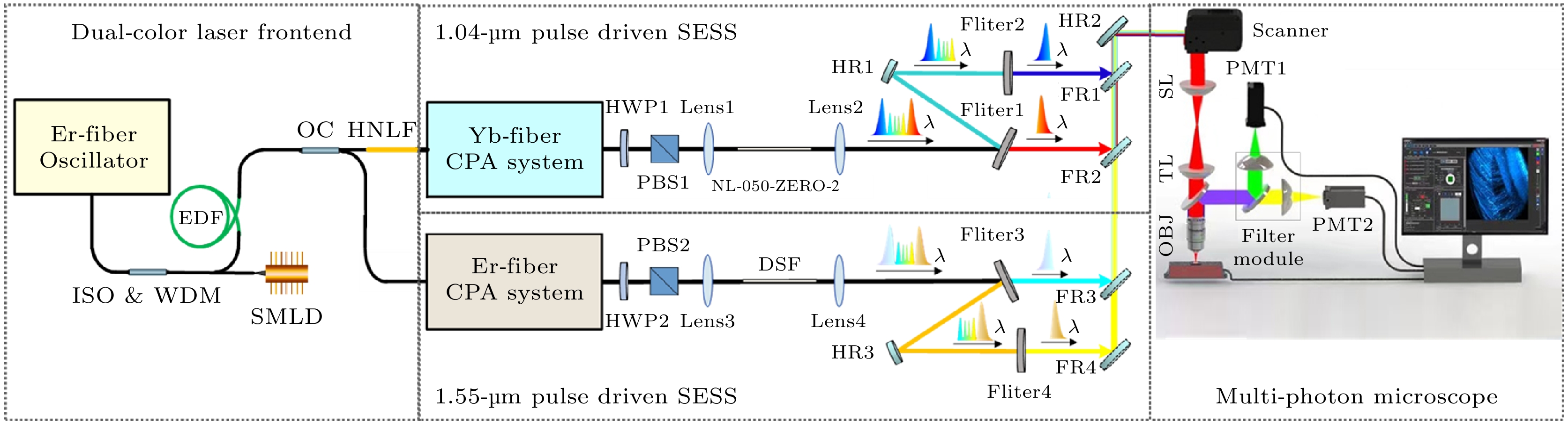
图 1 基于双波长泵浦SESS光源的多光子显微镜实验装置. ISO, 光隔离器; WDM, 波分复用器; EDF, 掺铒光纤; SMLD, 半导体激光二极管; OC, 输出耦合器; HNLF, 高非线性光纤; CPA, 啁啾脉冲放大; HWP, 半波片; PBS, 偏振分束器; HR, 高反射镜; SL, 扫描透镜; TL, 管镜; OBJ, 物镜; PMT: 光电倍增管
Fig. 1. Schematic setup of the multi-photon microscopy driven by dual-color laser enabled SESS source. ISO, isolator; WDM, wavelength-division multiplexing; EDF, erbium-doped fiber; SMLD, semiconductor laser diode; OC, output coupler; HNLF, highly nonlinear fiber; CPA, chirped-pulse amplification; HWP, half-wave plate; PBS, polarization beam splitter; HR, high reflectance mirror; SL, scan lens; TL, tube lens; OBJ, objective lens; PMT, photomultiplier tube.
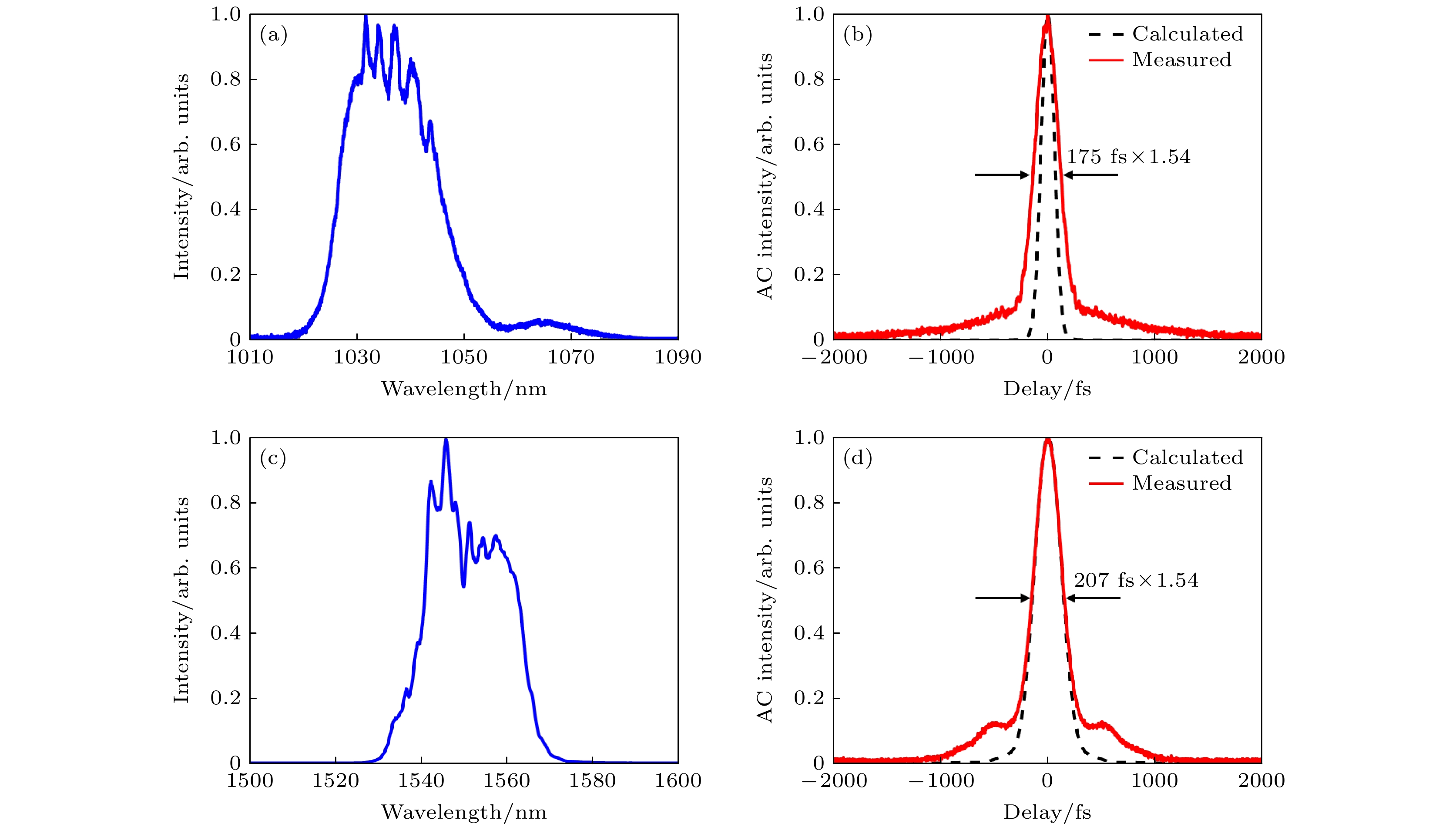
图 2 双波长激光放大后的光谱和自相关轨迹 (a) 1040 nm分支的输出光谱; (b) 测量的自相关曲线(红色)和计算的自相关曲线, 对应由1040 nm输出光谱(黑色)计算出的变换极限脉冲; (c) 1550 nm分支的输出光谱; (d) 测量的自相关曲线(红色)和计算的自相关曲线, 对应由1550 nm输出光谱(黑色)给出的变换极限脉冲
Fig. 2. Spectrum and corresponding autocorrelation of the dual color source after CPA: (a) 1040 nm branch output spectrum; (b) measured autocorrelation trace (red) and calculated autocorrelation trace corresponding to the transform-limited pulse given by the 1040 nm output spectrum (black); (c) 1550 nm branch output spectrum; (d) measured autocorrelation trace (red) and calculated autocorrelation trace corresponding to the transform-limited pulse given by the 1550 nm output spectrum (black).
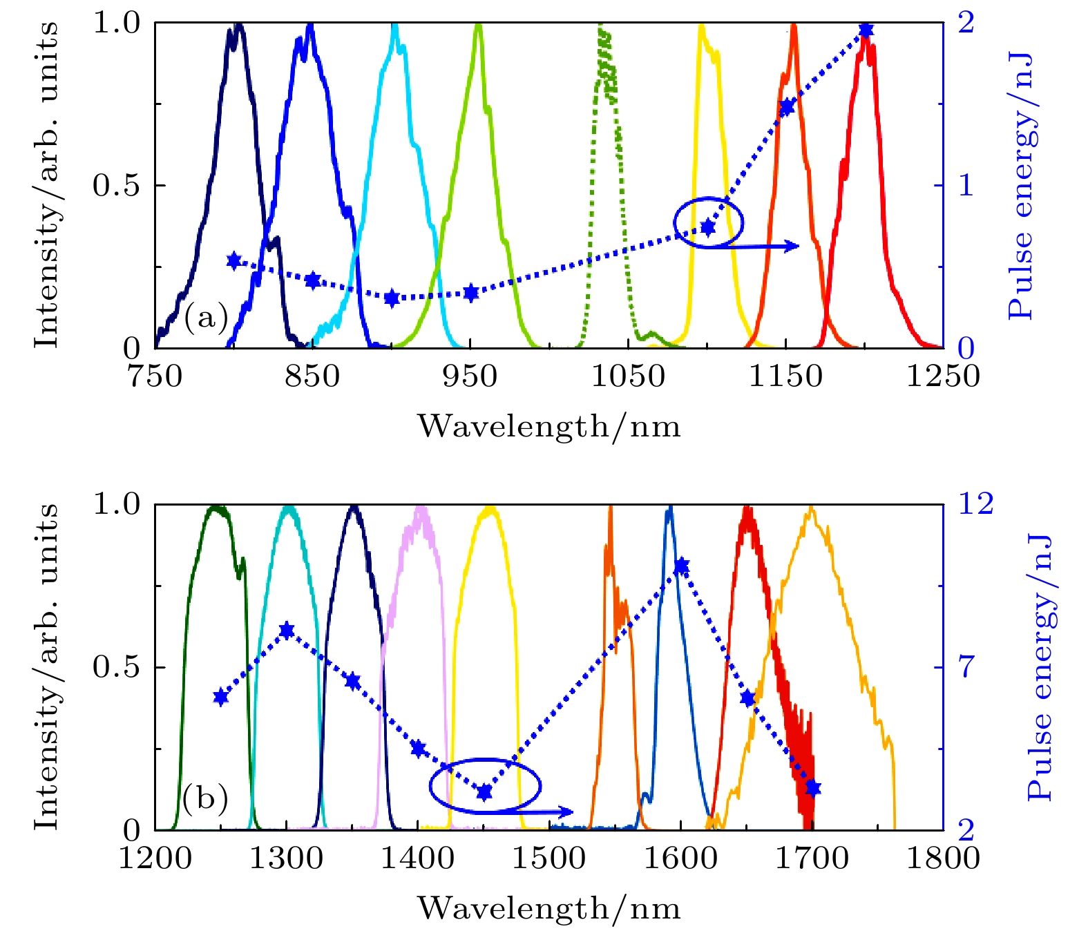
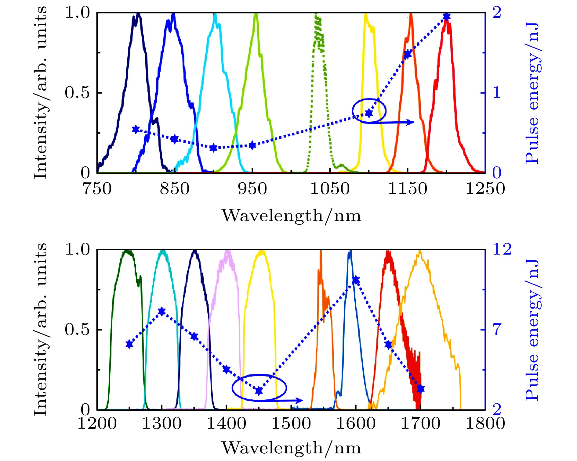
图 3 滤波得到的最左/最右的光谱波瓣和相应的脉冲能量 (a) 由4.3 cm的HNLF(NL-1050-ZERO-2)输出的滤波后光谱和相应的脉冲能量; (b) 由9.5 cm的DSF输出的滤波后光谱和相应的脉冲能量
Fig. 3. Filtered leftmost/rightmost spectral lobes and the corresponding pulse energy: (a) Filtered spectrum and the corresponding pulse energy from 4.3 cm HNLF (NL-1050-ZERO-2); (b) filtered spectrum and the corresponding pulse energy from 9.5 cm DSF.
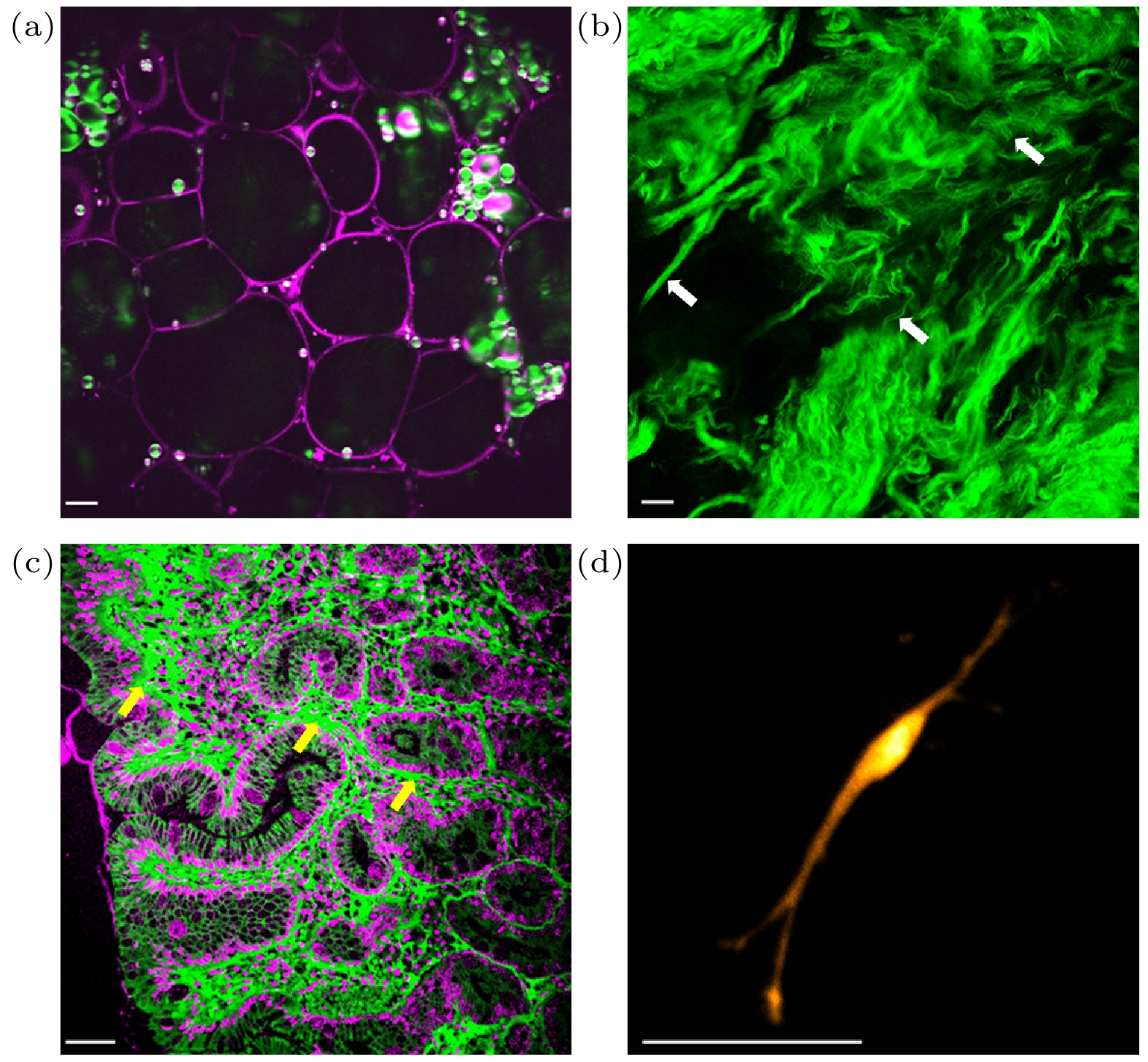
图 4 双波长泵浦SESS光源驱动的多光子显微镜的倍频及荧光成像结果 (a) 土豆块茎的SHG/THG图像; (b) 人类胃部活检组织SHG图像; (c) 人类胃部H&E染色切片SHG/THG图像; (d) 人类神经细胞TPEF图像. 白色箭头: 胶原纤维束; 黄色箭头: 胶原纤维. 比例尺: 50 μm
Fig. 4. Harmonic generation and fluorescence imaging based on multiphoton microscope driven by dual-color laser enabled SESS source: (a) SHG/THG image of potato tuber; (b) SHG image of human gastric biopsy tissue; (c) SHG/THG image of a H&E-stained section of human gastric tissue; (d) TPEF image of a human neuron. White arrows: collagen bundles; yellow arrows: collagen fibers. Scale bar: 50 μm.
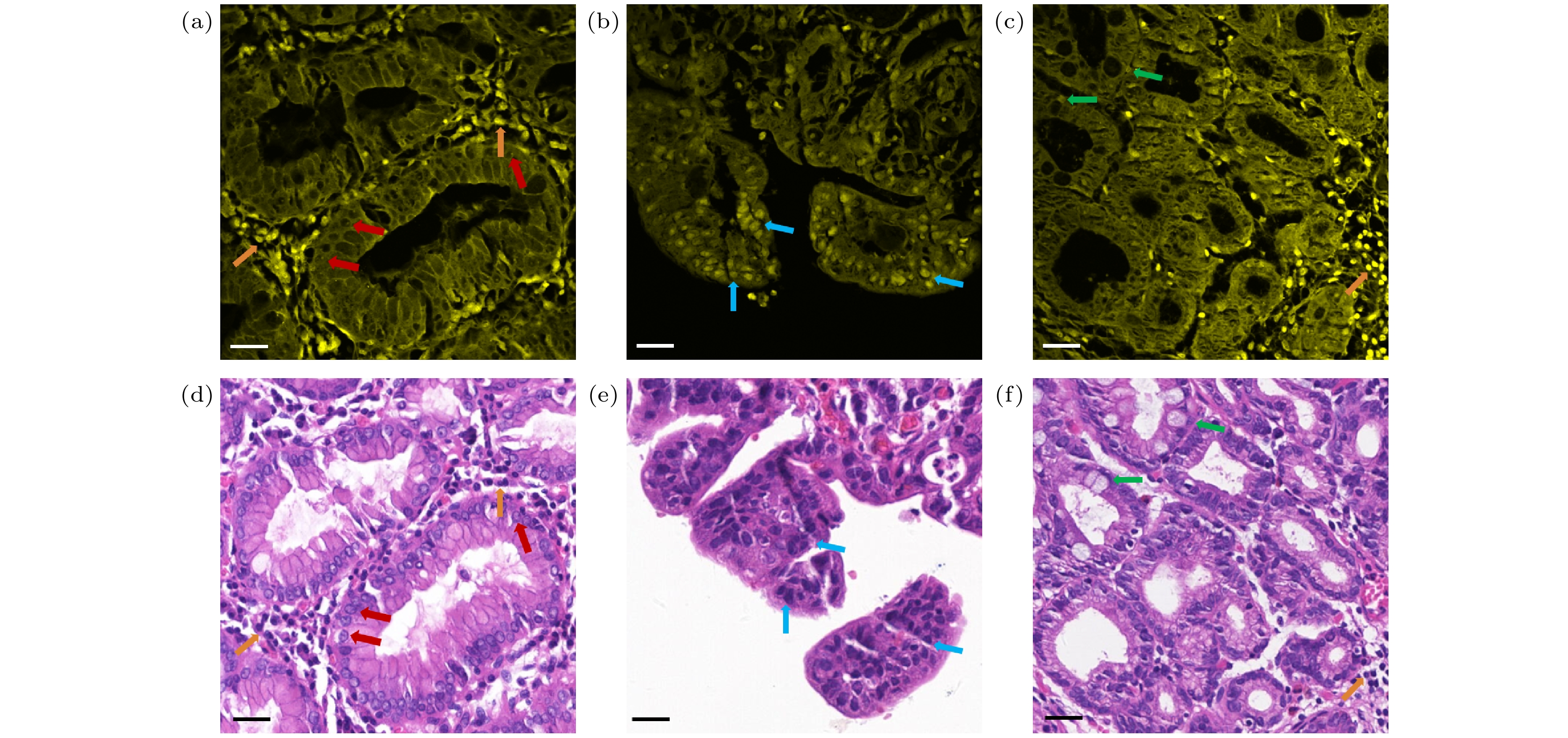
图 5 不同类型胃黏膜的双光子成像结果和相应的H&E染色图像 (a) 正常胃黏膜的TPEF图像; (b) 胃癌的TPEF图像; (c) 萎缩性胃炎的TPEF图像; (d) 正常胃黏膜的H&E染色图像; (e) 胃癌的H&E染色图像; (f) 萎缩性胃炎的H&E染色图像. 红色箭头: 正常胃黏膜上皮细胞核; 橙色箭头: 炎症细胞; 蓝色箭头: 癌变胃黏膜上皮细胞核; 绿色箭头: 杯状细胞核. 比例尺: 30 μm
Fig. 5. TPEF images and corresponding H&E-stained images of different gastric mucosa: (a) TPEF image of normal gastric mucosa; (b) TPEF image of gastric carcinoma, (c) TPEF image of atrophic gastritis; (d) H&E-stained image of normal gastric mucosa; (e) H&E-stained image of gastric carcinoma; (f) H&E-stained image of atrophic gastritis. Red arrows: normal gastric epithelial cell nuclei; orange arrows: inflammatory cells; blue arrows: cancerous gastric epithelial cell nuclei; green arrows: goblet cell nuclei. Scale bar: 30 μm.
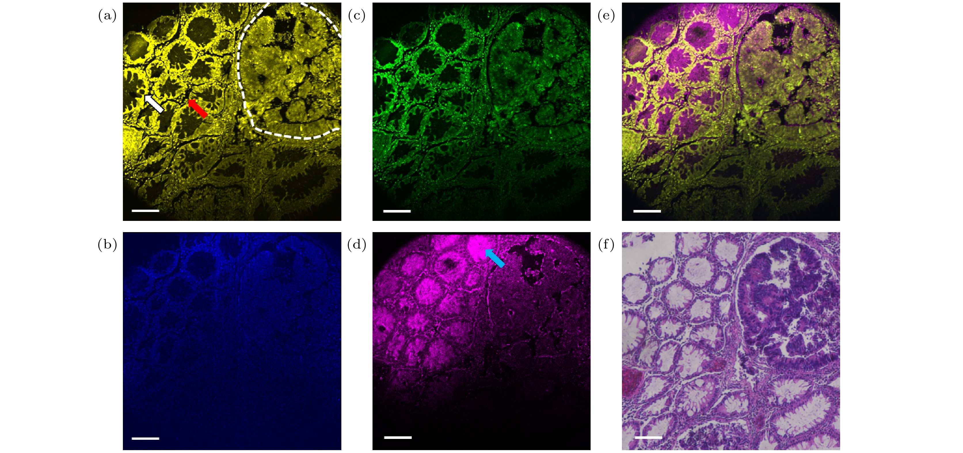
图 6 肠腺癌组织的多模态成像结果 (a) TPEF图像; (b) TrPEF图像; (c) SHG图像; (d) THG图像; (e) TPEF/SHG/TrPEF/THG叠加图像; (f) 相应的H&E图像. 白色箭头: 肠腺; 红色箭头: 炎症细胞; 蓝色箭头: 杯状细胞分泌的黏液; 白色虚线: 癌变区域. 比例尺: 100 μm
Fig. 6. Multimodal imaging of intestinal adenocarcinoma tissue: (a) TPEF image; (b) TrPEF image; (c) SHG image; (d) THG image; (e) merging of TPEF/SHG/TrPEF/THG images; (f) corresponding H&E-stained image. White arrow: intestinal glands; red arrow: inflammatory cells; blue arrow: mucus secreted by goblet cells; white dotted line: cancerous area. Scale bar: 100 μm.
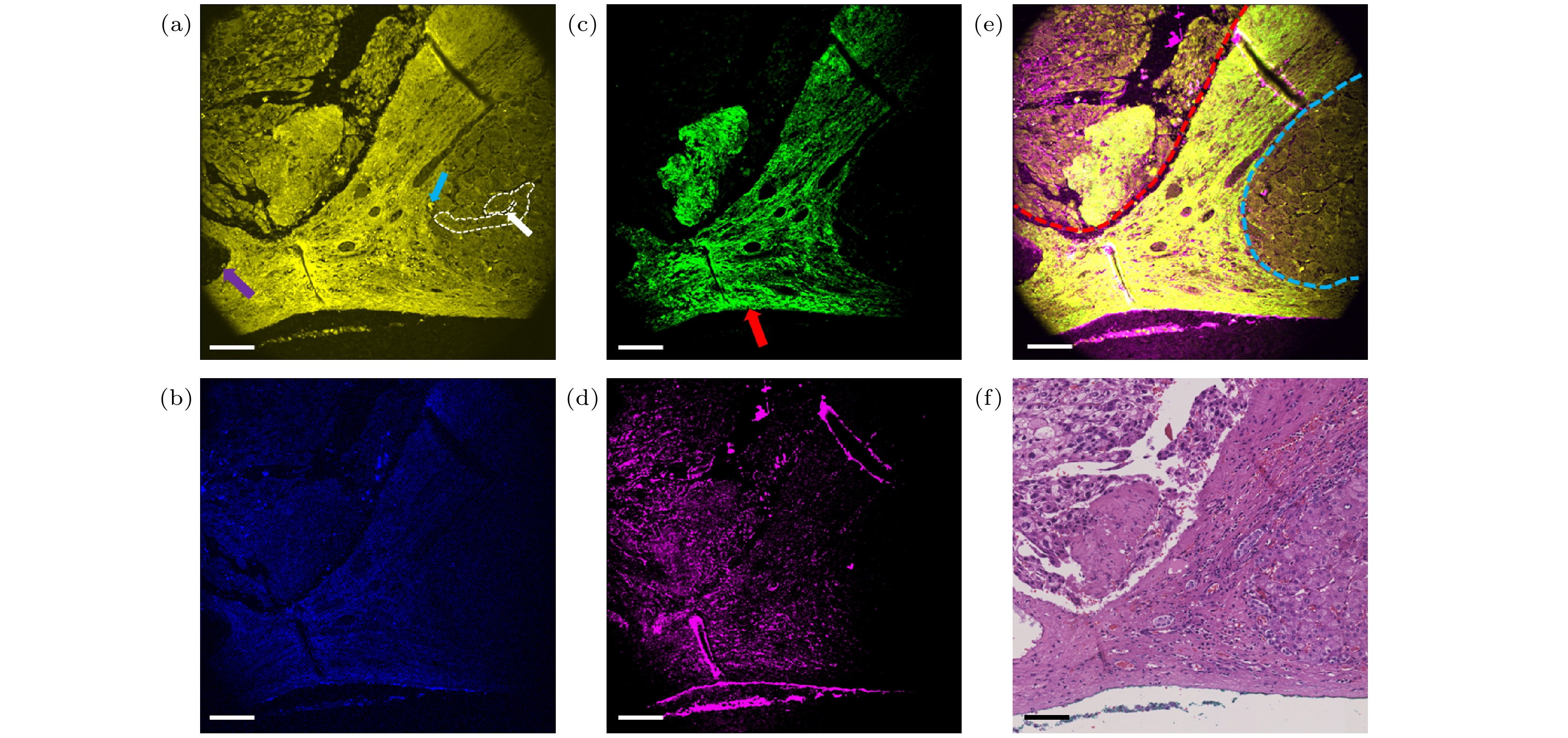
图 7 肝细胞癌组织的多模态成像结果 (a) TPEF图像; (b) TrPEF图像; (c) SHG图像; (d) THG图像; (e) TPEF/SHG/TrPEF/THG叠加图像; (f) 相应的H&E图像. 白色箭头: 肝血窦; 蓝色箭头: 巨噬细胞; 紫色箭头: 小叶间静脉; 红色箭头: 结缔组织; 白色虚线: 肝索; 蓝色虚线: 正常肝小叶; 红色虚线: 癌变区域. 比例尺: 100 μm
Fig. 7. Multimodal imaging of hepatocellular carcinoma tissue: (a) TPEF image; (b) TrPEF image; (c) SHG image; (d) THG image; (e) merging of TPEF/SHG/TrPEF/THG images; (f) corresponding H&E-stained image. White arrow: hepatic sinusoid; blue arrow: macrophage; purple arrow: interlobular vein; red arrow: connective tissue; white dotted line: hepatic cords; blue dotted line: normal hepatic lobule; red dotted line: cancerous area. Scale bar: 100 μm.
-
[1] Adur J, Carvalho H F, Cesar C L, Casco V H 2014 Cancer Inf. 13 67
Google Scholar
[2] Perry S W, Burke R M, Brown E B 2012 Ann. Biomed. Eng. 40 277
Google Scholar
[3] You S, Tu H, Chaney E J, Sun Y, Zhao Y, Bower A J, Liu Y, Marjanovic M, Sinha S, Pu Y, Boppart S A 2018 Nat. Commun. 9 2125
Google Scholar
[4] Huang S H, Heikal A A, Webb W W 2002 Biophys. J. 82 2811
Google Scholar
[5] Zipfel W R, Williams R M, Christie R, Nikitin A Y, Hyman B T, Webb W W 2003 Proc. Natl. Acad. Sci. U.S.A. 100 7075
Google Scholar
[6] Chu S, Chen I, Liu T, Cheng P, Sun C, Lin B 2001 Opt. Lett. 26 1909
Google Scholar
[7] Zhang H, Chen Y, Cao D, Wang Y, Zhang Y, Zhao J 2021 Biomed. Opt. Express 12 1308
Google Scholar
[8] Sordillo L A, Pu Y, Pratavieira S, Budansky Y, Alfano R R 2014 J. Biomed. Opt. 19 056004
Google Scholar
[9] Shi L, Sordillo L A, Rodríguez-Contreras A, Alfano R 2016 J. Biophotonics 9 38
Google Scholar
[10] Wang K, Horton N G, Charan K, Mirocha J D, Gaeta A L 2013 IEEE J. Sel. Top. Quantum Electron. 20 50
Google Scholar
[11] Ouzounov D G, Wang T, Wang M, Feng D D, Horton N G, Cruz-Hernández J C, Cheng Y T, Reimer J, Tolias A S, Nishimura N, Xu C 2017 Nat. Methods 14 388
Google Scholar
[12] Horton N G, Wang K, Kobat D, Clark C G, Wise F W, Schaffer C B, Xu C 2013 Nat. Photonics 7 205
Google Scholar
[13] Buttolph M L, Mejooli M A, Sidorenko P, Eom C Y, Schaffer C B, Wise F W 2022 Opt. Lett. 47 545
Google Scholar
[14] Travers J 2010 J. Opt. 12 113001
Google Scholar
[15] Liu Y, Tu H, Benalcazar W A, Boppart S A 2011 IEEE J. Sel. Top. Quantum Electron. 18 1209
Google Scholar
[16] Tu H, Liu Y, Turchinovich D, Marjanovic M, Lyngsø J K, Lægsgaard J, Chaney E J, Zhao Y, You S, Wilson W L, Xu B, Dantus M, Boppart S A 2016 Nat. Photonics 10 534
Google Scholar
[17] Dudley J M, Genty G, Coen S 2006 Rev. Mod. Phys. 78 1135
Google Scholar
[18] Birks T, Wadsworth W, Russell P S J 2000 Opt. Lett. 25 1415
Google Scholar
[19] Liu W, Li C, Zhang Z, Kärtner F X, Chang G Q 2016 Opt. Express 24 15328
Google Scholar
[20] Liu W, Chia S H, Chung H Y, Greinert R, Kärtner F X, Chang G Q 2017 Opt. Express 25 6822
Google Scholar
[21] Chung H Y, Liu W, Cao Q, Greinert R, Kärtner F X, Chang G Q 2019 IEEE J. Sel. Top. Quantum Electron. 25 6800708
Google Scholar
[22] Chung H Y, Greinert R, Kärtner F X, Chang G Q 2019 Biomed. Opt. Express 10 514
Google Scholar
[23] Boppart S A, You S, Li L H, Chen J, Tu H 2019 APL Photonics 4 100901
Google Scholar

 下载:
下载:
计量
- 文章访问数: 1063
- PDF下载量: 32
- 被引次数: 0
