


-
The Hefei Advanced Light Facility is the fourth-generation diffraction-limited storage ring light source, scheduled to begin operation in 2028. With its high-brightness and highly coherent X-rays, it will break through the current spatiotemporal resolution bottlenecks of X-ray techniques in studying correlated electron systems, providing crucial information for understanding the nature and microscopic origins of novel physical properties in these materials. This article introduces the main scientific goals and technical advantages of the Hefei Advanced Light Facility, focusing on the application perspectives of advanced technologies such as angle-resolved photoemission spectroscopy, magnetic circular dichroism, coherent X-ray scattering, and coherent X-ray imaging in researches of quantum materials and correlated electron systems. These techniques will enable the detailed analysis of the distribution and dynamics of electronic/spin/orbital states, reveal various novel quantum phenomena, and elucidate the fluctuations of order parameters in correlated electron systems. The completion of the Hefei Advanced Light Facility will provide advanced technical supports for decoding complex quantum states and non-equilibrium properties, ultimately promoting the application of quantum materials and correlated electron systems in frontier fields such as energy and information.
-
Keywords:
- X-ray spectroscopy /
- coherent X-ray scattering /
- coherent X-ray imaging /
- correlated electron systems
[1] Jens Als-Nielsen, Des McMorrow 著 (封东来 译) 2015 现代X光物理原理 (上海: 复旦大学出版社)
Als-Nielsen J, McMorrow D (translated by Feng D L) 2015 Modern Elements of X-ray Physics (Shanghai: Fudan University Press
[2] 麦振洪2013同步辐射光源及其应用 (上卷和下卷) (北京: 科学出版社)
Mai Z H 2013 Synchrotron Radiation Sources and Applications (Vol. 1 and 2) (Beijing: Science Press
[3] Eberhardt W 2015 J. Electron Spectrosc. 200 31
 Google Scholar
[4] Eriksson M, van der Veen J F, Quitmann C 2014 J. Synchrotron Radiat. 21 837
Google Scholar
[5] Sobota J A, He Y, Shen Z X 2021 Rev. Mod. Phys. 93 025006
Google Scholar
[6] Iwasawa H 2020 Electron. Struct. 2 043001
Google Scholar
[7] Lisi S, Lu X B, Benschop T, de Jong T A, Stepanov P, Duran J R, Margot F, Cucchi I, Cappelli E, Hunter A, Tamai A, Kandyba V, Giampietri A, Barinov A, Jobst J, Stalman V, Leeuwenhoek M, Watanabe K, Taniguchi T, Rademaker L, van der Molen S J, Allan M P, Efetov D K, Baumberger F 2021 Nat. Phys. 17 189
Google Scholar
[8] Cattelan M, Fox N A 2018 Nanomaterials-Basel 8 284
Google Scholar
[9] Mo S K 2017 Nano Converg. 4 6
Google Scholar
[10] Chen C T, Sette F, Ma Y, Modesti S 1990 Phys. Rev. B 42 7262
Google Scholar
[11] van der Laan G, Figueroa A I 2014 Coordin. Chem. Rev. 277 95
Google Scholar
[12] Klewe C, Qian L, Mengmeng Y, N’Diaye A T, Burn D M, Hesjedal T, Figueroa A I, Chanyong H, Jia L, Hicken R J, Shafer P, Arenholz E, van der Laan G, Qian Z 2020 Synchrotron Radiat. News 33 12
Google Scholar
[13] Purbawati A, Coraux J, Vogel J, Hadj-Azzem A, Wu N J, Bendiab N, Jegouso D, Renard J, Marty L, Bouchiat V, Sulpice A, Aballe L, Foerster M, Genuzio F, Locatelli A, Mentes T O, Han Z V, Sun X D, Núñez-Regueiro M, Rougemaille N 2020 ACS Appl. Mater. Inter. 12 30702
Google Scholar
[14] Barinov A, Dudin P, Gregoratti L, Locatelli A, Mentes T O, Niño M A, Kiskinova M 2009 Nucl. Instrum. Meth. A 601 195
Google Scholar
[15] Sutton M, Mochrie S G J, Greytak T, Nagler S E, Berman L E, Held G A, et al. 1991 Nature 352 608
Google Scholar
[16] Bluschke M, Basak R, Barbour A, Warner A N, Fürsich K, Wilkins S, Roy S, Lee J, Christiani G, Logvenov G, Minola M, Keimer B, Mazzoli C, Benckiser E, Frano A 2022 Sci. Adv. 8 eabn6882
Google Scholar
[17] Shpyrko O G 2014 J. Synchrotron Radiat. 21 1057
Google Scholar
[18] Sandy A R, Zhang Q T, Lurio L B 2018 Annu. Rev. Mater. Res. 48 167
Google Scholar
[19] Zhang Q T, Dufresne E M, Sandy A R 2018 Curr. Opin. Solid St. M. 22 202
Google Scholar
[20] Shpyrko O G, Isaacs E D, Logan J M, Feng Y J, Aeppli G, Jaramillo R, Kim H C, Rosenbaum T F, Zschack P, Sprung M, Narayanan S, Sandy A R 2007 Nature 447 68
Google Scholar
[21] Grübel G, Madsen A, Robert A 2008 Soft Matter Characterization (Dordrecht: Springer) p953
[22] 范家东, 江怀东 2012 物理学报 61 218702
Google Scholar
Fan J D, Jiang H D 2012 Acta Phys. Sin. 61 218702
Google Scholar
[23] Miao J W, Ishikawa T, Robinson I K, Murnane M M 2015 Science 348 530
Google Scholar
[24] Rau C 2017 SRN 30 19
Google Scholar
[25] Tripathi A, Mohanty J, Dietze S H, Shpyrko O G, Shipton E, Fullerton E E, Kim S S, McNulty I 2011 Proc. Natl. Acad. Sci. U. S. A. 108 13393
Google Scholar
[26] Prosekov P A, Nosik V L, Blagov A E 2021 Crystallogr. Rep. 66 867
Google Scholar
[27] Pfeiffer F 2018 Nat. Photonics 12 9
Google Scholar
[28] Donnelly C, Scagnoli V 2020 J. Phys. : Condens. Matter 32 213001
Google Scholar
[29] Lo Y H, Zhao L, Gallagher-Jones M, Rana A, Lodico J J, Xiao W, Regan B C, Miao J 2018 Nat. Commun. 9 1826
Google Scholar
-
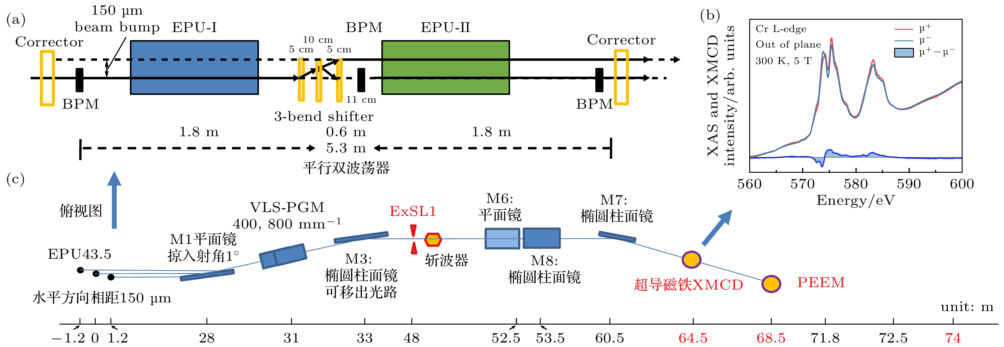
图 1 合肥先进光源XMCD线站配置 (a) 双波荡器光源示意图; (b) XMCD谱; (c) 双光路设计的俯视图
Figure 1. Configuration of the XMCD beamline at the Hefei Advanced Light Source: (a) Schematic diagram of the twin undulator sources; (b) XMCD spectrum; (c) top view of the dual beam path design.
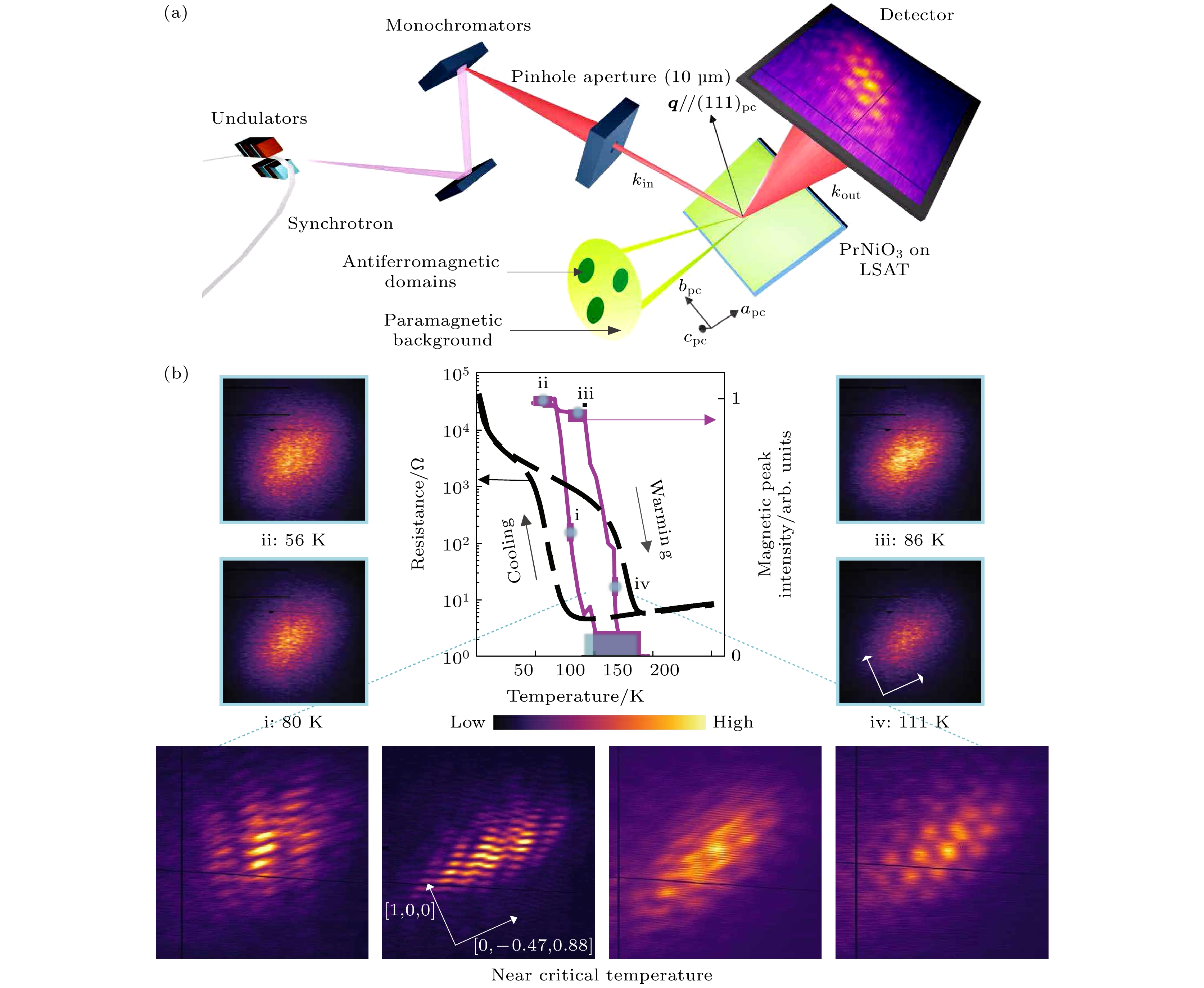
图 2 相干X射线散射实验设置(a)和反铁磁衍射图案的演化(b) (得到文献[16]的授权重印, 版权归©2022美国科学促进协会所有)
Figure 2. Coherent X-ray scattering experimental setup (a) and evolution of antiferromagnetic diffraction patterns (b) (Reproduced with permission of Ref. [16], Copyright of ©2022 The American Association for the Advancement of Science).
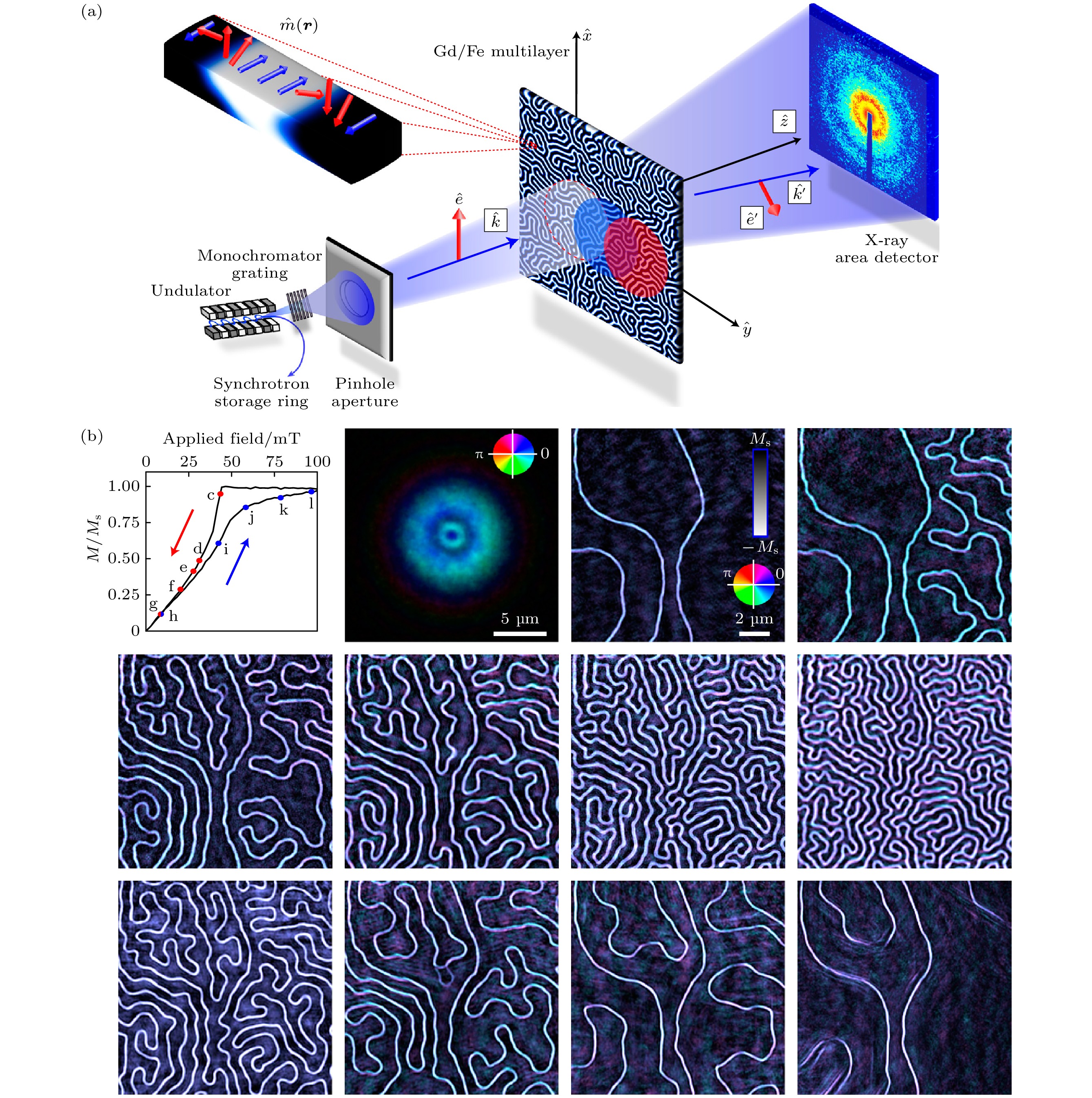
图 3 相干衍射成像技术对多层Gd/Fe薄膜中的铁磁畴的成像和原位磁场调控研究 (a) X射线扫描相干X射线成像测量的示意图, 对比度主要来自X射线磁圆二色性(XMCD)效应, 在远场用X射线面探测器记录衍射图案; (b) 样品磁化强度随外加磁场变化时, 重建图像中Gd的磁构型演化(得到文献[25]的授权重印, 版权归©2011美国国家科学院所有)
Figure 3. Coherent diffraction imaging of ferromagnetic domains in multilayer Gd/Fe thin films and their in-situ magnetic field manipulation study: (a) Schematic diagram of X-ray scanning coherent X-ray imaging measurement, where the contrast primarily arises from the XMCD effect, and diffraction patterns are recorded in the far-field using an X-ray area detector; (b) evolution of the Gd magnetic configuration in reconstructed images as the sample magnetization changes with the applied external magnetic field (Reproduced with permission of Ref. [25], Copyright of ©2011 National Academy of Sciences).
-
[1] Jens Als-Nielsen, Des McMorrow 著 (封东来 译) 2015 现代X光物理原理 (上海: 复旦大学出版社)
Als-Nielsen J, McMorrow D (translated by Feng D L) 2015 Modern Elements of X-ray Physics (Shanghai: Fudan University Press
[2] 麦振洪2013同步辐射光源及其应用 (上卷和下卷) (北京: 科学出版社)
Mai Z H 2013 Synchrotron Radiation Sources and Applications (Vol. 1 and 2) (Beijing: Science Press
[3] Eberhardt W 2015 J. Electron Spectrosc. 200 31
Google Scholar
[4] Eriksson M, van der Veen J F, Quitmann C 2014 J. Synchrotron Radiat. 21 837
Google Scholar
[5] Sobota J A, He Y, Shen Z X 2021 Rev. Mod. Phys. 93 025006
Google Scholar
[6] Iwasawa H 2020 Electron. Struct. 2 043001
Google Scholar
[7] Lisi S, Lu X B, Benschop T, de Jong T A, Stepanov P, Duran J R, Margot F, Cucchi I, Cappelli E, Hunter A, Tamai A, Kandyba V, Giampietri A, Barinov A, Jobst J, Stalman V, Leeuwenhoek M, Watanabe K, Taniguchi T, Rademaker L, van der Molen S J, Allan M P, Efetov D K, Baumberger F 2021 Nat. Phys. 17 189
Google Scholar
[8] Cattelan M, Fox N A 2018 Nanomaterials-Basel 8 284
Google Scholar
[9] Mo S K 2017 Nano Converg. 4 6
Google Scholar
[10] Chen C T, Sette F, Ma Y, Modesti S 1990 Phys. Rev. B 42 7262
Google Scholar
[11] van der Laan G, Figueroa A I 2014 Coordin. Chem. Rev. 277 95
Google Scholar
[12] Klewe C, Qian L, Mengmeng Y, N’Diaye A T, Burn D M, Hesjedal T, Figueroa A I, Chanyong H, Jia L, Hicken R J, Shafer P, Arenholz E, van der Laan G, Qian Z 2020 Synchrotron Radiat. News 33 12
Google Scholar
[13] Purbawati A, Coraux J, Vogel J, Hadj-Azzem A, Wu N J, Bendiab N, Jegouso D, Renard J, Marty L, Bouchiat V, Sulpice A, Aballe L, Foerster M, Genuzio F, Locatelli A, Mentes T O, Han Z V, Sun X D, Núñez-Regueiro M, Rougemaille N 2020 ACS Appl. Mater. Inter. 12 30702
Google Scholar
[14] Barinov A, Dudin P, Gregoratti L, Locatelli A, Mentes T O, Niño M A, Kiskinova M 2009 Nucl. Instrum. Meth. A 601 195
Google Scholar
[15] Sutton M, Mochrie S G J, Greytak T, Nagler S E, Berman L E, Held G A, et al. 1991 Nature 352 608
Google Scholar
[16] Bluschke M, Basak R, Barbour A, Warner A N, Fürsich K, Wilkins S, Roy S, Lee J, Christiani G, Logvenov G, Minola M, Keimer B, Mazzoli C, Benckiser E, Frano A 2022 Sci. Adv. 8 eabn6882
Google Scholar
[17] Shpyrko O G 2014 J. Synchrotron Radiat. 21 1057
Google Scholar
[18] Sandy A R, Zhang Q T, Lurio L B 2018 Annu. Rev. Mater. Res. 48 167
Google Scholar
[19] Zhang Q T, Dufresne E M, Sandy A R 2018 Curr. Opin. Solid St. M. 22 202
Google Scholar
[20] Shpyrko O G, Isaacs E D, Logan J M, Feng Y J, Aeppli G, Jaramillo R, Kim H C, Rosenbaum T F, Zschack P, Sprung M, Narayanan S, Sandy A R 2007 Nature 447 68
Google Scholar
[21] Grübel G, Madsen A, Robert A 2008 Soft Matter Characterization (Dordrecht: Springer) p953
[22] 范家东, 江怀东 2012 物理学报 61 218702
Google Scholar
Fan J D, Jiang H D 2012 Acta Phys. Sin. 61 218702
Google Scholar
[23] Miao J W, Ishikawa T, Robinson I K, Murnane M M 2015 Science 348 530
Google Scholar
[24] Rau C 2017 SRN 30 19
Google Scholar
[25] Tripathi A, Mohanty J, Dietze S H, Shpyrko O G, Shipton E, Fullerton E E, Kim S S, McNulty I 2011 Proc. Natl. Acad. Sci. U. S. A. 108 13393
Google Scholar
[26] Prosekov P A, Nosik V L, Blagov A E 2021 Crystallogr. Rep. 66 867
Google Scholar
[27] Pfeiffer F 2018 Nat. Photonics 12 9
Google Scholar
[28] Donnelly C, Scagnoli V 2020 J. Phys. : Condens. Matter 32 213001
Google Scholar
[29] Lo Y H, Zhao L, Gallagher-Jones M, Rana A, Lodico J J, Xiao W, Regan B C, Miao J 2018 Nat. Commun. 9 1826
Google Scholar

 DownLoad:
Catalog
Metrics
- Abstract views: 4644
- PDF Downloads: 141
- Cited By: 0
