


-
多光子成像技术由于具有低侵入性、强穿透力、高空间分辨率等优点, 自问世以来便成为生物医学研究的有力工具, 在癌症病理、神经疾病及脑功能成像等方面取得了一系列较好的研究成果. 目前, 应用较为广泛的多光子成像技术是双光子激发荧光显微成像技术, 其在生物医学应用中具有较大的发展潜力. 本文详细阐述了多光子成像技术在多色成像、功能成像及成像深度等方面的生物医学应用新进展, 包括多色双光子激发荧光显微成像、双光子激发荧光寿命显微成像、双光子光纤内窥成像和三光子显微成像技术, 并简要介绍这几种多光子成像技术的原理与特性, 最后展望其未来发展前景.
-
关键词:
- 多色双光子成像 /
- 双光子激发荧光寿命成像 /
- 双光子光纤内窥 /
- 三光子成像
In contrast to single photon excitation fluorescence imaging, laser scanning confocal imaging, and wide-field imaging, the multi-photon imaging has advantages of minimal invasion and deeper penetration by using near-infrared (NIR) laser source. Moreover, it can carry out three-dimensional high-spatial-resolution imaging of biological tissues due to its natural optical tomography capability. Since its advent, multi-photon imaging has become a powerful tool in biomedicine and achieved a series of significant discoveries in cancer pathology, neurological diseases and brain functional imaging. In the past decade, as a major form of multi-photon imaging techonoogy, two-photon excited fluorescence microscopy imaging has a great potential in biomedical applications. In order to satisfy the practical biomedical applications, multi-photon imaging technologies have made significant breakthroughs in improving the deficiencies of traditional 2PEF in multi-color imaging, functional imaging, live imaging and imaging depth, such as multicolor two-photon excitation fluorescence microscopy, two-photon fluorescence lifetime imaging microscopy, two-photon fiber endoscopic imaging, and three-photon microscopy imaging technology. For example, multicolor two-photon excitation fluorescence microscopy is demonstrated to achieve simultaneous imaging of multiple fluorophores with multiple wavelenth excitation lasers or continuous spectrum. In addition, the two-photon fluorescence lifetime microscopic imaging provides a method to achieve high-resolution three-dimensional imaging of biological tissue with multi-dimensional information including fluorescence intensity and lifetime. In addition, two-photon optical fiber endoscopic imaging with small system size and mimal invasion is developed and used to image the tissue inside the deep organ. Finally, two-photon excitation fluorescence microscopy technique still has relatively strong scattering for brain functional imaging in vivo. Therefore, the imaging depth is limited by the signal-to-background ratio. Three-photon microscopic imaging technique can achieve higher imaging depth and a desired signal-to-noise ratio by extending the wavelength from 1600 nm to 1820 nm because the attenuation of the excitation light in this wavelenth range is much smaller. In this article, we briefly introduce the principles and applications of these multi-photon imaging technologies, and finally provide our view for their future development.-
Keywords:
- multicolor two-photon excitation fluorescence microscopy /
- two-photon fluorescence lifetime imaging microscopy /
- two-photon fiber endoscopic imaging /
- three-photon microscopy imaging
[1] Goppert M M 1931 Ann. Phys-Berlin. 9 273
[2] Denk W, Strickler J H, Webb W W 1990 Science 248 73
 Google Scholar
[3] Wang K, Horton N G, Charan K, Xu C 2014 Ieee J. Sel. Top. in Quant. 20 6800311
[4] 崔权, 陈忠云, 张智红, 骆清铭, 付玲 2017 激光与光电子学进展 06 16
Cui Q, Chen Z Y, Zhang Z H, Luo Q M, Fu L 2017 Las. Opto-elect. Prog. 06 16
[5] 杨洪权 2011 硕士学位论文 (福建: 福建师范大学)
Yang H Q 2011 M. S. Thesis (Fujian: Fujian Normal University) (in Chinese)
[6] Drobizhev M, Makarov N S, Tillo S E, Hughes T E, Rebane A 2011 Nat. Methods 8 393
Google Scholar
[7] Tragardh J, Murtagh M, Robb G, Parsons M, Lin J P, Spence D J, McConnell G 2016 Microsc. and Microanal. 22 803
Google Scholar
[8] Herz J, Siffrin V, Hauser A E, Brandt A U, Leuenberger T, Radbruch H, Zipp F, Niesner R A 2010 Biophys. J. 98 715
Google Scholar
[9] Xi P, Andegeko Y, Weisel L R, Lozovoy V, Dantus M 2008 Opt. Commun. 281 1841
Google Scholar
[10] Liang X B, Hu W Y, Fu L 2010 Opt. Express 18 14893
Google Scholar
[11] Zhao Z, Wu B, Wang X, Pan Z, Liu Z, Zhang P, Shen X, Nie Q, Dai S, Wang R 2017 Laser Photonics Rev. 11 1700005
Google Scholar
[12] Li D, Zheng W, Qu J A Y 2009 Opt. Lett. 34 202
Google Scholar
[13] Li C, Pastila R K, Lin C P 2016 J. Innov. Opt. Heal. Sci. 9 1640003
Google Scholar
[14] Le Dévédec S E, Lalai R, Pont C, de Bont H, van de Water B 2011 Mol. Imaging Biol. 13 67
Google Scholar
[15] Entenberg D, Wyckoff J, Gligorijevic B, Roussos E T, Verkhusha V, Pollard J W, Condeelis J 2011 Nat. Protoc. 6 1500
Google Scholar
[16] Piatkevich K D, Hulit J, Subach O M, Wu B, Abdulla A, Segall J E, Verkhusha V 2010 P. Natl. Acad. Sci. U. S. A. 107 5369
Google Scholar
[17] Collot M, Fam T K, Ashokkumar P, Faklaris O, Galli T, Danglot L, Klymchenko A S 2018 J. Am. Chem. Soc. 140 5401
Google Scholar
[18] Garaschuk O, Milos R-I, Konnerth A 2006 Nat. Protoc. 1 380
Google Scholar
[19] Hayakawa Y, Nemoto T, Iino M, Kasai H 2005 Cell Calcium. 37 359
Google Scholar
[20] Mahou P, Zimmerley M, Loulier K, Matho K S, Labroille G, Morin X, Supatto W, Livet J, Debarre D, Beaurepaire E 2012 Nat. Methods 9 815
Google Scholar
[21] Mahou P, Vermot J, Beaurepaire E, Supatto W 2014 Nat. Methods 11 600
Google Scholar
[22] Stringari C, Abdeladim L, Malkinson G, Mahou P, Solinas X, Lamarre I, Brizion S, Galey J B, Supatto W, Legouis R, Pena A M, Beaurepaire E 2017 Sci. Rep-UK. 7 3792
Google Scholar
[23] Abdeladim L, Matho K S, Clavreul S, et al. 2019 Nat. Commun. 10 1662
Google Scholar
[24] Lin D Y, Luo T, Liu L W, Lu Y, Liu S X, Yuan Z, Qu J L 2017 Chin. Opt. Lett. 15 090006
Google Scholar
[25] 刘雄波, 林丹樱, 吴茜茜, 严伟, 罗腾, 杨志刚, 屈军乐 2017 物理学报 67 178701
Liu X B, Lin D Y, Wu Q Q, Wei Y, Luo T, Yang Z G, Qu J L 2017 Acta Phys. Sin. 67 178701
[26] Liu X B, Lin D Y, Becker W, Niu J, Yu B, Liu L W, Qu J L 2019 J. Innov. Opt. Heal. Sci. 12 1930003
Google Scholar
[27] 李慧, 夏先园, 陈廷爱, 余佳, 李曦, 郑炜 2018 中国激光 45 0207010
Google Scholar
Li H, Xia X Y, Chen T A, Yu J, Li X, Zheng W 2018 Chin. J. Lasers 45 0207010
Google Scholar
[28] Ranawat H, Pal S, Mazumder N 2019 Biomed. Eng. Lett. 9 293
Google Scholar
[29] Li H, Yu J, Zhang R, Li X, Zheng W 2019 J. Innov. Opt. Heal. Sci. 12 1930009
Google Scholar
[30] Anh C, Pimenta R M L, Lee H B, Mereddy V, Holy J, Heikal A 2019 Cytom. Part A 95A 80
[31] Luo T, Lu Y, Liu S X, Lin D Y, Qu J L 2017 Anal. Chem. 89 8104
Google Scholar
[32] Chen B L, Lu Y, Pan W H, Xiong J, Yang Z G, Yan W, Liu L W, Qu J L 2019 Anal. Chem. 91 10640
Google Scholar
[33] Shen B L, Yan J S, Wang S Q, Zhou F, Zhao Y H, Hu R, Qu J L, Liu L W 2020 Theranostics 10 1849
Google Scholar
[34] Alam S R, Wallrabe H, Svindrych Z, et al. 2017 Sci. Rep-UK. 7 10451
Google Scholar
[35] Poulon F, Pallud J, Varlet P, et al. 2018 Sci. Rep-UK. 8 14888
Google Scholar
[36] Kantelhardt S R, Kalasauskas D, Konig K, Kim E, Weinigel M, Uchugonova A, Giese A 2016 J. Neuro-Oncol. 127 473
Google Scholar
[37] Teh S K, Zheng W, Li S X, Li D, Zeng Y, Yang Y Q, Qu J A Y 2013 J. Biomed. Opt. 18 036001
Google Scholar
[38] Rück A, Hauser C, Mosch S, Kalinina S 2014 J. Biomed. Opt. 19 096005
Google Scholar
[39] Shen Y F, Tsai M R, Chen S C, et al. 2015 Anal. Chem. 87 7575
Google Scholar
[40] Li X, Li H, He X Z, Chen T G, Xia X Y, Yang C X, Zheng W 2018 Biomed. Opt. Express 9 453
Google Scholar
[41] Tyurikova O, Zheng K Y, Rings A, Drews A, Klenerman D, Rusakov D A 2018 Brain Res. Bull. 136 85
Google Scholar
[42] Zheng K Y, Jensen T P, Rusakov D A 2018 Nat. Protoc. 13 581
Google Scholar
[43] Feeks J A, Hunter J 2017 Biomed. Opt. Express 8 2483
Google Scholar
[44] Chakraborty S, Nian F S, Tsai J W, Karmenyan A, Chiou A 2016 Sci. Rep-UK. 6 19145
Google Scholar
[45] Looney M R, Thornton E, Sen D, Lamm W J, Glenny R W, Krummel M F 2011 Nat. Methods 8 91
Google Scholar
[46] 石玉洁, 张广杰, 陆政元, 应亚宸, 贾荟琳, 席鹏 2018 中国光学 11 296
Google Scholar
Shi Y J, Zhang G J, Lu Z Y, Ying Y C, Jia H L, Xi P 2018 Chin. Opt. 11 296
Google Scholar
[47] Bird D, Gu M 2002 Opt. Lett. 27 1031
Google Scholar
[48] Bao H, Allen J, Pattie R, Vance R, Gu M 2008 Opt. Lett. 33 1333
Google Scholar
[49] Bao H, Ryu S Y, Lee B H, Tao W, Gu M 2010 Opt. Lett. 35 995
Google Scholar
[50] Tang S, Jung W, McCormick D, Xie T, Su J, Ahn Y C, Tromberg B J, Chen Z 2009 J. Biomed. Opt. 14 034005
Google Scholar
[51] Piyawattanametha W, Cocker E D, Burns L D, Barretto R P J, Jung J C, Ra H, Solgaard O, Schnitzer M J 2009 Opt. Lett. 34 2309
Google Scholar
[52] Wu Y C, Leng Y X, Xi J F, Li X D 2009 Opt. Express 17 7907
Google Scholar
[53] Lee C M, Engelbrecht C J, Soper T D, Helmchen F, Seibel E J 2010 J. Biophotonics 3 385
Google Scholar
[54] Peyrot D A, Lefort C, Steffenhagen M, Mansuryan T, Ducourthial G, Abi-Haidar D, Sandeau N, Vever-Bizet C, Kruglik S G, Thiberville L, Louradour F, Bourg-Heckly G 2012 Biomed. Opt. Express 3 840
Google Scholar
[55] Ducourthial G, Leclerc P, Mansuryan T, Fabert M, Brevier J, Habert R, Braud F, Batrin R, Vever-Bizet C, Bourg-Heckly G, Thiberville L, Druilhe A, Kudlinski A, Louradour F 2015 Sci. Rep-UK. 5 18303
Google Scholar
[56] Hage C H, Leclerc P, Brevier J, Fabert M, Le Nezet C, Kudlinski A, Heliot L, Louradour F 2018 Biomed. Opt. Express 9 142
Google Scholar
[57] Sibai M, Mehidine H, Poulon F, Ibrahim A, Varlet P, Juchaux M, Pallud J, Devaux B, Kudlinski A, Abi Haidar D 2018 Sci. Rep-UK. 8 11124
Google Scholar
[58] Gu M, Bao H, Gan X, Stokes N, Wu J 2014 Light-Sci. Appl. 3 e126
Google Scholar
[59] Xu C, Zipfel W, Shear J B, Williams R M, Webb W 1996 P. Natl. Acad. Sci. U. S. A. 93 10763
Google Scholar
[60] Horton N G, Wang K, Kobat D, Clark C G, Wise F W, Schaffer C B, Xu C 2013 Nat. Photonics 7 205
Google Scholar
[61] Huland D M, Charan K, Ouzounov D G, Jones J S, Nishimura N, Xu C 2013 Biomed. Opt. Express 4 652
Google Scholar
[62] Ouzounov D G, Horton N, Wang T Y, Feng D, Nishimura N, Xu C 2014 Conference on Lasers and Electro-Optics San Jose, America, June 8–13, 2014 p2.
[63] Horton N G, Xu C 2015 Biomed. Opt. Express 6 1392
Google Scholar
[64] Ouzounov D G, Wang T, Wang M, Feng D, Horton N G, Cruz-Hernandez J C, Cheng Y T, Reimer J, Tolias A S, Nishimura N, Xu C 2017 Nat. Methods 14 388
Google Scholar
[65] Li B, Wang M, Wu C, Charan K, Xu C 2018 Conference on Lasers and Electro-Optics San Jose, America, May 13-18, 2018 pJTh5 C.5
[66] Wang T, Ouzounov D G, Wu C, Horton N G, Zhang B, Wu C H, Zhang Y, Schnitzer M J, Xu C 2018 Nat. Methods 15 789
Google Scholar
[67] Li B, Wu C, Wang M, Charan K, Xu C 2020 Nat. Methods 17 163
Google Scholar
[68] Chow D M, Sinefeld D, Kolkman K E, Ouzounov D G, Akbari N, Tatarsky R, Bass A, Xu C, Fetcho J R 2020 Nat. Methods 17 605
Google Scholar
[69] Wang S, Xi W, Cai F, Zhao X, Xu Z, Qian J, He S 2015 Theranostics 5 251
Google Scholar
[70] Zhu Z, Leung C W, Zhao X, Wang Y, Qian J, Tang B Z, He S 2015 Sci. Rep-UK. 5 15189
Google Scholar
[71] Li D, Zhao X, Qin W, Zhang H, Fei Y, Liu L, Yong K T, Chen G, Tang B Z, Qian J 2016 Nano Res. 9 1921
Google Scholar
[72] Wang Y, Chen M, Alifu N, Li S, Qin W, Qin A, Tang B Z, Qian J 2017 ACS Nano 11 10452
Google Scholar
[73] Wang Y, Han X, Xi W, Li J, Roe A W, Lu P, Qian J 2017 Adv. Healthc. Mater. 6 1700685
Google Scholar
[74] Liu W, Wang Y, Han X, Lu P, Zhu L, Sun C, Qian J, He S 2018 Nanoscale 10 10025
Google Scholar
[75] Ni H, Xu Z, Li D, Chen M, Tang B Z, Qian J 2019 J. Innov. Opt. Heal. Sci. 12 1940005
Google Scholar
[76] Qin W, Alifu N, Lam J W Y, Cui Y, Su H, Liang G, Qian J, Tang B Z 2020 Adv. Mater. 32 e2000364
Google Scholar
[77] Liu H, Du Y, Peng X, Zhou X, Qiu P, Wang K 2017 IEEE Photonics J. 9 1
[78] Tong S, Liu H, Cheng H, He C, Du Y, Zhuang Z, Qiu P, Wang K 2019 J. Biophotonics 12 e201800423
[79] Liu H, Deng X, Tong S, He C, Cheng H, Zhuang Z, Gan M, Li J, Xie W, Qiu P, Wang K 2019 Nano. Lett. 19 5260
Google Scholar
[80] Tong S, Gan M Y, Zhuang Z W, Liu H J, Cheng H, Li J, Qiu P, Wang K 2020 J. Lightwave Technol. 38 2450
Google Scholar
[81] Weisenburger S, Tejera F, Demas J, Chen B, Manley J, Sparks F T, Traub F M, Daigle T, Zeng H, Losonczy A, Vaziri A 2019 Cell 177 1050
Google Scholar
[82] Klioutchnikov A, Wallace D J, Frosz M H, Zeltner R, Sawinski J, Pawlak V, Voit K M, Russell P S J, Kerr J N D 2020 Nat. Methods 17 509
Google Scholar
-

图 1 2PEF、3PEF过程能级图. 2PEF和3PEF都是非直接激发辐射过程, 存在非辐射能量转移. 图中
$ {\rm{\nu }}_{\rm{p}} $ 为吸收光子频率,$ {\rm{\nu }}_{\rm{f}} $ 为发射荧光频率,$ {\rm{\nu }}_{\rm{NR}} $ 为非辐射能量转移.Fig. 1. Energy level diagram of 2PEF and 3PEF process. Both 2PEF and 3PEF are indirect excitation radiation processes, and there is non-radiative energy transfer. In the figure,
$ {\rm{\nu }}_{\rm{p}} $ is the frequency of absorbed photons,$ {\rm{\nu }}_{\rm{f}} $ is the frequency of emitted fluorescence, and$ {\rm{\nu }}_{\rm{NR}} $ is the non-radiative energy transfer.
图 2 激光扫描多色双光子激发荧光显微镜系统示意图. 图中各部分为: Femtosecond Laser, 飞秒激光器; Optical isolator, 光隔离器; Mirror, 反射镜; HWP (half-wave plate), 半波片; Lens, 透镜; PCF (photonic crystal fiber), 光子晶体光纤; FL (fiber launcher), 光纤耦合器; Filter, 滤光片; Scanners, 扫描振镜; DM (dichroic mirror), 二向色镜; PMT (photomultiplier tube), 光电倍增管探测器; Monochromator, 单色仪; Obj (objective), 物镜
Fig. 2. Schematic diagram of laser scanning multicolor two-photon fluorescence microscope system. The abbreviations in the figure are as follows: HWP, half-wave plate; PCF, photonic crystal fiber; FL, fiber launcher; DM, dichroic mirror; PMT, photomultiplier tube; Obj, objective.
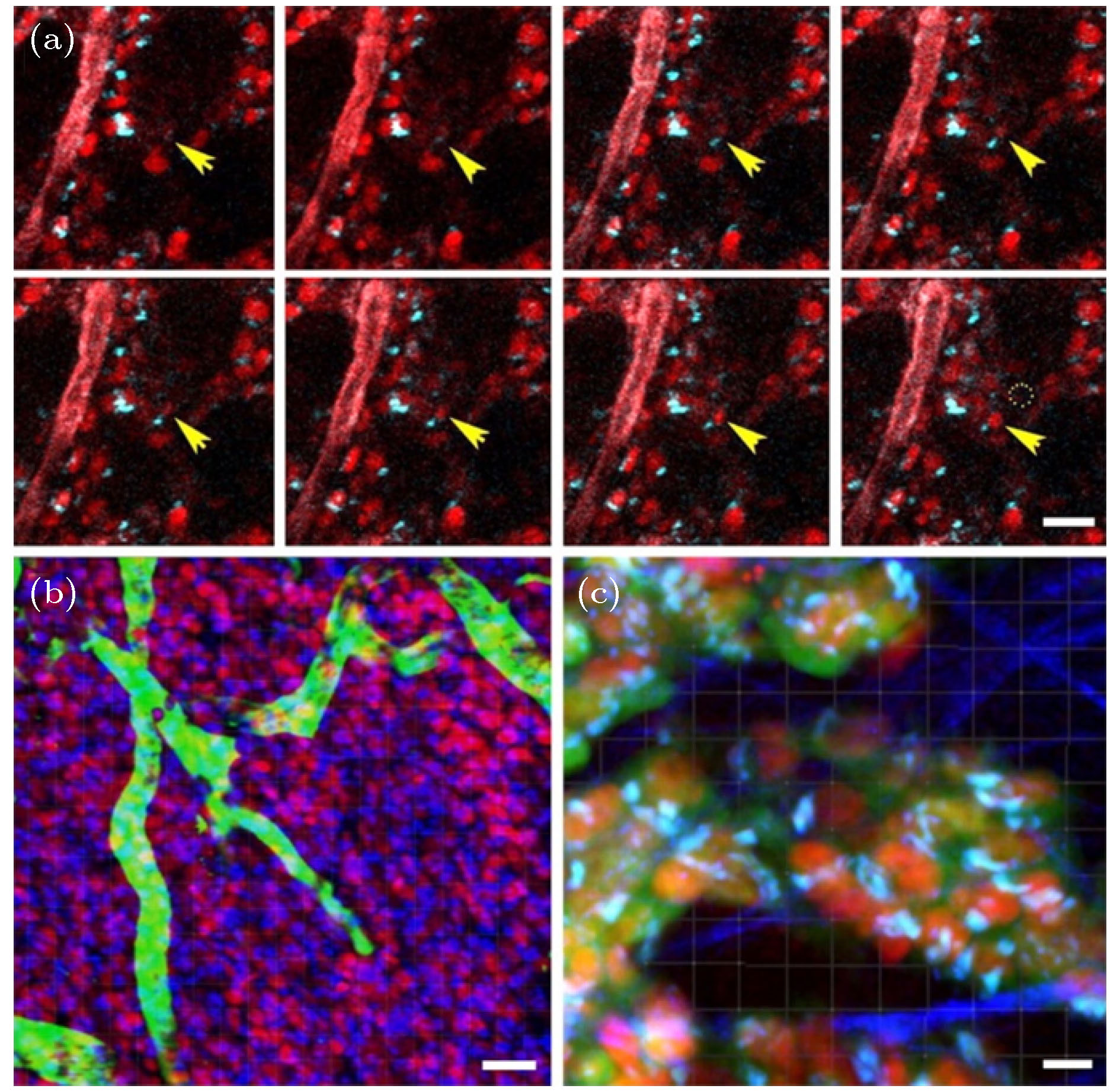
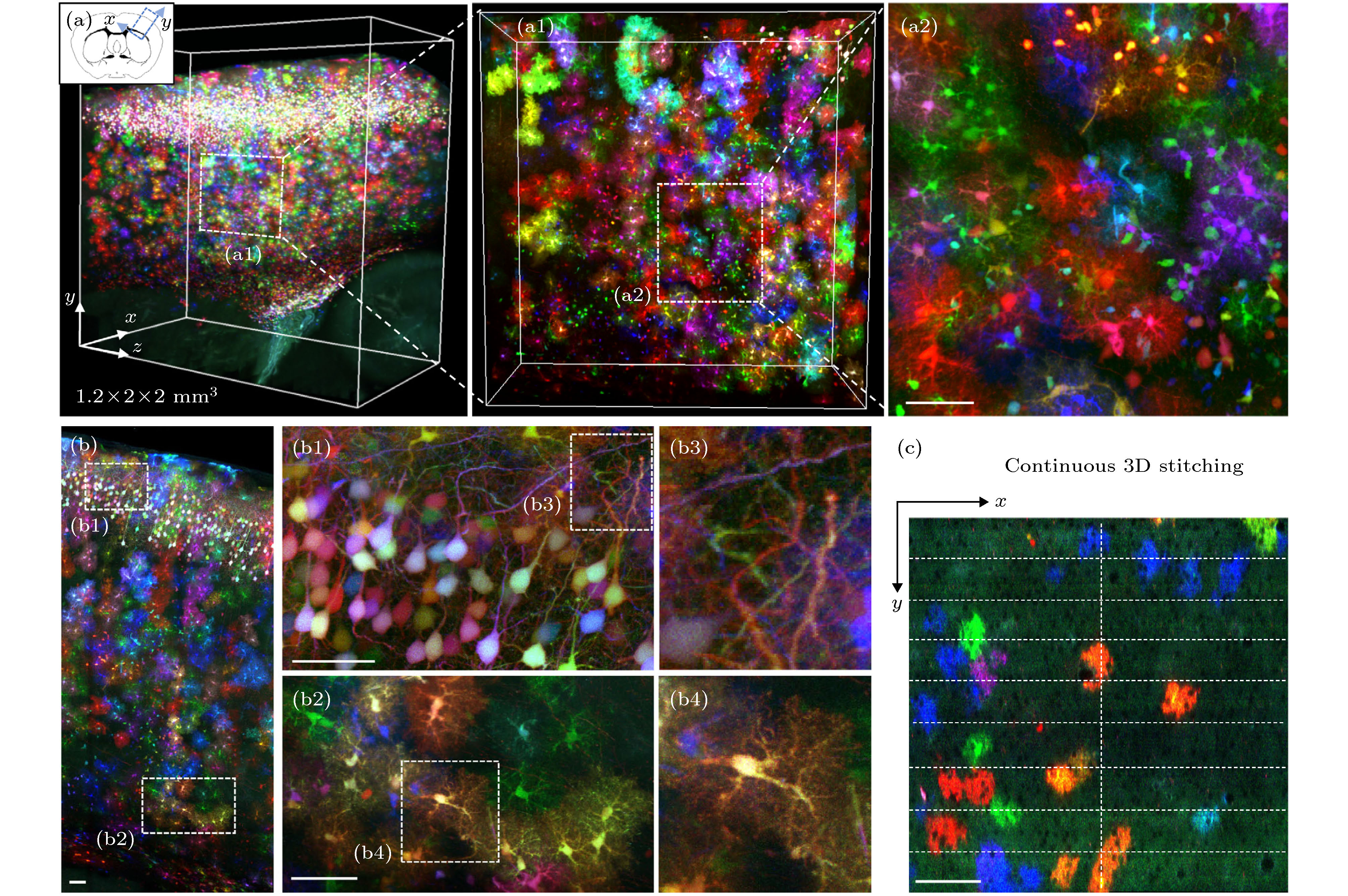
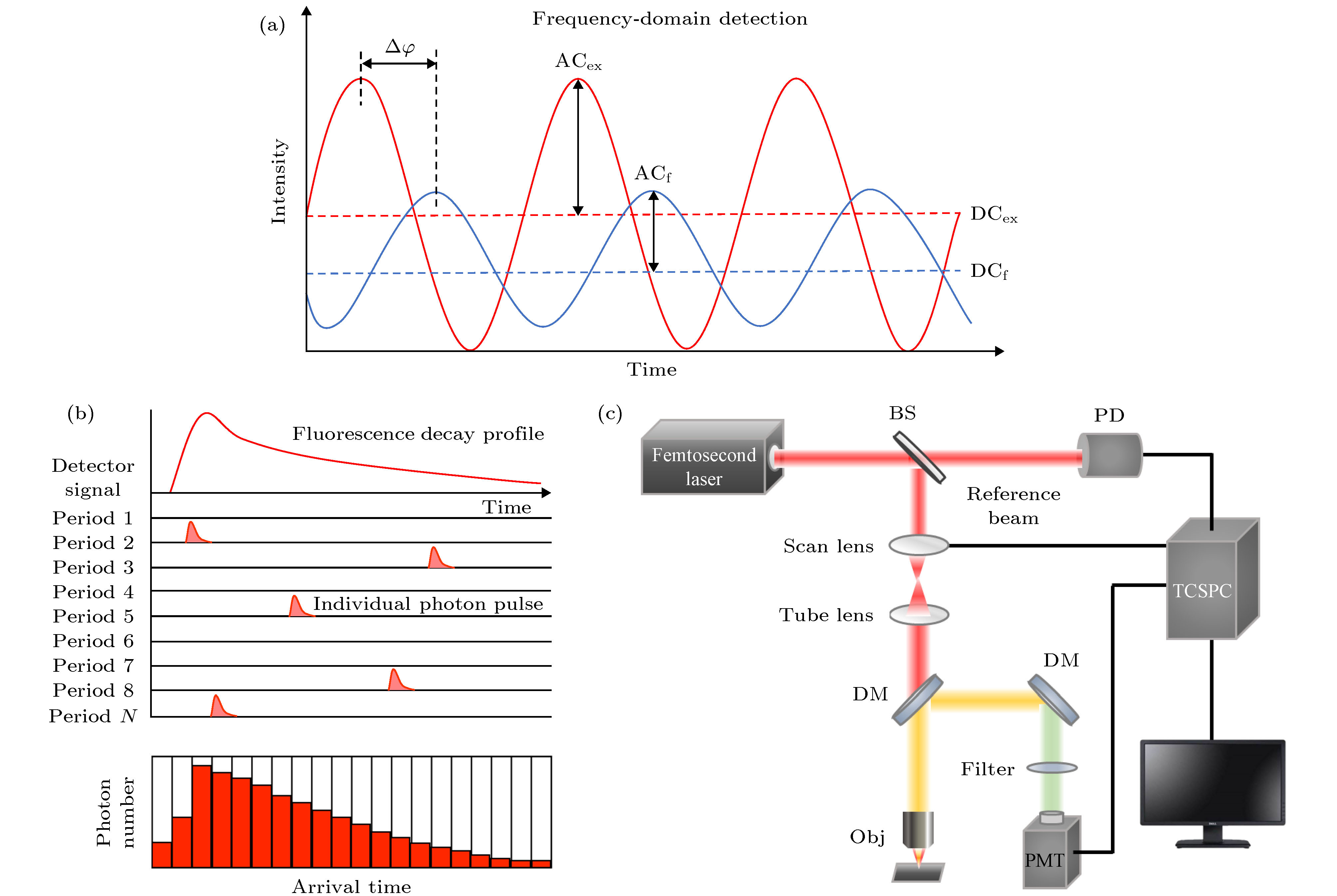
图 5 常见的荧光寿命测量方法及双光子TCSPC-FLIM成像系统示意图 (a) 频域法; (b) TCSPC法; (c) 基于TCSPC的双光子FLIM成像系统示意图. 图中各部分为: Femtosecond Laser, 飞秒激光器; BS (beam splitter), 分光镜; Scan Lens, 扫描镜; Tube Lens, 镜筒透镜; DM (dichroic mirror), 二向色镜; Obj (objective), 物镜; Filter, 滤光片; PMT (photomultiplier tube), 光电倍增管探测器; Reference Beam, 参考光; PD (photodiode), 光电二极管; TCSPC, 时间相关单光子计数法
Fig. 5. Schematic diagram of common fluorescence lifetime measurement methods and imaging systems: (a) Frequency domain method; (b) TCSPC method; (c) schematic diagram of a two-photon FLIM imaging system based on TCSPC. The abbreviations in the figure are as follows: BS, beam splitter; DM, dichroic mirror; Obj, objective; PMT, photomultiplier tube; PD, photodiode; TCSPC, time-correlated single photon counting.


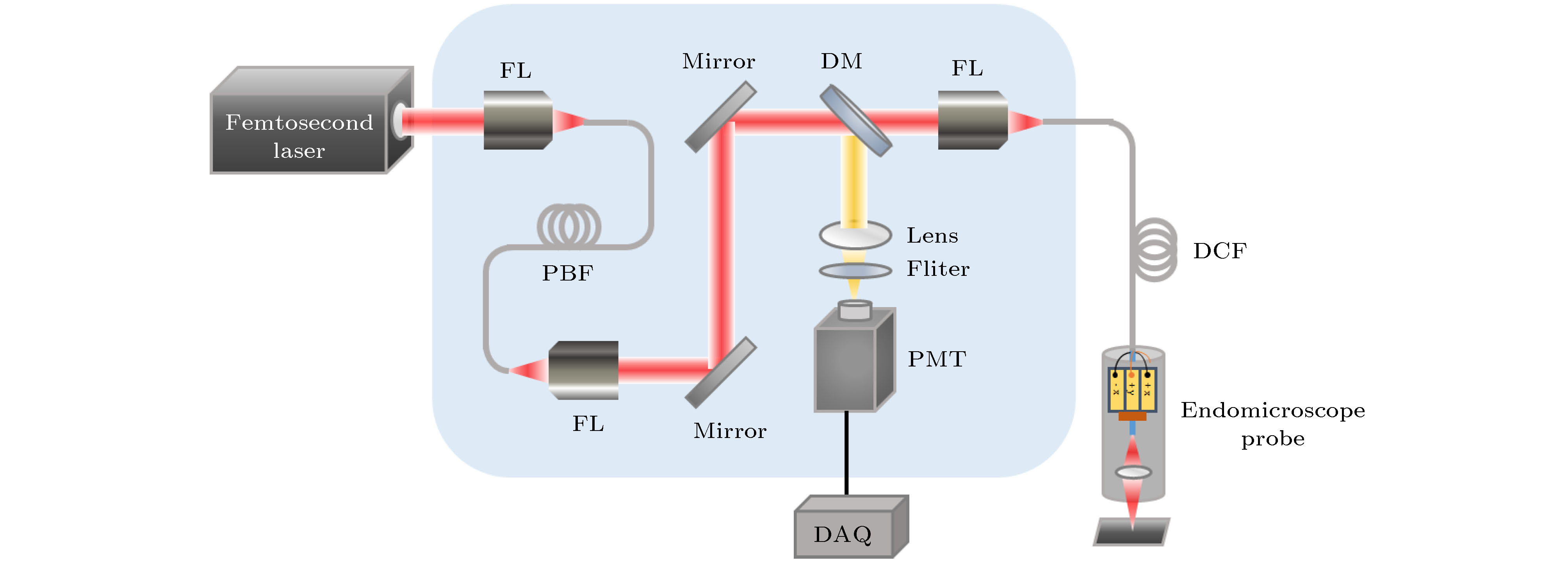
图 8 双光子光纤内窥系统示意图. 图中各部分为: Femtosecond Laser, 飞秒激光器; FL (fiber launcher), 光纤耦合器; PBF (photonic band-gap fiber), 光子带隙光纤; Mirror, 反射镜; DM (dichroic mirror), 二向色镜; Lens, 透镜; Filter, 滤光片; PMT (Photomultiplier tube), 光电倍增管探测器; DAQ (data acquisition), 数据采集; DCF (double-clad fiber), 双包层光纤; Endomicroscope Probe, 内窥镜探头
Fig. 8. Schematic diagram of a two-photon fiber endoscopic system. The abbreviations in the figure are as follows: FL, fiber launcher; PBF, photonic band gap light; DM, dichroic mirror; PMT, Photomultiplier tube; DAQ, data acquisition; DCF, double-clad fiber.
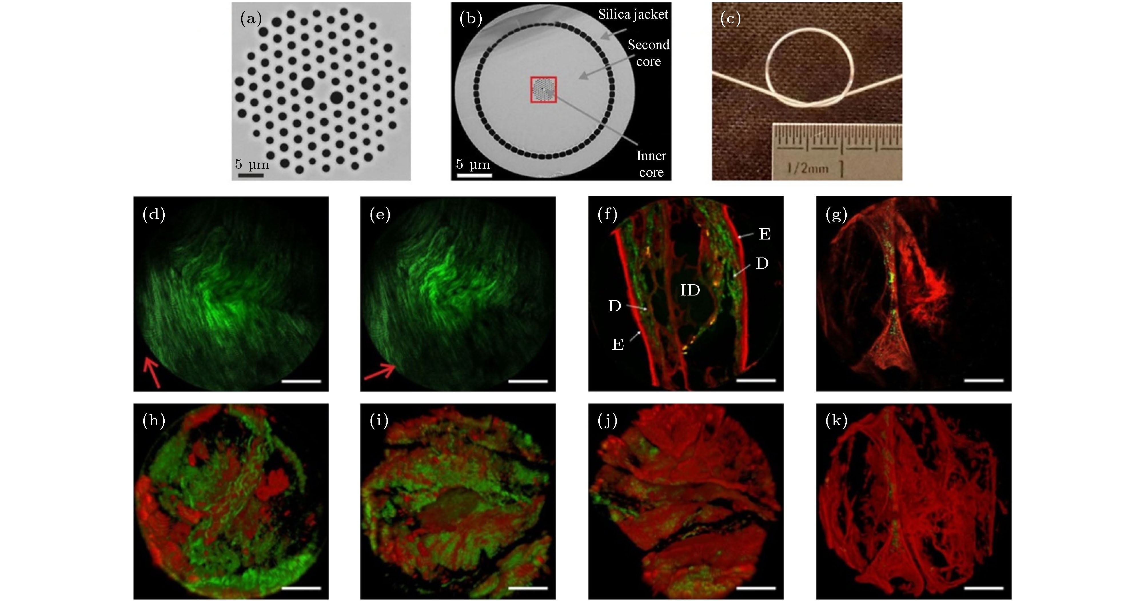
图 9 用于双光子内窥镜的空气-二氧化硅DC-PCF设计及系统成像图[55] (a) 光纤纤芯示意图, 二氧化硅部分为灰色, 空气部分为黑色; (b) 双包层光纤纤芯截面示意图; (c) DC-PCF具有灵活性; (d)−(k) 组织样本的无标记双光子光纤内窥成像, 红色为TPEF信号, 绿色为SHG信号. (d), (e) 大鼠尾肌腱; (f) 鼠耳. D: 真皮; E: 表皮; IC: 内部软骨; (g)健康人类肺部样品(肺泡区域); (h) 小鼠动脉; (i)−(k)健康人类肺部样品里3个位置的细胞外基质. 比例尺: 50 µm
Fig. 9. Design and system imaging diagram of an Air-silica DC-PCF for a two-photon endoscope[55]: (a) Schematic diagram of the optical fiber core, the silica part is gray, and the air part is black; (b) the cross-sectional schematic view of the double-clad fiber core; (c) DC-PCF is flexible; (d)−(k) Unlabeled two-photon fiber endoscopy imaging of tissue samples, red is TPEF signal, green is SHG signal; (d), (e) rat tail tendon; (f) mouse ear. D: dermis; E: epidermis; IC: internal cartilage; (g) healthy human lung samples (alveolar regions); (h) mouse arteries; (i)−(k) extracellular matrix at 3 locations in healthy human lung samples. Scale bar: 50 µm.

图 10 激光扫描三光子显微镜示意图. 图中各部分为: Fiber Laser, 光纤激光器; HWP (half-wave plate), 半波片; PBS (polarization beam splitter), 偏振分束器; Mirror, 反射镜; Lens, 透镜; PCF (photonic crystal fiber), 光子晶体光纤; Scan Mirror, 扫描镜; Scan Lens, 扫描透镜; Tube Lens, 镜筒透镜; DM (dichroic mirror), 二向色镜; Filter, 滤光片; Obj (objective), 物镜; PMT (photomultiplier tube), 光电倍增管探测器
Fig. 10. Schematic diagram of a laser scanning three-photon microscope. The abbreviations in the figure are as follows: HWP, half-wave plate; PBS, polarization beam splitter; PCF, photonic crystal fiber; DM, dichroic mirror; Obj, objective; PMT, photomultiplier tube.

图 11 小鼠组织模型衰减谱及活体成像[60] (a) 基于米氏散射和吸水率的组织模型的衰减谱; (b) FVB/N小鼠脑血管三光子图像的三维重构; (c) B6.Cg-Tg(Thy1-Brainbow1.0)HLich/J小鼠脑内神经元三光子图像的三维重构
Fig. 11. Attenuation spectrum and in vivo imaging of mouse tissue model[60]: (a) Attenuation spectrum of tissue model based on Mie scattering and water absorption; (b) three-dimensional reconstruction of three-photon image of FVB/N mouse cerebrovascular; (c) B6.Cg-Tg (Thy1-Brainbow1.0) three-dimensional reconstruction of three-photon images of neurons in the brain of HLich/J mice.
表 1 双光子FLIM监测NADH和FAD的工作原理[28]
Table 1. Working principle of NADH and FAD monitoring by two-photon FLIM[28].
名称 本质 主要分布 工作原理 与FLIM联系 生理功能 NADH 烟酰胺腺嘌呤二核苷酸(NAD)的还原态, 一种还原型辅酶 线粒体和
细胞质在氧化还原反应中, NADH作为氢和电子
的供体, NAD作为氢
和电子的受体NAD与脱氢酶结合时, 激发和发射都有
蓝移, 同时荧光量子产率增加. NADH跟蛋
白质结合后显示较长的寿命成分. FLIM
还可以计算这些物质的相对含量改善能量水平、保护细胞、促进神经递质的产生等 FAD 黄素腺嘌呤二核
苷酸, 某些氧化还
原酶的辅基线粒体 FAD参与体内各种氧
化还原反应, 在生物氧
化系统中起传递氢的作用游离的FAD分子显示较长的寿命成
分, 和蛋白质结合的FAD
分子显示较短的寿命成分可做成活性型维生素B2, 用于神经性耳鸣、脑动脉硬化等  下载: 导出CSV
-
[1] Goppert M M 1931 Ann. Phys-Berlin. 9 273
[2] Denk W, Strickler J H, Webb W W 1990 Science 248 73
Google Scholar
[3] Wang K, Horton N G, Charan K, Xu C 2014 Ieee J. Sel. Top. in Quant. 20 6800311
[4] 崔权, 陈忠云, 张智红, 骆清铭, 付玲 2017 激光与光电子学进展 06 16
Cui Q, Chen Z Y, Zhang Z H, Luo Q M, Fu L 2017 Las. Opto-elect. Prog. 06 16
[5] 杨洪权 2011 硕士学位论文 (福建: 福建师范大学)
Yang H Q 2011 M. S. Thesis (Fujian: Fujian Normal University) (in Chinese)
[6] Drobizhev M, Makarov N S, Tillo S E, Hughes T E, Rebane A 2011 Nat. Methods 8 393
Google Scholar
[7] Tragardh J, Murtagh M, Robb G, Parsons M, Lin J P, Spence D J, McConnell G 2016 Microsc. and Microanal. 22 803
Google Scholar
[8] Herz J, Siffrin V, Hauser A E, Brandt A U, Leuenberger T, Radbruch H, Zipp F, Niesner R A 2010 Biophys. J. 98 715
Google Scholar
[9] Xi P, Andegeko Y, Weisel L R, Lozovoy V, Dantus M 2008 Opt. Commun. 281 1841
Google Scholar
[10] Liang X B, Hu W Y, Fu L 2010 Opt. Express 18 14893
Google Scholar
[11] Zhao Z, Wu B, Wang X, Pan Z, Liu Z, Zhang P, Shen X, Nie Q, Dai S, Wang R 2017 Laser Photonics Rev. 11 1700005
Google Scholar
[12] Li D, Zheng W, Qu J A Y 2009 Opt. Lett. 34 202
Google Scholar
[13] Li C, Pastila R K, Lin C P 2016 J. Innov. Opt. Heal. Sci. 9 1640003
Google Scholar
[14] Le Dévédec S E, Lalai R, Pont C, de Bont H, van de Water B 2011 Mol. Imaging Biol. 13 67
Google Scholar
[15] Entenberg D, Wyckoff J, Gligorijevic B, Roussos E T, Verkhusha V, Pollard J W, Condeelis J 2011 Nat. Protoc. 6 1500
Google Scholar
[16] Piatkevich K D, Hulit J, Subach O M, Wu B, Abdulla A, Segall J E, Verkhusha V 2010 P. Natl. Acad. Sci. U. S. A. 107 5369
Google Scholar
[17] Collot M, Fam T K, Ashokkumar P, Faklaris O, Galli T, Danglot L, Klymchenko A S 2018 J. Am. Chem. Soc. 140 5401
Google Scholar
[18] Garaschuk O, Milos R-I, Konnerth A 2006 Nat. Protoc. 1 380
Google Scholar
[19] Hayakawa Y, Nemoto T, Iino M, Kasai H 2005 Cell Calcium. 37 359
Google Scholar
[20] Mahou P, Zimmerley M, Loulier K, Matho K S, Labroille G, Morin X, Supatto W, Livet J, Debarre D, Beaurepaire E 2012 Nat. Methods 9 815
Google Scholar
[21] Mahou P, Vermot J, Beaurepaire E, Supatto W 2014 Nat. Methods 11 600
Google Scholar
[22] Stringari C, Abdeladim L, Malkinson G, Mahou P, Solinas X, Lamarre I, Brizion S, Galey J B, Supatto W, Legouis R, Pena A M, Beaurepaire E 2017 Sci. Rep-UK. 7 3792
Google Scholar
[23] Abdeladim L, Matho K S, Clavreul S, et al. 2019 Nat. Commun. 10 1662
Google Scholar
[24] Lin D Y, Luo T, Liu L W, Lu Y, Liu S X, Yuan Z, Qu J L 2017 Chin. Opt. Lett. 15 090006
Google Scholar
[25] 刘雄波, 林丹樱, 吴茜茜, 严伟, 罗腾, 杨志刚, 屈军乐 2017 物理学报 67 178701
Liu X B, Lin D Y, Wu Q Q, Wei Y, Luo T, Yang Z G, Qu J L 2017 Acta Phys. Sin. 67 178701
[26] Liu X B, Lin D Y, Becker W, Niu J, Yu B, Liu L W, Qu J L 2019 J. Innov. Opt. Heal. Sci. 12 1930003
Google Scholar
[27] 李慧, 夏先园, 陈廷爱, 余佳, 李曦, 郑炜 2018 中国激光 45 0207010
Google Scholar
Li H, Xia X Y, Chen T A, Yu J, Li X, Zheng W 2018 Chin. J. Lasers 45 0207010
Google Scholar
[28] Ranawat H, Pal S, Mazumder N 2019 Biomed. Eng. Lett. 9 293
Google Scholar
[29] Li H, Yu J, Zhang R, Li X, Zheng W 2019 J. Innov. Opt. Heal. Sci. 12 1930009
Google Scholar
[30] Anh C, Pimenta R M L, Lee H B, Mereddy V, Holy J, Heikal A 2019 Cytom. Part A 95A 80
[31] Luo T, Lu Y, Liu S X, Lin D Y, Qu J L 2017 Anal. Chem. 89 8104
Google Scholar
[32] Chen B L, Lu Y, Pan W H, Xiong J, Yang Z G, Yan W, Liu L W, Qu J L 2019 Anal. Chem. 91 10640
Google Scholar
[33] Shen B L, Yan J S, Wang S Q, Zhou F, Zhao Y H, Hu R, Qu J L, Liu L W 2020 Theranostics 10 1849
Google Scholar
[34] Alam S R, Wallrabe H, Svindrych Z, et al. 2017 Sci. Rep-UK. 7 10451
Google Scholar
[35] Poulon F, Pallud J, Varlet P, et al. 2018 Sci. Rep-UK. 8 14888
Google Scholar
[36] Kantelhardt S R, Kalasauskas D, Konig K, Kim E, Weinigel M, Uchugonova A, Giese A 2016 J. Neuro-Oncol. 127 473
Google Scholar
[37] Teh S K, Zheng W, Li S X, Li D, Zeng Y, Yang Y Q, Qu J A Y 2013 J. Biomed. Opt. 18 036001
Google Scholar
[38] Rück A, Hauser C, Mosch S, Kalinina S 2014 J. Biomed. Opt. 19 096005
Google Scholar
[39] Shen Y F, Tsai M R, Chen S C, et al. 2015 Anal. Chem. 87 7575
Google Scholar
[40] Li X, Li H, He X Z, Chen T G, Xia X Y, Yang C X, Zheng W 2018 Biomed. Opt. Express 9 453
Google Scholar
[41] Tyurikova O, Zheng K Y, Rings A, Drews A, Klenerman D, Rusakov D A 2018 Brain Res. Bull. 136 85
Google Scholar
[42] Zheng K Y, Jensen T P, Rusakov D A 2018 Nat. Protoc. 13 581
Google Scholar
[43] Feeks J A, Hunter J 2017 Biomed. Opt. Express 8 2483
Google Scholar
[44] Chakraborty S, Nian F S, Tsai J W, Karmenyan A, Chiou A 2016 Sci. Rep-UK. 6 19145
Google Scholar
[45] Looney M R, Thornton E, Sen D, Lamm W J, Glenny R W, Krummel M F 2011 Nat. Methods 8 91
Google Scholar
[46] 石玉洁, 张广杰, 陆政元, 应亚宸, 贾荟琳, 席鹏 2018 中国光学 11 296
Google Scholar
Shi Y J, Zhang G J, Lu Z Y, Ying Y C, Jia H L, Xi P 2018 Chin. Opt. 11 296
Google Scholar
[47] Bird D, Gu M 2002 Opt. Lett. 27 1031
Google Scholar
[48] Bao H, Allen J, Pattie R, Vance R, Gu M 2008 Opt. Lett. 33 1333
Google Scholar
[49] Bao H, Ryu S Y, Lee B H, Tao W, Gu M 2010 Opt. Lett. 35 995
Google Scholar
[50] Tang S, Jung W, McCormick D, Xie T, Su J, Ahn Y C, Tromberg B J, Chen Z 2009 J. Biomed. Opt. 14 034005
Google Scholar
[51] Piyawattanametha W, Cocker E D, Burns L D, Barretto R P J, Jung J C, Ra H, Solgaard O, Schnitzer M J 2009 Opt. Lett. 34 2309
Google Scholar
[52] Wu Y C, Leng Y X, Xi J F, Li X D 2009 Opt. Express 17 7907
Google Scholar
[53] Lee C M, Engelbrecht C J, Soper T D, Helmchen F, Seibel E J 2010 J. Biophotonics 3 385
Google Scholar
[54] Peyrot D A, Lefort C, Steffenhagen M, Mansuryan T, Ducourthial G, Abi-Haidar D, Sandeau N, Vever-Bizet C, Kruglik S G, Thiberville L, Louradour F, Bourg-Heckly G 2012 Biomed. Opt. Express 3 840
Google Scholar
[55] Ducourthial G, Leclerc P, Mansuryan T, Fabert M, Brevier J, Habert R, Braud F, Batrin R, Vever-Bizet C, Bourg-Heckly G, Thiberville L, Druilhe A, Kudlinski A, Louradour F 2015 Sci. Rep-UK. 5 18303
Google Scholar
[56] Hage C H, Leclerc P, Brevier J, Fabert M, Le Nezet C, Kudlinski A, Heliot L, Louradour F 2018 Biomed. Opt. Express 9 142
Google Scholar
[57] Sibai M, Mehidine H, Poulon F, Ibrahim A, Varlet P, Juchaux M, Pallud J, Devaux B, Kudlinski A, Abi Haidar D 2018 Sci. Rep-UK. 8 11124
Google Scholar
[58] Gu M, Bao H, Gan X, Stokes N, Wu J 2014 Light-Sci. Appl. 3 e126
Google Scholar
[59] Xu C, Zipfel W, Shear J B, Williams R M, Webb W 1996 P. Natl. Acad. Sci. U. S. A. 93 10763
Google Scholar
[60] Horton N G, Wang K, Kobat D, Clark C G, Wise F W, Schaffer C B, Xu C 2013 Nat. Photonics 7 205
Google Scholar
[61] Huland D M, Charan K, Ouzounov D G, Jones J S, Nishimura N, Xu C 2013 Biomed. Opt. Express 4 652
Google Scholar
[62] Ouzounov D G, Horton N, Wang T Y, Feng D, Nishimura N, Xu C 2014 Conference on Lasers and Electro-Optics San Jose, America, June 8–13, 2014 p2.
[63] Horton N G, Xu C 2015 Biomed. Opt. Express 6 1392
Google Scholar
[64] Ouzounov D G, Wang T, Wang M, Feng D, Horton N G, Cruz-Hernandez J C, Cheng Y T, Reimer J, Tolias A S, Nishimura N, Xu C 2017 Nat. Methods 14 388
Google Scholar
[65] Li B, Wang M, Wu C, Charan K, Xu C 2018 Conference on Lasers and Electro-Optics San Jose, America, May 13-18, 2018 pJTh5 C.5
[66] Wang T, Ouzounov D G, Wu C, Horton N G, Zhang B, Wu C H, Zhang Y, Schnitzer M J, Xu C 2018 Nat. Methods 15 789
Google Scholar
[67] Li B, Wu C, Wang M, Charan K, Xu C 2020 Nat. Methods 17 163
Google Scholar
[68] Chow D M, Sinefeld D, Kolkman K E, Ouzounov D G, Akbari N, Tatarsky R, Bass A, Xu C, Fetcho J R 2020 Nat. Methods 17 605
Google Scholar
[69] Wang S, Xi W, Cai F, Zhao X, Xu Z, Qian J, He S 2015 Theranostics 5 251
Google Scholar
[70] Zhu Z, Leung C W, Zhao X, Wang Y, Qian J, Tang B Z, He S 2015 Sci. Rep-UK. 5 15189
Google Scholar
[71] Li D, Zhao X, Qin W, Zhang H, Fei Y, Liu L, Yong K T, Chen G, Tang B Z, Qian J 2016 Nano Res. 9 1921
Google Scholar
[72] Wang Y, Chen M, Alifu N, Li S, Qin W, Qin A, Tang B Z, Qian J 2017 ACS Nano 11 10452
Google Scholar
[73] Wang Y, Han X, Xi W, Li J, Roe A W, Lu P, Qian J 2017 Adv. Healthc. Mater. 6 1700685
Google Scholar
[74] Liu W, Wang Y, Han X, Lu P, Zhu L, Sun C, Qian J, He S 2018 Nanoscale 10 10025
Google Scholar
[75] Ni H, Xu Z, Li D, Chen M, Tang B Z, Qian J 2019 J. Innov. Opt. Heal. Sci. 12 1940005
Google Scholar
[76] Qin W, Alifu N, Lam J W Y, Cui Y, Su H, Liang G, Qian J, Tang B Z 2020 Adv. Mater. 32 e2000364
Google Scholar
[77] Liu H, Du Y, Peng X, Zhou X, Qiu P, Wang K 2017 IEEE Photonics J. 9 1
[78] Tong S, Liu H, Cheng H, He C, Du Y, Zhuang Z, Qiu P, Wang K 2019 J. Biophotonics 12 e201800423
[79] Liu H, Deng X, Tong S, He C, Cheng H, Zhuang Z, Gan M, Li J, Xie W, Qiu P, Wang K 2019 Nano. Lett. 19 5260
Google Scholar
[80] Tong S, Gan M Y, Zhuang Z W, Liu H J, Cheng H, Li J, Qiu P, Wang K 2020 J. Lightwave Technol. 38 2450
Google Scholar
[81] Weisenburger S, Tejera F, Demas J, Chen B, Manley J, Sparks F T, Traub F M, Daigle T, Zeng H, Losonczy A, Vaziri A 2019 Cell 177 1050
Google Scholar
[82] Klioutchnikov A, Wallace D J, Frosz M H, Zeltner R, Sawinski J, Pawlak V, Voit K M, Russell P S J, Kerr J N D 2020 Nat. Methods 17 509
Google Scholar


 下载:
下载:
计量
- 文章访问数: 27775
- PDF下载量: 780
- 被引次数: 0
