


-
Lipid rafts are small biomembrane functional units, resulting from the lateral phase separation of phospholipids. The phospholipid phase separation plays a crucial role in spatially organizing the biomolecules in life activities. Here, we study the kinetics of multi-component phospholipid phase separation quantitatively by using the single domain characterization methods including the movement tracking and radial fluctuation analyses, which provide valuable information about the physical and mechanical properties of the bulks and domains. The study is carried out in a low line tension condition similar to that in cells. The order of magnitude of line tension is ~0.1 pN as estimated from the radial fluctuation analysis. Fluorescence microscopy characterization shows that domains mainly coarsen through the coalescence pathways, while the evaporation-condensation is negligible. Through the tracking of domains, it is found that the bulk viscosity dominates the dynamics of domain coalescence. The coalescence of domains produces strong hydrodynamic flows in low viscosity bulk, which promotes the non-Brownian motion of surrounding domains, accelerating the lateral diffusion and coalescence of the domains. However, these hydrodynamic flows decrease significantly in high viscosity bulk. The domains rely mainly on Brownian motion to diffuse in this highly viscous medium, resulting in the slow lateral diffusion and low coalescence. Picking the domains following Brownian motion, the viscosities of liquid ordered bulk and liquid disordered bulk are determined to be, respectively, in a range of 10–8–10–7 Pa⋅s⋅m and 10–9 Pa⋅s⋅m from the Hughes-Pailthorpe-White empirical relation. Furthermore, we observe a bulk-viscosity-dependent scaling relation between the domain size and coarsening time experimentally. A theoretical model of domain diffusion and coalescence is established to understand the scaling relation. If the bulk viscosity is low, the hydrodynamic flow produces a high power exponent of 1.0. And if the bulk viscosity is high, the Brownian diffusion produces a low power exponent of 0.5. In addition, we demonstrate that the bulk viscosity can be regulated through the relative content of cholesterol. The 1,6-Diphenyl-1,3,5-hexatriene fluorescence anisotropy characterization exhibits that the increase of cholesterol in liquid ordered and liquid disordered bulks disorders and orders the phospholipid packing, thus reducing and increasing the bulk viscosity, respectively. It is expected that this viscosity regulation strategy can be used to control the multicomponent phospholipid phase separation. All in all, our study deepens the understanding of the physical mechanism behind the formation of lipid rafts. It also provides a reference for regulating the biomolecule distribution in cell membranes.
-
Keywords:
- phospholipid phase separation /
- single domain study /
- diffusion and coalescence /
- membrane viscosity
[1] Sezgin E, Levental I, Mayor S, Eggeling C 2017 Nat. Rev. Mol. Cell Biol. 18 361
 Google Scholar
[2] Wang X J, Tian L F, Ren Y S, Zhao Z Y, Du H, Zhang Z Z, Drinkwater B W, Mann S, Han X J 2020 Small 16 1906394
Google Scholar
[3] Zhou R H, Weikl T, Ma Y Q 2020 Nanoscale 12 10426
Google Scholar
[4] Fu M F, Li J B 2018 Angew. Chem. Int. Ed. 57 11404
Google Scholar
[5] 梁燚然, 梁清 2019 物理学报 68 028701
Google Scholar
Liang Y R, Liang Q 2019 Acta Phys. Sin. 68 028701
Google Scholar
[6] Maekawa T, Chin H, Nyu T, Sut T N, Ferhan A R, Hayashi T, Cho N J 2019 PCCP 21 16686
Google Scholar
[7] Veatch S L, Keller S L 2003 Biophys. J. 85 3074
Google Scholar
[8] Baumgart T, Hess S T, Webb W W 2003 Nature 425 821
Google Scholar
[9] Li W W, Lin Z, Yuan B, Yang K 2020 Chin. Phys. B 29 128701
Google Scholar
[10] Ding H M, Yin Y W, Ni S D, Sheng Y J, Ma Y Q 2021 Chin. Phys. Lett. 38 018701
Google Scholar
[11] Ye X Q, Hao C C, Yang J J, Sun R G 2018 Colloids Surf., B 172 480
Google Scholar
[12] Chen J X, Chen Y G, Kapral R 2018 Adv. Sci. 5 1800028
Google Scholar
[13] Chen J X, Yuan R, Cui R F, Qiao L Y 2021 Nanoscale 13 1055
Google Scholar
[14] Stanich C A, Honerkamp-Smith A R, Putzel G G, Warth C S, Lamprecht A K, Mandal P, Mann E, Hua T A D, Keller S L 2013 Biophys. J. 105 444
Google Scholar
[15] Garcia-Saez A J, Chiantia S, Schwille P 2007 J. Biol. Chem. 282 33537
Google Scholar
[16] Rozovsky S, Kaizuka Y, Groves J T 2005 JACS 127 36
Google Scholar
[17] Li J F, Zhang H D, Qiu F 2013 J. Phys. Chem. B 117 843
Google Scholar
[18] Talbot E L, Parolini L, Kotar J, Di Michele L, Cicuta P 2017 PNAS 114 846
Google Scholar
[19] Wongsirojkul N, Shimokawa N, Opaprakasit P, Takagi M, Hamada T 2020 Langmuir 36 2937
Google Scholar
[20] Zhou Q, Wang P, Ma B B, Jiang Z Y, Zhu T 2022 Chin. Phys. B 31 098701
Google Scholar
[21] Zhu T, Jiang Z Y, Ma Y Q, Hu Y 2016 ACS Appl. Mater. Interfaces 8 5857
Google Scholar
[22] Veatch S L, Keller S L 2005 Phys. Rev. Lett. 94 148101
Google Scholar
[23] Hormel T T, Reyer M A, Parthasarathy R 2015 Biophys. J. 109 732
Google Scholar
[24] Duan Y, Mahault B, Ma Y Q, Shi X Q, Chate H 2021 Phys. Rev. Lett. 126 178001
Google Scholar
[25] Cicuta P, Keller S L, Veatch S L 2007 J. Phys. Chem. B 111 3328
Google Scholar
[26] Bhuyan N N, Pattnaik G P, Mishra A, Chakraborty H 2021 J. Mol. Liq. 325 115152
Google Scholar
[27] Liang X Y, Li L, Qiu F, Yang Y L 2010 Physica A 389 3965
Google Scholar
[28] Usery R D, Enoki T A, Wickramasinghe S P, Weiner M D, Tsai W C, Kim M B, Wang S, Torng T L, Ackerman D G, Heberle F A, Katsaras J, Feigenson G W 2017 Biophys. J. 112 1431
Google Scholar
[29] Yanagisawa M, Imai M, Masui T, Komura S, Ohta T 2007 Biophys. J. 92 115
Google Scholar
[30] Almeida P F F, Vaz W L C, Thompson T E 1992 Biochem. 31 6739
Google Scholar
[31] Nagao M, Kelley E G, Faraone A, Saito M, Yoda Y, Kurokuzu M, Takata S, Seto M, Butler P D 2021 Phys. Rev. Lett. 127 078102
Google Scholar
[32] Petrov E P, Schwille P 2008 Biophys. J. 94 L41
Google Scholar
[33] Sakuma Y, Kawakatsu T, Taniguchi T, Imai M 2020 Biophys. J. 118 1576
Google Scholar
[34] Chakraborty S, Doktorova M, Molugu T R, Heberle F A, Scott H L, Dzikovski B, Nagao M, Stingaciu L R, Standaert R F, Barrera F N, Katsaras J, Khelashvili G, Brown M F, Ashkar R 2020 PNAS 117 21896
Google Scholar
[35] Kim K, Choi S Q, Zell Z A, Squires T M, Zasadzinski J A 2013 PNAS 110 E3054
Google Scholar
[36] Tayebi L, Ma Y, Vashaee D, Chen G, Sinha S K, Parikh A N 2012 Nat. Mater. 11 1074
Google Scholar
[37] Saeki D, Hamada T, Yoshikawa K 2006 J. Phys. Soc. Jpn. 75 013602
Google Scholar
[38] Taniguchi T 1996 Phys. Rev. Lett. 76 4444
Google Scholar
-
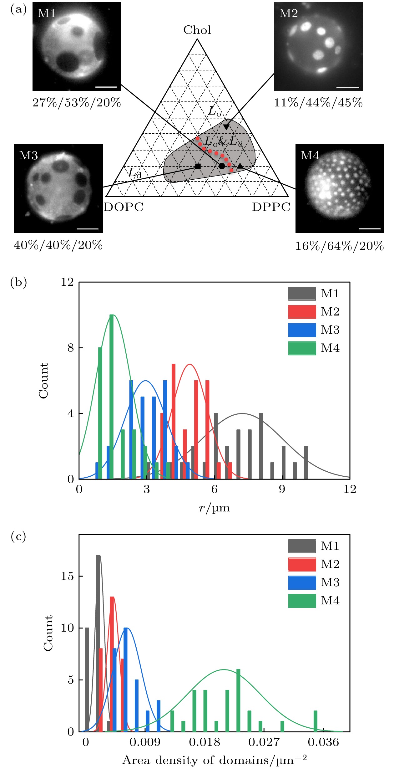
图 1 DOPC/DPPC/Chol三组分相分离 (a) 32 °C的相图, 红色点线表示Lo与Ld面积相同的组分, 标尺为 10 μm; (b) 微畴尺寸分布图; (c) 微畴面密度分布图
Figure 1. Phase diagram for vesicles of DOPC/DPPC/Chol: (a) Phase diagram at 32 °C. Red dotted line denotes the composition whose Lo and Ld phases occupy the same surface area. Scale bar is 10 μm. (b) Size distribution of the domains. (c) Surface density of the domains.
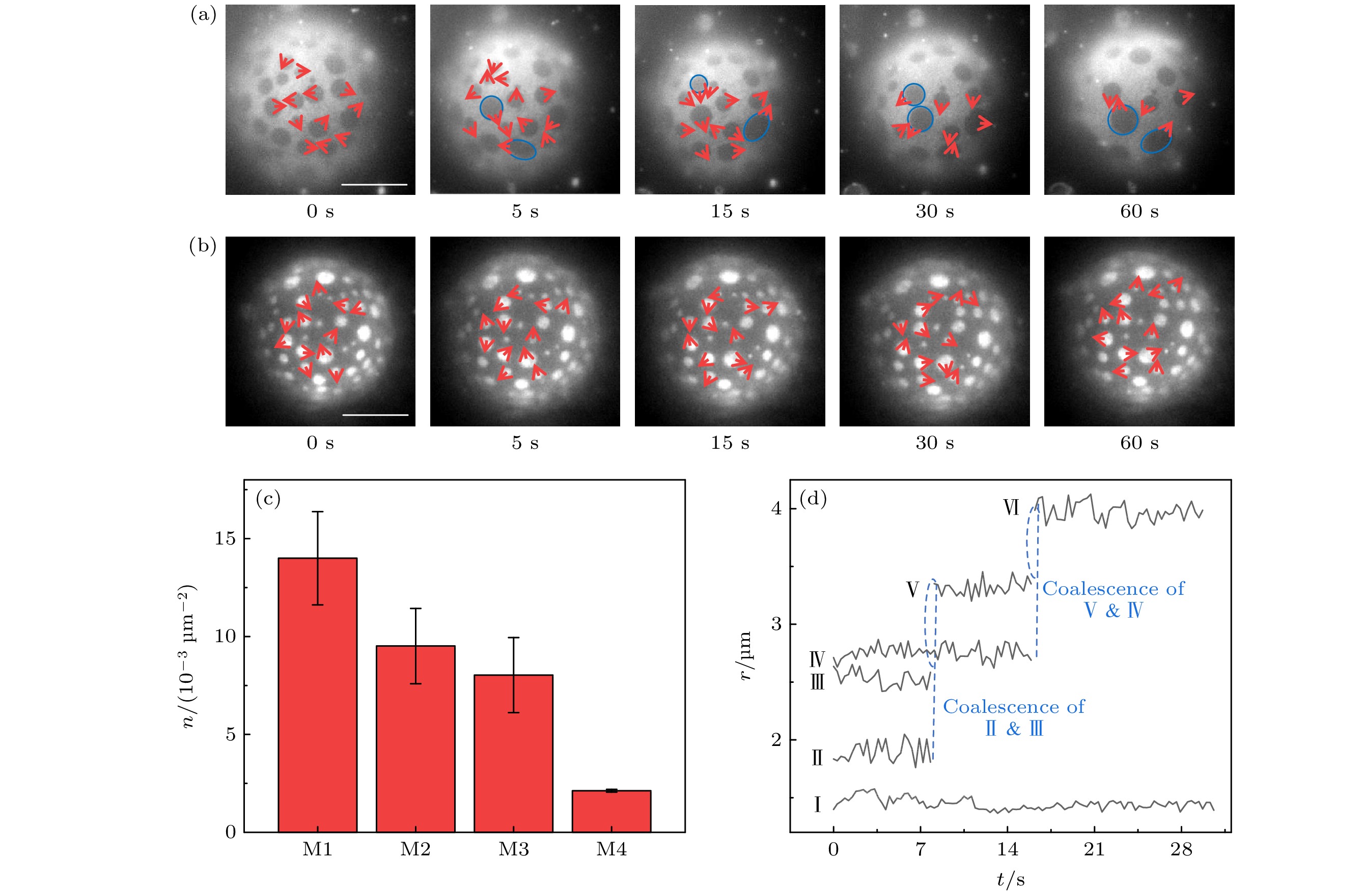
图 2 微畴的粗化 (a) M1和(b) M4的微畴融合粗化. 部分典型微畴的扩散和融合被标记出来. 其中绿色圆环表示发生融合, 红色箭头表示相邻时刻间微畴的扩散方向. (c) 2.5—4.0 min中M1—M4单位膜面积的融合次数. (d) 微畴的尺寸演变. 微畴Ⅱ, Ⅲ, Ⅳ融合生成微畴Ⅵ, 产生了显著的微畴粗化. 而未发生融合的微畴Ⅰ尺寸没有发生变化. 标尺为10 μm
Figure 2. Coarsening of domains. Coarsening by coalescence of domains in (a) M1 and (b) M4. The diffusion and coalescence of some typical domains are marked. The green circles denote the coalescence. The red arrows denote the diffusion direction of domains between the adjacent images. (c) Number of coalescences in unit surface area between 2.5–4.0 min. (d) Time evolution of domain size. Coarsening of domains is produced by the coalescence of the domains Ⅱ, Ⅲ, Ⅳ to form domain Ⅵ. Size of the domain Ⅰ remains unchanged without coalescence. Scale bar is 10 μm.
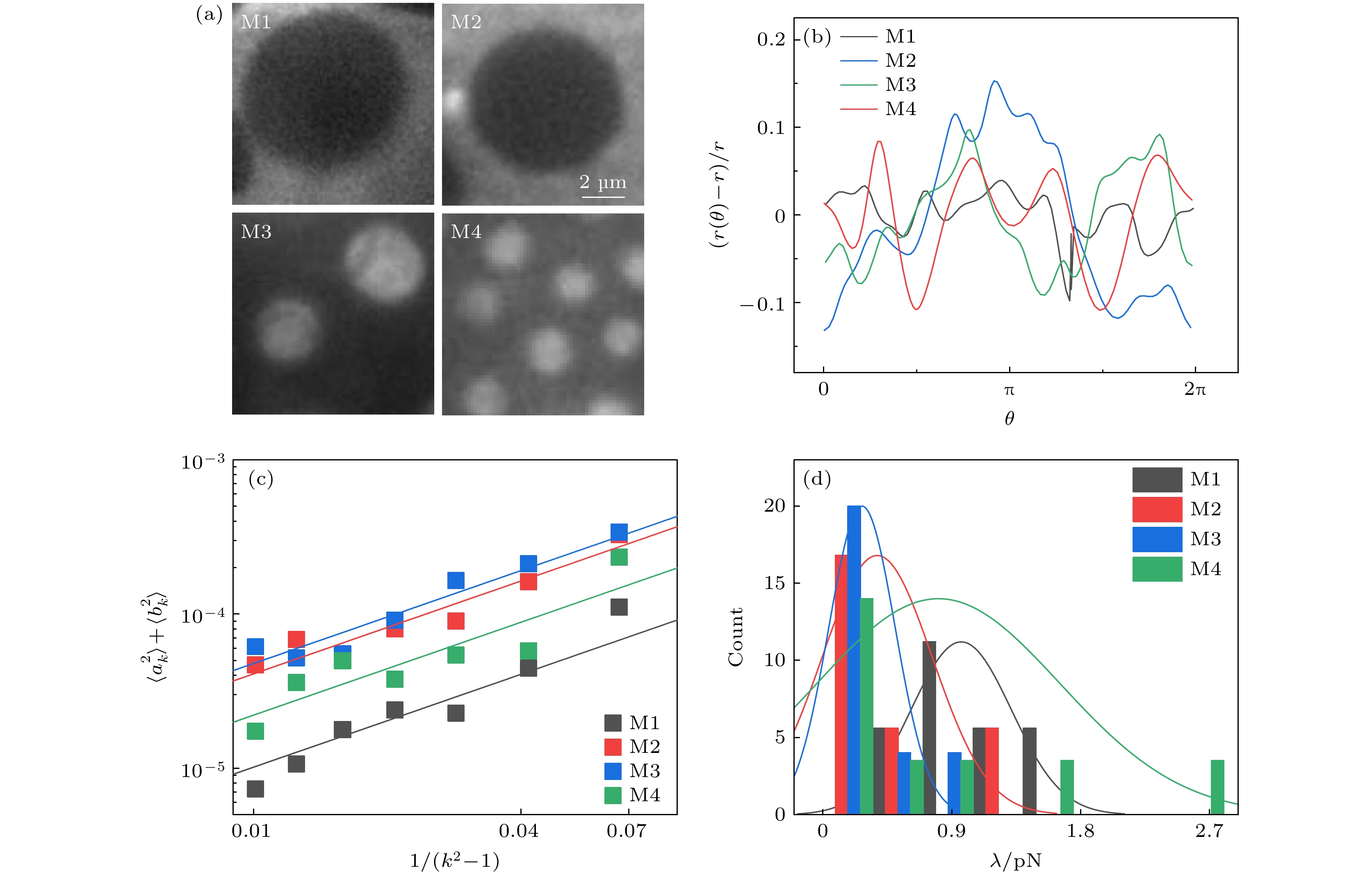
图 3 基于微畴径向波动性计算的线张力 (a) M1—M4微畴的荧光显微图; (b) 微畴极角(θ)对应的径向波动性; (c) 由傅里叶级数展开的波数(k)和系数(ak, bk)计算的
$ \left\langle{{a}_{k}^{2}}\right\rangle+\left\langle{{b}_{k}^{2}}\right\rangle $ 与$1/({k}^{2}-1)$ 关系; (d) 磷脂组分依赖的线张力Figure 3. Line tension calculated by domain boundary fluctuation: (a) Fluorescence microscopy of domains in M1-M4; (b) radial fluctuation as a function of polar angle (θ); (c) relationship between
$ \left\langle{{a}_{k}^{2}}\right\rangle+\left\langle{{b}_{k}^{2}}\right\rangle $ and$1/(k^2-1)$ calculated from the Fourier coefficients (ak, bk) and mode number (k); (d) dependence of line tension on lipid composition.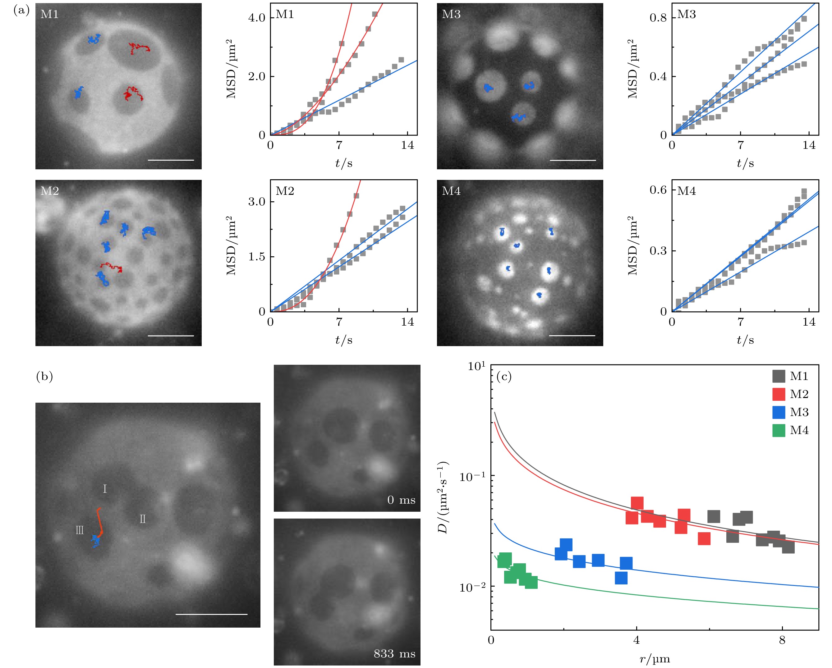
图 4 微畴在膜内的扩散 (a) 典型的运动轨迹与MSD, 红、蓝色分别标记了布朗运动与非布朗运动. (b) 融合促进的微畴扩散, 在微畴Ⅰ和Ⅱ融合前, 微畴Ⅲ呈布朗运动(蓝色轨迹); 在微畴Ⅰ和Ⅱ融合后, 微畴Ⅲ以近线性轨迹(红色)向Ⅰ, Ⅱ方向迁移. 微畴Ⅲ在833 ms内扩散迁移了约3 μm (右图). (c) 基于布朗运动的微畴统计获得的扩散速率-微畴尺寸关系. 标尺为10 μm
Figure 4. Diffusion of domains in lipid membranes: (a) Typical trajectories and MSD. Brownian and non-Brownian motions are marked by blue and red colors. (b) Coalescence-induced domain diffusion. Before the coalescence of domains Ⅰ and Ⅱ, domain Ⅲ underwent Brownian motion (blue trajectory). After the coalescence, domain Ⅲ diffused to Ⅰ and Ⅱ through a nearly straight line (red trajectory). The diffusion distance of domain Ⅲ is around 3 μm in 833 ms (images at right). (c) Plot of diffusion coefficient versus domain size obtained from the Brownian motion of the domains. Scale bar is 10 μm.
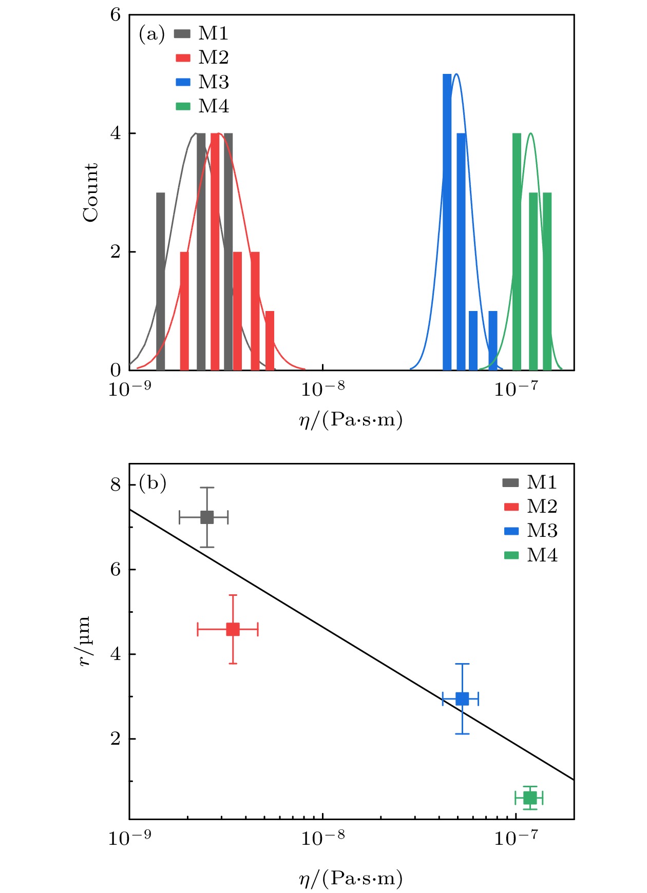
图 5 大相的黏滞性 (a) 数值求解的大相黏滞系数; (b) 大相黏滞系数与降温5.0 min的M1 —M4的微畴尺寸
Figure 5. Bulk viscosity: (a) Numerically computed bulk viscosity; (b) plot of the bulk viscosity versus domain size in M1− M4 (5.0 min after the temperature quench).
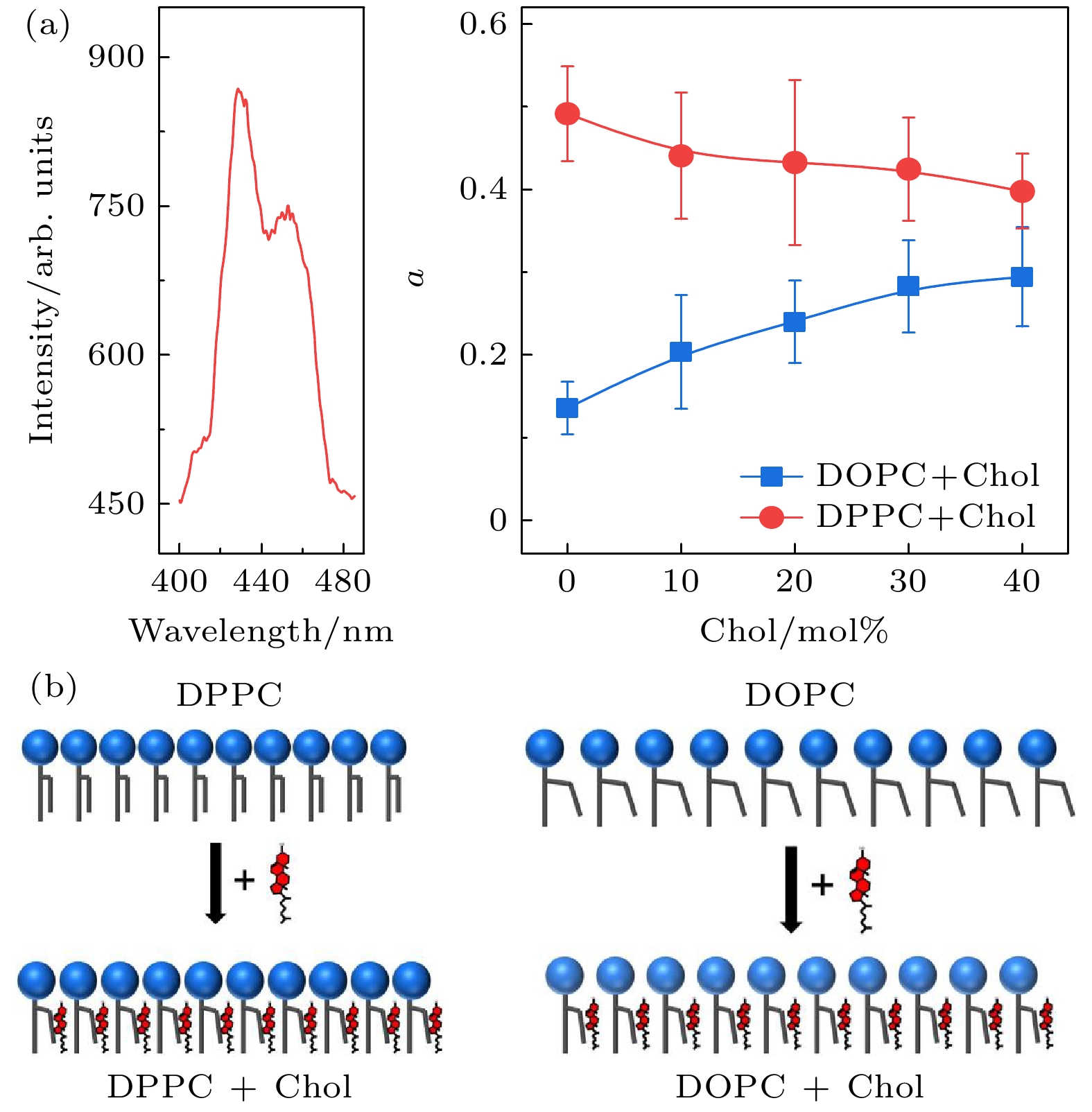
图 6 Chol影响的磷脂膜黏滞性 (a) 掺杂DPH的DPPC/Chol与 DOPC/Chol组分GUVs的荧光光谱与各向异性(32 °C); (b) 黏滞性变化对应的分子排布示意图. 磷脂排布有序度的提高造成膜黏滞性提高[34]
Figure 6. Membrane viscosity influenced by Chol: (a) Fluorescence spectrum and anisotropy of DPH-doped DPPC/Chol and DOPC/Chol GUVs (32 °C); (b) schematic illustration for the molecular packing associated with the different membrane viscosity. The increase of the packing order increases the viscosity[34].
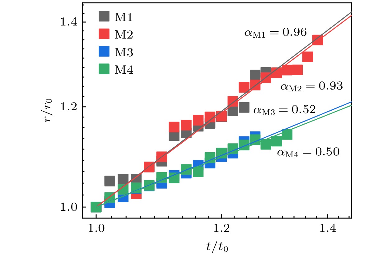
图 7 大相黏滞性依赖的微畴尺寸与时间标度律. r与t被归一化以方便比较(下标0代表初始时刻)
Figure 7. Bulk viscosity-depended scaling relation between domain size and time. r and t are normalized for comparison of the data (the subscript 0 denotes the initial).
表 1 微畴与大相的混合磷脂含量
Table 1. Lipid composition in domains and bulks.
组分 微畴 大相 DOPC DPPC Chol DOPC DPPC Chol M1 4% 67% 29% 52% 39% 9% M2 2% 66% 32% 60% 24% 16% M3 41% 26% 33% 5% 48% 47% M4 62% 26% 12% 10% 70% 20%  DownLoad: CSV
表 2 实验条件下
$ \eta $ 与$ {\eta }_{\mathrm{m}}r $ 的估算值(降温后2.5—4.0 min)Table 2. Estimated values of
$ \eta $ and$ {\eta }_{\mathrm{m}}r $ at the experiments (2.5−4.0 min after the temperature quench).r/μm $ {\eta }_{\mathrm{m}}r $/(10–9 Pa⋅s⋅m) $ \eta $/(10–9 Pa⋅s⋅m) M1 1.9—10.5 2.6—14.0 2.5 M2 1.7—6.6 2.3—8.8 3.4 M3 0.7—4.5 0.9—6.0 52.8 M4 0.4—2.9 0.5—3.9 118.0
DownLoad: CSV
-
[1] Sezgin E, Levental I, Mayor S, Eggeling C 2017 Nat. Rev. Mol. Cell Biol. 18 361
Google Scholar
[2] Wang X J, Tian L F, Ren Y S, Zhao Z Y, Du H, Zhang Z Z, Drinkwater B W, Mann S, Han X J 2020 Small 16 1906394
Google Scholar
[3] Zhou R H, Weikl T, Ma Y Q 2020 Nanoscale 12 10426
Google Scholar
[4] Fu M F, Li J B 2018 Angew. Chem. Int. Ed. 57 11404
Google Scholar
[5] 梁燚然, 梁清 2019 物理学报 68 028701
Google Scholar
Liang Y R, Liang Q 2019 Acta Phys. Sin. 68 028701
Google Scholar
[6] Maekawa T, Chin H, Nyu T, Sut T N, Ferhan A R, Hayashi T, Cho N J 2019 PCCP 21 16686
Google Scholar
[7] Veatch S L, Keller S L 2003 Biophys. J. 85 3074
Google Scholar
[8] Baumgart T, Hess S T, Webb W W 2003 Nature 425 821
Google Scholar
[9] Li W W, Lin Z, Yuan B, Yang K 2020 Chin. Phys. B 29 128701
Google Scholar
[10] Ding H M, Yin Y W, Ni S D, Sheng Y J, Ma Y Q 2021 Chin. Phys. Lett. 38 018701
Google Scholar
[11] Ye X Q, Hao C C, Yang J J, Sun R G 2018 Colloids Surf., B 172 480
Google Scholar
[12] Chen J X, Chen Y G, Kapral R 2018 Adv. Sci. 5 1800028
Google Scholar
[13] Chen J X, Yuan R, Cui R F, Qiao L Y 2021 Nanoscale 13 1055
Google Scholar
[14] Stanich C A, Honerkamp-Smith A R, Putzel G G, Warth C S, Lamprecht A K, Mandal P, Mann E, Hua T A D, Keller S L 2013 Biophys. J. 105 444
Google Scholar
[15] Garcia-Saez A J, Chiantia S, Schwille P 2007 J. Biol. Chem. 282 33537
Google Scholar
[16] Rozovsky S, Kaizuka Y, Groves J T 2005 JACS 127 36
Google Scholar
[17] Li J F, Zhang H D, Qiu F 2013 J. Phys. Chem. B 117 843
Google Scholar
[18] Talbot E L, Parolini L, Kotar J, Di Michele L, Cicuta P 2017 PNAS 114 846
Google Scholar
[19] Wongsirojkul N, Shimokawa N, Opaprakasit P, Takagi M, Hamada T 2020 Langmuir 36 2937
Google Scholar
[20] Zhou Q, Wang P, Ma B B, Jiang Z Y, Zhu T 2022 Chin. Phys. B 31 098701
Google Scholar
[21] Zhu T, Jiang Z Y, Ma Y Q, Hu Y 2016 ACS Appl. Mater. Interfaces 8 5857
Google Scholar
[22] Veatch S L, Keller S L 2005 Phys. Rev. Lett. 94 148101
Google Scholar
[23] Hormel T T, Reyer M A, Parthasarathy R 2015 Biophys. J. 109 732
Google Scholar
[24] Duan Y, Mahault B, Ma Y Q, Shi X Q, Chate H 2021 Phys. Rev. Lett. 126 178001
Google Scholar
[25] Cicuta P, Keller S L, Veatch S L 2007 J. Phys. Chem. B 111 3328
Google Scholar
[26] Bhuyan N N, Pattnaik G P, Mishra A, Chakraborty H 2021 J. Mol. Liq. 325 115152
Google Scholar
[27] Liang X Y, Li L, Qiu F, Yang Y L 2010 Physica A 389 3965
Google Scholar
[28] Usery R D, Enoki T A, Wickramasinghe S P, Weiner M D, Tsai W C, Kim M B, Wang S, Torng T L, Ackerman D G, Heberle F A, Katsaras J, Feigenson G W 2017 Biophys. J. 112 1431
Google Scholar
[29] Yanagisawa M, Imai M, Masui T, Komura S, Ohta T 2007 Biophys. J. 92 115
Google Scholar
[30] Almeida P F F, Vaz W L C, Thompson T E 1992 Biochem. 31 6739
Google Scholar
[31] Nagao M, Kelley E G, Faraone A, Saito M, Yoda Y, Kurokuzu M, Takata S, Seto M, Butler P D 2021 Phys. Rev. Lett. 127 078102
Google Scholar
[32] Petrov E P, Schwille P 2008 Biophys. J. 94 L41
Google Scholar
[33] Sakuma Y, Kawakatsu T, Taniguchi T, Imai M 2020 Biophys. J. 118 1576
Google Scholar
[34] Chakraborty S, Doktorova M, Molugu T R, Heberle F A, Scott H L, Dzikovski B, Nagao M, Stingaciu L R, Standaert R F, Barrera F N, Katsaras J, Khelashvili G, Brown M F, Ashkar R 2020 PNAS 117 21896
Google Scholar
[35] Kim K, Choi S Q, Zell Z A, Squires T M, Zasadzinski J A 2013 PNAS 110 E3054
Google Scholar
[36] Tayebi L, Ma Y, Vashaee D, Chen G, Sinha S K, Parikh A N 2012 Nat. Mater. 11 1074
Google Scholar
[37] Saeki D, Hamada T, Yoshikawa K 2006 J. Phys. Soc. Jpn. 75 013602
Google Scholar
[38] Taniguchi T 1996 Phys. Rev. Lett. 76 4444
Google Scholar

 DownLoad:


Catalog
Metrics
- Abstract views: 6912
- PDF Downloads: 118
- Cited By: 0
