


-
基于晶体外延生长技术构建稀土掺杂的微纳米核壳结构, 不仅有利于增强稀土离子的发射强度, 且可通过对离子的空间分离实现其发光颜色的精细调控. 为此, 本工作巧妙地借助外延生长技术构建了一系列掺杂不同离子浓度的NaYF4@NaYF4核壳结构微米晶体. 在980 nm近红外光激发下, 借助共聚焦显微光谱测试系统, 通过改变激发位置, 研究了掺杂离子在单颗粒核壳微米晶体中的能量传递特性. 研究表明, 当改变其激发位置时, 核壳微米盘不同区域掺杂的离子均展现出了不同的光谱特性, 其原因主要是由于在核壳结构中激发能传递方向不同所致. 基于其不同位置发射光谱及功率变化光谱, 证实微米核壳的激发能量主要是由外向内传递. 同时依据相应的光波导模型, 揭示了核壳结构微米盘多彩发射图案的产生. 由此可见, 通过构建不同微米核壳结构, 不仅能实现微米晶体发光特性的可控调节, 而且为进一步拓展微米晶体在光电子器件、光学编码及多色显示等领域中的应用提供重要的实验参考.The rare-earth doped micro/nano core-shell structure not only is beneficial to enhancing the upconversion emission intensity, but also can realize the fine control of luminescence color through the spatial separation of ions. In this work, a series of NaYF4@NaYF4 core-shell (CS) microcrystals doped with different ion concentrations is constructed by using the epitaxial growth technology. The structure and morphology for each of the prepared microcrystals are characterized by X-ray diffractometer (XRD) and scanning electron microscope (SEM). The experimental results show that the prepared CS structures each have a pure hexagonal-phase crystal structure, and exhibit a disk-like shape. Under the excitation of 980 nm laser, the energy transfer characteristics of doped ions in single CS microcrystal are carefully studied by using a confocal microscope spectroscopy test system and changing the excitation position. The study shows that the ions doped in different regions of the CS microdisks exhibit different spectral characteristics when the excitation position is changed, which is mainly due to the different directions of excitation energy transfer in the CS structure. Based on the emission spectra of different positions and power variation spectra, it is proved that the excitation energy of the micron CS is mainly transmitted from outside to inside. Meanwhile, the colorful emission pattern of the CS microdisk is revealed by the corresponding optical waveguide model, which is mainly due to the optical waveguide effect. Therefore, by constructing different micron core-shell structures, the luminescence characteristics of microcrystals can be controlled and adjusted, which can provide important experimental reference for the applications of microcrystals in optoelectronic devices, optical coding and multicolor display.
-
Keywords:
- single particle /
- micron core-shell structure /
- upconversion luminescence regulation /
- energy transfer /
- optical waveguide effect
[1] Gnach A, Bednarkiewicz A 2012 Nano Today 7 532
 Google Scholar
[2] Bettinelli M, Carlos L, Liu X G 2015 Phys. Today 68 38
[3] Mandl G A, Cooper D R, Hirsch T, Seuntjens J, Capobianco J A 2019 Methods Appl. Fluoresc. 7 012004
Google Scholar
[4] Zhang H X, Chen Z H, Liu X, Zhang F 2020 J. Nano Res. 13 1795
Google Scholar
[5] Han Q Y, Zhao B C, Gao W, Li Y X, Sun Z Y, Wang C, Chen Y, Wang Y K, Yan X W, Dong J 2022 Phys. Chem. Chem. Phys. 24 13730
Google Scholar
[6] Xiang Y, Zheng S S, Yuan S S, Wang J, Wu Y H, Zhu X H 2022 Mikrochim. Acta 189 120
Google Scholar
[7] Li Y, Chen C, Liu F F, Liu J L 2022 Mikrochim. Acta 189 109
Google Scholar
[8] Xu M M, Ge W Y, Zhang X M, Li Y X 2022 Opt. Laser Technol. 145 107529
Google Scholar
[9] Alkahtani M, Almuqhim A A, Qasem H, Alsofyani N, Alfahd A, Alenzi S M, Aljuwayr A, Alzahrani Y A, Al-Badri A, Alotaibi M H, Bagabas A, AlHazaa A N, Hemmer P R 2021 Nanomaterials 11 2909
Google Scholar
[10] Liu B T, Huang T H, Wang T L, Hsu C C 2021 Sol Energy 227 1
Google Scholar
[11] Dong H, Sun L D, Yan C H 2021 J. Am. Chem. Soc. 143 20546
Google Scholar
[12] Wang J, Sun X Y, Han Y D, Cheng Z Z, Liu T G 2021 Opt. Commun. 483 126663
Google Scholar
[13] 董军, 张晨雪, 程小同, 邢宇, 韩庆艳, 严学文, 祁建霞, 刘继红, 杨祎, 高伟 2021 物理学报 70 154208
Google Scholar
Dong J, Zhang C X, Cheng X T, Xing Y, Han Q Y, Yan X W, Qi J X, Liu J H, Yang Y, Gao W 2021 Acta Phys. Sin. 70 154208
Google Scholar
[14] Wang Y, Low J X, Bi Y F, Bai Y, Jiang Y W, Wang H H, Liu W Y, Ma Y Q, Chen Y N, Long R, Xiong Y J 2022 Chin. Chem. Lett. 33 1087
Google Scholar
[15] Jia H, Li D G, Zhang D, Dong Y H, Ma S T, Zhou M, Di W H, Qin W P 2021 ACS Appl. Mater. Inter. 13 4402
Google Scholar
[16] Chen T, Hao S W, Azimbay A, Shang Y F, Pi Q L, Hou Y D, Yang C H 2019 J. Power Sources 430 43
Google Scholar
[17] Jiao X F, Ye W H, Huang Q Y, Luo G, Yu L L, Liu X T 2020 J. Rare Earths 38 697
Google Scholar
[18] Ju D D, Gao X L, Zhang S C, Li Y, Cui W J, Yang Y H, Luo M Y, Liu S J 2021 CrystEngComm 23 3892
Google Scholar
[19] Gao W, Sun Z Y, Han Q Y, Han S S, Cheng X T, Wang Y K, Yan X W, Dong J 2021 J. Alloys Compd. 857 157578
Google Scholar
[20] Gao W, Zhang C X, Han Q Y, Lu Y R, Yan X W, Wang Y K, Yang Y, Liu J H, Dong J 2022 J. Lumin. 241 118501
Google Scholar
[21] Felsted R G, Pant A, Bard A B, Xia X J, Luntz-Martin D R, Dadras S, Zhang S, Vamivakas A N, Pauzauskie P J 2022 Cryst. Growth Des. 22 3605
Google Scholar
[22] Zhang Y H, Huang L, Liu X G 2016 Angew. Chem. Int. Ed. 55 5718
Google Scholar
[23] He E J, Yu J J, Wang C, Jiang Y, Zuo X Z, Xu B, Wen J, Qin Y F, Wang Z J 2020 Mater Res Bull. 121 110613
[24] Yang D D, Peng Z X, Zhan Q Q, Huang X J, Peng X Y, Guo X, Dong G P, Qiu J R 2019 Small 15 1904298
Google Scholar
[25] Zhou Z Q, Xue J B, Zhang B P, Wang C, Yang X C, Fan W, Ying L Y, Zheng Z W, Xie Y J, Wu Y F, Yang X D, Zhang D 2021 Appl. Phys. Lett. 118 173301
Google Scholar
[26] Mehrdel B, Nikbakht A, Aziz A A, Jameel M S, Dheyab M A, Khaniabadi P M 2022 Nanotechnology 33 082001
Google Scholar
[27] Wu Y F S, Lai F Q, Liu B, Li Z B, Liang T X, Qiang Y C, Huang J H, Ye X Y, You W X 2020 J. Rare Earths 38 130
Google Scholar
[28] Yan L, Zhou B, Song N, Liu X L, Huang J S, Wang T, Tao L L, Zhang Q Y 2018 Nanoscale 10 17949
Google Scholar
[29] Wu Q X, Xu Z, Wageh S, Al-Ghamdia A, Zhao S L 2022 J. Alloys Compd. 891 162067
Google Scholar
[30] 赵越, 杨帆, 孙佳石, 李香萍, 张金苏, 张希珍, 徐赛, 程丽红, 陈宝玖 2019 物理学报 68 213301
Google Scholar
Zhao Y, Yang F, Sun J S, Li X P, Zhang J S, Zhang X Z, Xu S, Cheng L H, Chen B J 2019 Acta Phys. Sin. 68 213301
Google Scholar
[31] 张翔宇, 王丹, 石焕文, 王晋国, 侯兆阳, 张力东, 高当丽 2018 物理学报 67 084203
Google Scholar
Zhang X Y, Wang D, Shi H W, Wang J G, Hou Z Y, Zhang L D, Gao D L 2018 Acta Phys. Sin. 67 084203
Google Scholar
[32] 高伟, 孙泽煜, 郭立淳, 韩珊珊, 陈斌辉, 韩庆艳, 严学文, 王勇凯, 刘继红, 董军 2022 物理学报 71 034207
Google Scholar
Gao W, Sun Z Y, Guo L C, Han S S, Chen B H, Han Q Y, Yan X W, Wang Y K, Liu J H, Dong J 2022 Acta Phys. Sin. 71 034207
Google Scholar
[33] Kuang Y, Xu J T, Wang C, Li T Y, Gai S L, He F, Yang P P, Lin J 2019 Chem. Mater. 31 7898
Google Scholar
[34] Gao W, Wang B Y, Han Q Y, Gao L, Wang Z J, Sun Z Y, Zhang B, Dong J 2020 J. Alloys Compd. 818 152934
Google Scholar
[35] Gao D L, Wang D, Zhang X Y, Feng X J, Xin H, Yun S N, Tian D P 2018 J. Mater. Chem. C 6 622
Google Scholar
-

图 1 微/纳米晶体借助核壳结构实现上转换发射的模型示意图
Fig. 1. Model schematic diagram of micro/nanocrystals up-conversion (UC) emission via core-shell (CS) structure.
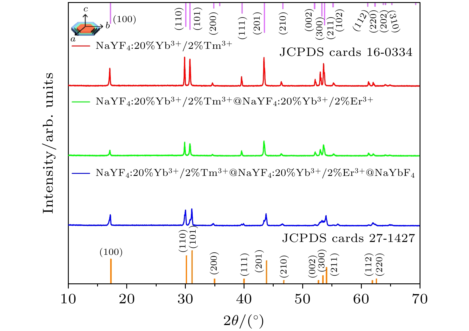
图 2 NaYF4:20%Yb3+/2%Tm3+微米晶体及其包覆不同核壳结构的XRD图谱
Fig. 2. The XRD patterns of NaYF4:20%Yb3+ /2%Tm3+ microcrystals and their coating with different CS structures.
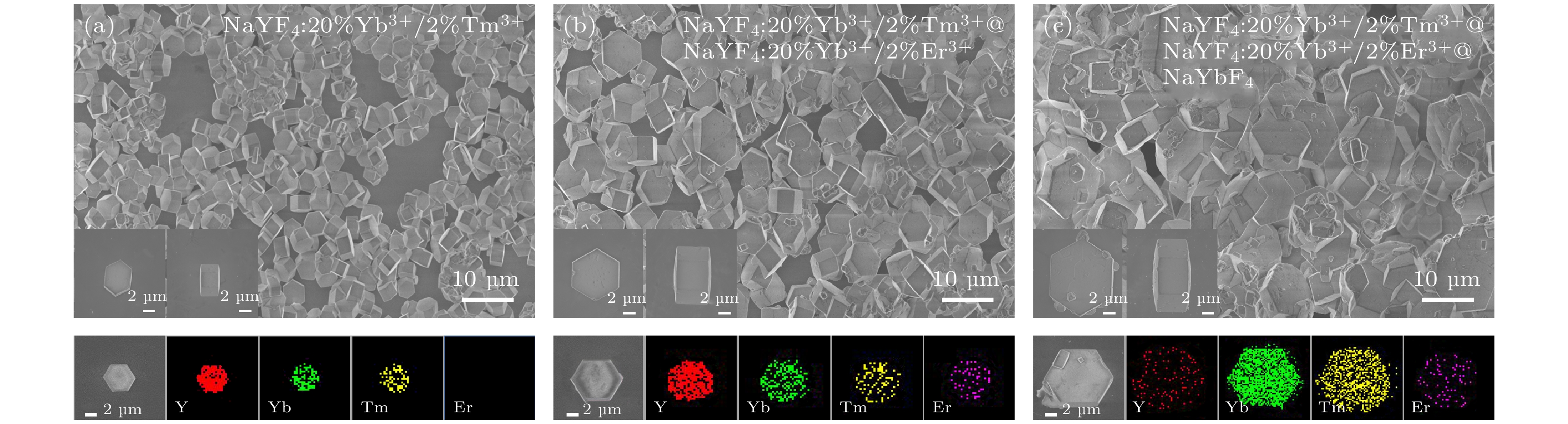
图 3 NaYF4:20%Yb3+/2%Tm3+微米晶体及包覆不同核壳结构的SEM及其相应的元素映射图 (a) NaYF4:20%Yb3+/2%Tm3+; (b) NaYF4:20%Yb3+/2%Tm3+ @ NaYF4:20%Yb3+/2%Er3+; (c) NaYF4:20%Yb3+/2%Tm3+ @ NaYF4:20%Yb3+/2%Er3+@ NaYbF4
Fig. 3. The SEM images and element mappings of NaYF4:20%Yb3+/2%Tm3+ microcrystals with corresponding CS structures: (a) NaYF4:20%Yb3+/2%Tm3+; (b) NaYF4:20%Yb3+/2%Tm3+ @ NaYF4:20%Yb3+/2%Er3+; (c) NaYF4:20%Yb3+/2%Tm3+ @ NaYF4:20%Yb3+/2%Er3+@ NaYbF4.
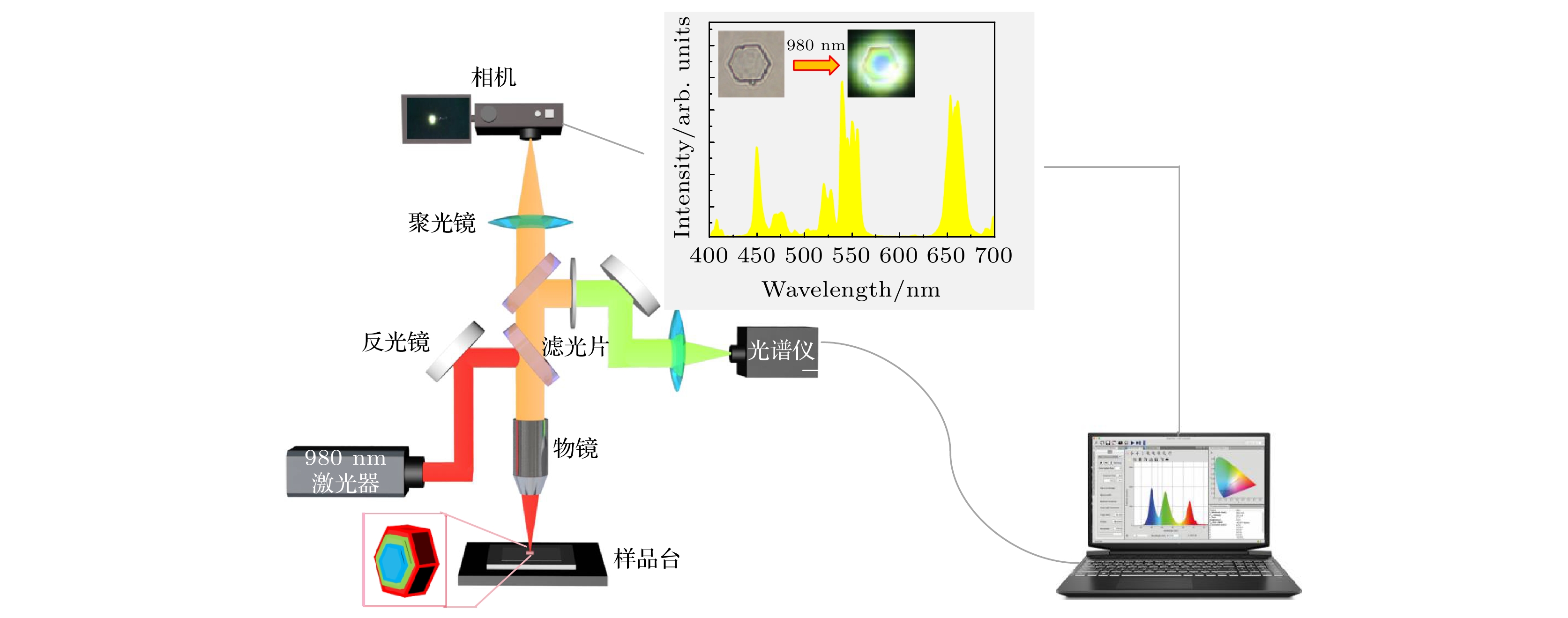
图 4 共聚焦显微光谱测试系统示意图
Fig. 4. Schematic diagram of the confocal microscope spectroscopic test system.

图 5 在980 nm激发下, 单个NaYF4:20%Yb3+/2%Tm3+@NaYF4:20%Yb3+/2%Er3+核壳微米盘在不同激发位置的 (a)上转换发射光谱(插图为不同激发位置下对应的发光照片), (b)蓝光、绿光和红光的发射峰面积, (c)增强倍数和(d)红绿比和红蓝比
Fig. 5. (a) The UC emission spectra (The insert is corresponding optical micrographs at different excitation positions), (b) the peak area of the bule, green and red emission intensity, (c) enhancement and (d) R/G ratio and R/B ratio of the single NaYF4:20%Yb3+/2%Tm3+@ NaYF4:20%Yb3+/2%Er3+ CS microdisk at different excitation positions under the excitation of a 980 nm near-infrared (NIR) laser.

图 6 在980 nm激发下, 单个NaYF4:20%Yb3+/2%Tm3+@NaYF4:20%Yb3+/2%Er3+@NaYbF4核-壳-壳微米盘在不同激发位置的 (a)上转换发射光谱(插图为不同激发位置下对应的发光照片), (b)蓝光、绿光和红光的发射峰面积, (c)增强倍数和(d)红绿比和红蓝比
Fig. 6. (a) The UC emission spectra (The insert is corresponding optical micrographs at different excitation positions), (b) the peak area of the bule, green and red emission intensity, (c) enhancement and (d) R/G ratio and R/B ratio of the single NaYF4:20%Yb3+/2%Tm3+@ NaYF4:20%Yb3+/2%Er3+@NaYbF4 core-shell-shell (CSS) microdisk at different excitation positions under the excitation of a 980 nm NIR laser.

图 7 在980 nm激发下, 单个NaYF4:20%Yb3+/2%Tm3+@NaYF4:20%Yb3+/2%Er3+@NaYbF4核-壳-壳微米盘中Yb3+, Tm3+和Er3+离子在不同核壳体系之间所对应的能级图及跃迁机制图(插图为核-壳-壳微米盘的模型, 白点、绿点、蓝点和紫点分别代表Yb3+离子、Er3+离子、Tm3+离子和表面猝灭点)
Fig. 7. The corresponding energy level diagrams and transition mechanism diagrams of Yb3+, Tm3+ and Er3+ ions between different CS systems in a single NaYF4:20%Yb3+/2%Tm3+@ NaYF4:20%Yb3+/2%Er3+@NaYbF4 CSS microdisk under the excitation of a 980 nm NIR laser. (The inset is a model of a CSS microdisk, and the white, green, blue, and purple dots represent Yb3+ ions, Er3+ ions, Tm3+ ions, and surface quenching points, respectively.)

图 8 在980 nm不同激发功率下, 单个(a) NaYF4:20%Yb3+/2%Tm3+和(c) NaYF4:20%Yb3+/2%Tm3+@NaYF4:20%Yb3+/2%Er3+微米盘在激发位置d的上转换发射光谱(插图为其对应的发光照片); (b), (d) 其蓝光, 绿光和红光发射强度与泵浦功率间的依赖关系
Fig. 8. The UC emission spectra of a single (a) NaYF4:20%Yb3+/2%Tm3+ and (c) NaYF4:20%Yb3+/2%Tm3+@NaYF4:20%Yb3+/2%Er3+ microdisk at excitation position d under different excitation powers of a 980 nm NIR laser (The insert is corresponding optical micrographs); (b), (d) the dependence of its blue, green and red emission intensity on pump power.

图 9 (a)—(d) 在980 nm激发下, 单个NaYF4:Yb3+/Tm3+@NaYF4:Yb3+/Er3+@NaYbF4核-壳-壳微米盘分别在激发位置a, b, c, d的发光图案及其光波导模型
Fig. 9. (a)–(d) The Luminescence patterns and optical waveguide models of a single NaYF4:20%Yb3+/2%Tm3+@NaYF4:20%Yb3+/2%Er3+@NaYbF4 CSS microdisk at excitation positions a, b, c, d respectively under the excitation of a 980 nm NIR laser.
表 1 水热法制备微米晶体的药品详细参数
Table 1. Detailed parameters of medicines for the preparation of microcrystals by hydrothermal method.
样品 核(核/壳)体积/mL m(EDTA-2Na)/g V(RE(NO3)3)/mL V(NaF)
/mLNaYF4:Yb3+/Tm3+ — 0.282 1.5 11.0 NaYF4:Yb3+/Tm3+@NaYF4:Yb3+/Er3+ 5.0 0.282 1.5 11.0 NaYF4:Yb3+/Tm3+@NaYF4:Yb3+/Er3+@NaYbF4 5.0 0.282 1.5 11.0 注: RE(NO3)3和NaF溶液均为0.5 mol/L水溶液.  下载: 导出CSV
-
[1] Gnach A, Bednarkiewicz A 2012 Nano Today 7 532
Google Scholar
[2] Bettinelli M, Carlos L, Liu X G 2015 Phys. Today 68 38
[3] Mandl G A, Cooper D R, Hirsch T, Seuntjens J, Capobianco J A 2019 Methods Appl. Fluoresc. 7 012004
Google Scholar
[4] Zhang H X, Chen Z H, Liu X, Zhang F 2020 J. Nano Res. 13 1795
Google Scholar
[5] Han Q Y, Zhao B C, Gao W, Li Y X, Sun Z Y, Wang C, Chen Y, Wang Y K, Yan X W, Dong J 2022 Phys. Chem. Chem. Phys. 24 13730
Google Scholar
[6] Xiang Y, Zheng S S, Yuan S S, Wang J, Wu Y H, Zhu X H 2022 Mikrochim. Acta 189 120
Google Scholar
[7] Li Y, Chen C, Liu F F, Liu J L 2022 Mikrochim. Acta 189 109
Google Scholar
[8] Xu M M, Ge W Y, Zhang X M, Li Y X 2022 Opt. Laser Technol. 145 107529
Google Scholar
[9] Alkahtani M, Almuqhim A A, Qasem H, Alsofyani N, Alfahd A, Alenzi S M, Aljuwayr A, Alzahrani Y A, Al-Badri A, Alotaibi M H, Bagabas A, AlHazaa A N, Hemmer P R 2021 Nanomaterials 11 2909
Google Scholar
[10] Liu B T, Huang T H, Wang T L, Hsu C C 2021 Sol Energy 227 1
Google Scholar
[11] Dong H, Sun L D, Yan C H 2021 J. Am. Chem. Soc. 143 20546
Google Scholar
[12] Wang J, Sun X Y, Han Y D, Cheng Z Z, Liu T G 2021 Opt. Commun. 483 126663
Google Scholar
[13] 董军, 张晨雪, 程小同, 邢宇, 韩庆艳, 严学文, 祁建霞, 刘继红, 杨祎, 高伟 2021 物理学报 70 154208
Google Scholar
Dong J, Zhang C X, Cheng X T, Xing Y, Han Q Y, Yan X W, Qi J X, Liu J H, Yang Y, Gao W 2021 Acta Phys. Sin. 70 154208
Google Scholar
[14] Wang Y, Low J X, Bi Y F, Bai Y, Jiang Y W, Wang H H, Liu W Y, Ma Y Q, Chen Y N, Long R, Xiong Y J 2022 Chin. Chem. Lett. 33 1087
Google Scholar
[15] Jia H, Li D G, Zhang D, Dong Y H, Ma S T, Zhou M, Di W H, Qin W P 2021 ACS Appl. Mater. Inter. 13 4402
Google Scholar
[16] Chen T, Hao S W, Azimbay A, Shang Y F, Pi Q L, Hou Y D, Yang C H 2019 J. Power Sources 430 43
Google Scholar
[17] Jiao X F, Ye W H, Huang Q Y, Luo G, Yu L L, Liu X T 2020 J. Rare Earths 38 697
Google Scholar
[18] Ju D D, Gao X L, Zhang S C, Li Y, Cui W J, Yang Y H, Luo M Y, Liu S J 2021 CrystEngComm 23 3892
Google Scholar
[19] Gao W, Sun Z Y, Han Q Y, Han S S, Cheng X T, Wang Y K, Yan X W, Dong J 2021 J. Alloys Compd. 857 157578
Google Scholar
[20] Gao W, Zhang C X, Han Q Y, Lu Y R, Yan X W, Wang Y K, Yang Y, Liu J H, Dong J 2022 J. Lumin. 241 118501
Google Scholar
[21] Felsted R G, Pant A, Bard A B, Xia X J, Luntz-Martin D R, Dadras S, Zhang S, Vamivakas A N, Pauzauskie P J 2022 Cryst. Growth Des. 22 3605
Google Scholar
[22] Zhang Y H, Huang L, Liu X G 2016 Angew. Chem. Int. Ed. 55 5718
Google Scholar
[23] He E J, Yu J J, Wang C, Jiang Y, Zuo X Z, Xu B, Wen J, Qin Y F, Wang Z J 2020 Mater Res Bull. 121 110613
[24] Yang D D, Peng Z X, Zhan Q Q, Huang X J, Peng X Y, Guo X, Dong G P, Qiu J R 2019 Small 15 1904298
Google Scholar
[25] Zhou Z Q, Xue J B, Zhang B P, Wang C, Yang X C, Fan W, Ying L Y, Zheng Z W, Xie Y J, Wu Y F, Yang X D, Zhang D 2021 Appl. Phys. Lett. 118 173301
Google Scholar
[26] Mehrdel B, Nikbakht A, Aziz A A, Jameel M S, Dheyab M A, Khaniabadi P M 2022 Nanotechnology 33 082001
Google Scholar
[27] Wu Y F S, Lai F Q, Liu B, Li Z B, Liang T X, Qiang Y C, Huang J H, Ye X Y, You W X 2020 J. Rare Earths 38 130
Google Scholar
[28] Yan L, Zhou B, Song N, Liu X L, Huang J S, Wang T, Tao L L, Zhang Q Y 2018 Nanoscale 10 17949
Google Scholar
[29] Wu Q X, Xu Z, Wageh S, Al-Ghamdia A, Zhao S L 2022 J. Alloys Compd. 891 162067
Google Scholar
[30] 赵越, 杨帆, 孙佳石, 李香萍, 张金苏, 张希珍, 徐赛, 程丽红, 陈宝玖 2019 物理学报 68 213301
Google Scholar
Zhao Y, Yang F, Sun J S, Li X P, Zhang J S, Zhang X Z, Xu S, Cheng L H, Chen B J 2019 Acta Phys. Sin. 68 213301
Google Scholar
[31] 张翔宇, 王丹, 石焕文, 王晋国, 侯兆阳, 张力东, 高当丽 2018 物理学报 67 084203
Google Scholar
Zhang X Y, Wang D, Shi H W, Wang J G, Hou Z Y, Zhang L D, Gao D L 2018 Acta Phys. Sin. 67 084203
Google Scholar
[32] 高伟, 孙泽煜, 郭立淳, 韩珊珊, 陈斌辉, 韩庆艳, 严学文, 王勇凯, 刘继红, 董军 2022 物理学报 71 034207
Google Scholar
Gao W, Sun Z Y, Guo L C, Han S S, Chen B H, Han Q Y, Yan X W, Wang Y K, Liu J H, Dong J 2022 Acta Phys. Sin. 71 034207
Google Scholar
[33] Kuang Y, Xu J T, Wang C, Li T Y, Gai S L, He F, Yang P P, Lin J 2019 Chem. Mater. 31 7898
Google Scholar
[34] Gao W, Wang B Y, Han Q Y, Gao L, Wang Z J, Sun Z Y, Zhang B, Dong J 2020 J. Alloys Compd. 818 152934
Google Scholar
[35] Gao D L, Wang D, Zhang X Y, Feng X J, Xin H, Yun S N, Tian D P 2018 J. Mater. Chem. C 6 622
Google Scholar

 下载:
下载:
计量
- 文章访问数: 7067
- PDF下载量: 99
- 被引次数: 0
