


-
离子辐照可以改变二氧化硅(SiO2)的晶体结构和光学性质. 采用 645 MeV Xe35+离子辐照SiO2单晶, 在辐照过程中, 利用光栅光谱仪测量在200—800 nm范围内的光发射. 在发射光谱中, 观测到中心位于461和631 nm的发射带. 这些发射带是弗伦克尔激子辐射退激产生的, 其强度与辐照离子能量和辐照离子剂量密切相关. 实验结果表明: 发射光强随离子在固体中的电子能损呈指数增加. 由于离子辐照对晶体造成损伤, 发射光谱强度随辐照剂量的增加而降低. 文中讨论了这些与晶体结构有关的发射带, 结合能量损失机制讨论了激子形成和退激过程. 快重离子辐照过程中发射光谱的原位测量对研究辐照改性具有重要意义, 有助于揭示离子辐照引起晶体损伤的物理机制.Silicon dioxide (SiO2) is an important component of nuclear reactor optical fiber and is also a candidate material for wast solidification. Owing to its special physical and chemical characteristics, it is used in many different technology fields like optics, electronics, energy orspace. Swift heavy ion irradiation can modify the crystal structure and optical property of optical material SiO2. Swift heavy ions deposit their energy mainly by inelastic interaction. Highly ionized lattice atoms may be formed along the trajectory, and a fraction of their electrical energy can be converted directly into the kinetic energy of the ions. The irradiation experiment is performed with Xeq+ ions at the irradiation terminal of the sector-focused cyclotron at heavy-ion research facility in Lanzhou (HIRFL). The on-line spectral measurement experiment is carried out during irradiation. In the darkroom, the UV-visible light emission from the target is focused into optical fiber by a collimating lens, and then is analyzed with the Sp-2558 spectrometer equipped with a 1200 g/mm optical grating blazed at 500 nm. In the present work, SiO2 single crystals are irradiated with 93–609 MeV Xeq+ ions with a dose in a range of 1×10¹¹–3×10¹¹ ions/cm2. During irradiation, the emission spectra, in a range of 200–800 nm, from SiO2 irradiated by 93, 245, 425 and 609 MeV Xeq+ ions, are obtained. Two emission bands centered at 461 and 631 nm are observed. These emission bands are produced by Frenkel exciton radiation de-excitation and their intensities are closely related to the irradiated ion energy and radiation dose. The results show that the light intensity increases with the electron energy loss index increasing. And owing to crystal damage caused by ion irradiation, the intensity of emission spectrum decreases with the augment of irradiation dose. Ion loses its energy throughout the ion track via Sn and Se interacting with target atoms and electrons respectively, and the energy lost by the ion is estimated by using SRIM code. The SRIM simulated ion ranges and recoil atom distribution, target ionization (energy loss to target electrons), damage production in SiO2 are presented. Based on the energy deposition process, the emission bands related to the crystal structure itself are discussed. It indicates that electron energy loss plays a leading role in the process of light emission. In-situ measurement of the optical emission is of great significance in studying the irradiation modification and can help to understand the process of crystal damage caused by ion irradiation.
-
Keywords:
- optical emission band /
- swift heavy ions /
- the electron energy loss /
- silicon dioxide
[1] Yang P, An YL, Yang D Y, Li Y H, Chen J M 2020 Ceram. Int. 46 21367
 Google Scholar
[2] Li Y H, Wen J, Wang Y Q, Wang Z G, Tang M, Valdez J A, Sickafus K E 2012 Nucl. Instrum. Methods Phys. Res. , Sect. B 287 130
Google Scholar
[3] Devine R A B 1994 Nucl. Instrum. Methods Phys. Res. , Sect. B 91 378
Google Scholar
[4] Zhu Z, Jung P, Langenscheidt E 1997 J. Non-Cryst. Solids 217 173
Google Scholar
[5] Zhu Z Y, Jung P 1994 Nucl. Instrum. Methods Phys. Res. , Sect. B 91 269
Google Scholar
[6] Saito K, Ikushima A J 2002 J. Appl. Phys. 91 4886
Google Scholar
[7] Wang R P, Tai N, Saitio K, Ikushima A J 2005 J. Appl. Phys. 98 023701
Google Scholar
[8] Xue S W, Zu X T, Su H Q, Zheng W G, Xia X, Hong D, Yang C R 2007 Chin. Phys. 16 1119
Google Scholar
[9] Imai H, Arai K, Imagawa H, Hosono H, Abe Y 1988 Phys. Rev. B 38 12772
Google Scholar
[10] Nishikawa H, Nakamura R, Tohmon R, Ohki Y, Sakurai Y, Nagasawa K, Hama Y 1990 Phys. Rev. B 41 7828
Google Scholar
[11] Ziegler J F 2004 Nucl. Instrum. Methods Phys. Res., Sect. B 219 1027
[12] Bettger K (姜东兴, 刘洪涛 译) 1982 重离子物理实验方法 (北京: 原子能出版社) 第149页
Bettger K (translated by Jiang Dongxing, Liu Hongtao) 1982 Experimental Methods in Heavy Ion Physics (Beijing: Atomic Energy Press) p149 (in Chinese)
[13] Stevens-Kalceff M A 2011 J. Phys. D: Appl. Phys. 44 255402
Google Scholar
[14] Kaddouri A, Ashraf I, El Fqih M A, Targaoui H, El Boujlaïdi A, Berrada K 2009 Appl. Surf. Sci. 256 116
Google Scholar
[15] Song Y, Zhang C H, Yang Y T, Gou J, Zhang L Q, He D Y 2013 Opt. Mater. 35 1057
Google Scholar
[16] Patra P, Shah S, Toulemonde M, Sulania I, Singh F 2022 Radiat. Eff. Defects Solids 177 513
Google Scholar
[17] Meftah A, Brisard F, Costantini J M, Dooryhee E, Hage-Ali M, Hervieu M, Stoquert J P, Studer F, Toulemonde M 1994 Phys. Rev. B 49 12457
Google Scholar
[18] Kluth P, Schnohr C S, Pakarinen O H, Djurabekova F, Sprouster D J, Giulian R, Ridgway M C, Byrne A P, Trautmann C, Cookson D J, Nordlund K, Toulemonde M 2008 Phys. Rev. Lett. 101 175503
Google Scholar
[19] Toulemonde M, Weber W J, Li G S, Shutthanandan V, Kluth P, Yang T F, Wang Y G, Zhang Y W 2011 Phys. Rev. B 83 054106
Google Scholar
[20] Schwartz K, Trautmann C, El-Said A S, Neumann R, Toulemonde M, Knolle W 2004 Phys. Rev. B 70 184104
Google Scholar
[21] Liu C B, Wang Z G 2011 Chin. J. Lumin. 32 608
Google Scholar
[22] Udelson B J, Creedon J E, French J C 1957 J. Appl. Phys. 28 717
Google Scholar
[23] Liao L S, Bao X M, Zheng X Q, Li N S, Min N B 1996 Chin. J. Semicond. 17 789
-
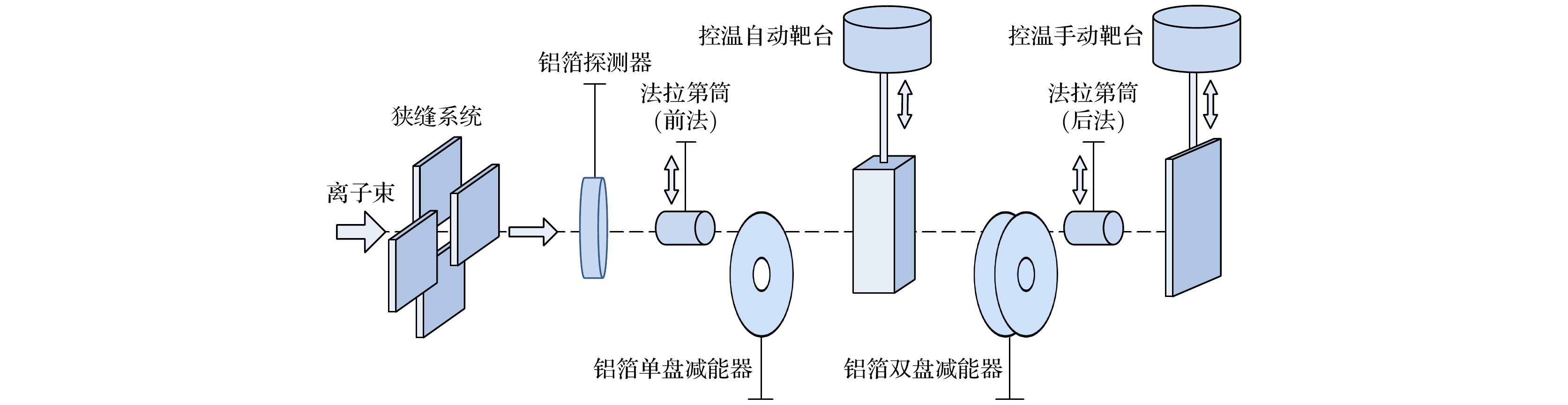
图 1 中能辐照终端束线系统示意图
Fig. 1. Schematic diagram of intermediate energy irradiation terminal beam system.

图 2 快重离子辐照固体引起光发射测量装置示意图
Fig. 2. A schematic diagram of the experimental setup for the measurement of optical emission from the solid induced by swift heavy ions.

图 3 245 MeV Xeq+离子辐照SiO2发射光谱
Fig. 3. The optical emission spectrum from SiO2 irradiated by 245 MeV Xeq+ ions.

图 4 93—609 MeV Xeq+离子辐照SiO2发射谱461 nm处的光强度随离子动能的变化
Fig. 4. The intensity of emission bands of centered at 461 nm from SiO2 irradiated by 93–609 MeV Xeq+ ions as a function of kinetic energy.
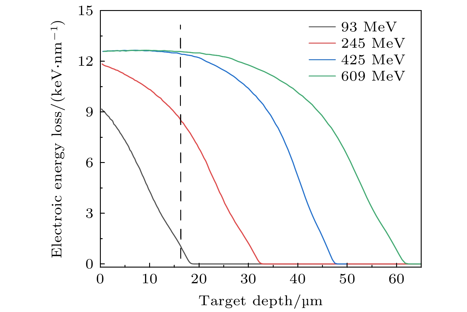
图 5 利用SRIM 程序计算93, 245, 425和609 MeV Xeq+离子在SiO2中的电子能损(Se)随辐照深度的变化
Fig. 5. Variation of electronic energy losses (Se) with the SiO2 depth for 93, 245, 425 and 609 MeV Xeq+ ion irradiation using SRIM code.
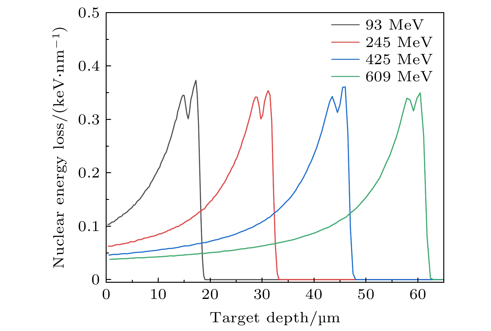
图 6 利用SRIM 程序计算93, 245, 425和609 MeV Xeq+ 离子在SiO2中的核能损(Sn)随辐照深度的变化
Fig. 6. Variation of nuclear electronic energy losses (Sn) with the SiO2 depth for 93, 245, 425 and 609 MeV Xeq+ ion irradiation using SRIM code.
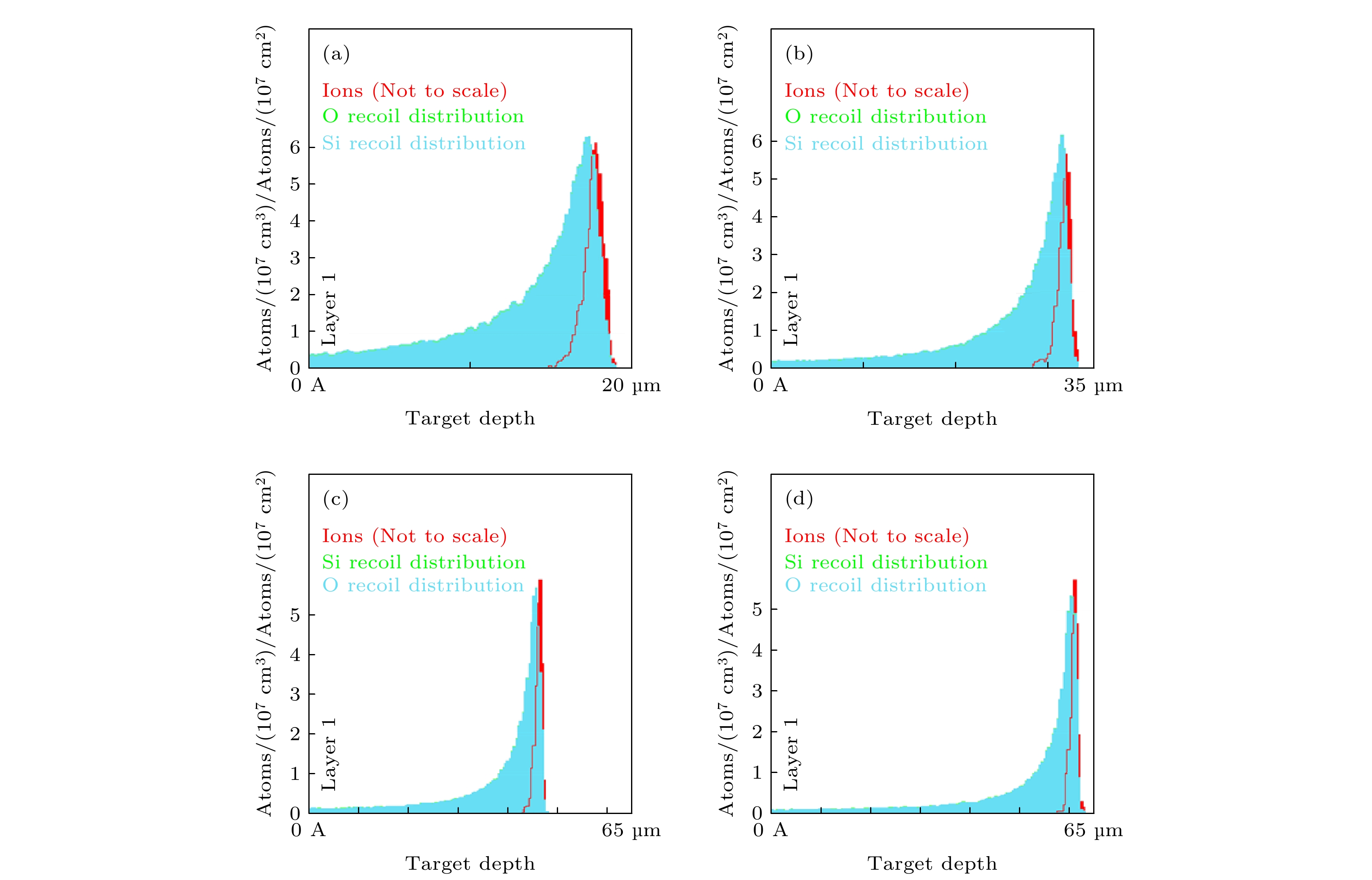
图 7 SRIM模拟93 (a), 245 (b), 425 (c)和 609 (d) MeV Xeq+ 离子在SiO2中的离子射程和反冲原子分布
Fig. 7. SRIM simulated plot of ion ranges and recoil atom distribution of SiO2 target by 93(a), 245(b), 425(c) and 609 (d) MeV Xeq+ ion

图 10 SRIM模拟93 (a), 245 (b), 425 (c) 和 609 (d) MeV Xeq+ 离子在SiO2中的移位损伤以及Si和O原子空位
Fig. 10. SRIM simulated plot of displacement damage of SiO2 target and vacancies of Si and O atoms with target depth by 93 (a), 245 (b), 425 (c) and 609 (d) MeV Xeq+ ion.

图 8 SRIM模拟93 (a), 245 (b), 425 (c)和609 (d) MeV Xeq+ 离子在SiO2中的电离
Fig. 8. SRIM simulated plot of target ionization (energy loss to target electrons) of SiO2 target by 93 (a), 245 (b), 425 (c) and 609(d) MeV Xeq+ ion.
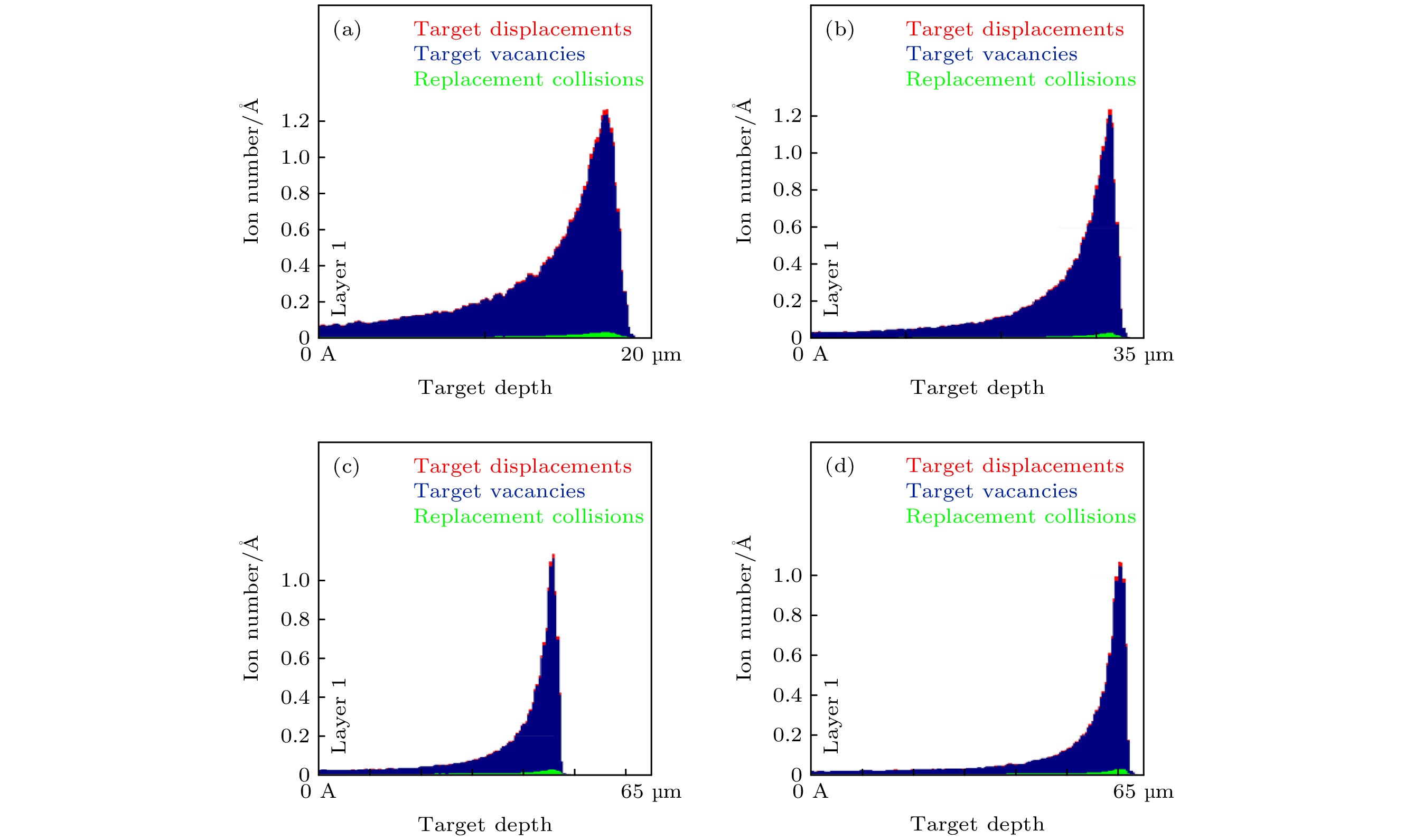
图 9 SRIM模拟93 (a), 245 (b), 425 (c)和 609 (d) MeV Xeq+ 离子在SiO2中的移位损伤
Fig. 9. SRIM simulated plot of displacement damage of SiO2 target by 93 (a), 245 (b), 425 (c) and 609 (d) MeV Xeq+ ion.

图 11 93—609 MeV Xeq+离子辐照SiO2发射谱461 nm处的光强度随电子能损的变化
Fig. 11. The intensity of emission bands of centered at 461 nm from SiO2 irradiated by 93–609 MeV Xeq+ ions as a function of electronic energy loss.

图 12 609 MeV Xeq+离子辐照SiO2发射谱
Fig. 12. The optical emission spectra from SiO2 irradiate by 609 MeV Xeq+ ions.
表 1 不同能量Xeq+离子辐照SiO2植入深度、电子能损和核能损
Table 1. the penetrating depth and, its electronic energy loss and nuclear energy loss of Xeq+ ion in SiO2.
Ion energy
/MeVProjected
range/μmElectronic energy
loss/(×104 keV·μm–1)Nuclear energy loss
/(×10 keV·μm–1)609 60.69 1.258 1.518 425 46.14 1.260 2.063 245 31.59 1.183 3.283 93 17.54 0.9225 7.271  下载: 导出CSV
-
[1] Yang P, An YL, Yang D Y, Li Y H, Chen J M 2020 Ceram. Int. 46 21367
Google Scholar
[2] Li Y H, Wen J, Wang Y Q, Wang Z G, Tang M, Valdez J A, Sickafus K E 2012 Nucl. Instrum. Methods Phys. Res. , Sect. B 287 130
Google Scholar
[3] Devine R A B 1994 Nucl. Instrum. Methods Phys. Res. , Sect. B 91 378
Google Scholar
[4] Zhu Z, Jung P, Langenscheidt E 1997 J. Non-Cryst. Solids 217 173
Google Scholar
[5] Zhu Z Y, Jung P 1994 Nucl. Instrum. Methods Phys. Res. , Sect. B 91 269
Google Scholar
[6] Saito K, Ikushima A J 2002 J. Appl. Phys. 91 4886
Google Scholar
[7] Wang R P, Tai N, Saitio K, Ikushima A J 2005 J. Appl. Phys. 98 023701
Google Scholar
[8] Xue S W, Zu X T, Su H Q, Zheng W G, Xia X, Hong D, Yang C R 2007 Chin. Phys. 16 1119
Google Scholar
[9] Imai H, Arai K, Imagawa H, Hosono H, Abe Y 1988 Phys. Rev. B 38 12772
Google Scholar
[10] Nishikawa H, Nakamura R, Tohmon R, Ohki Y, Sakurai Y, Nagasawa K, Hama Y 1990 Phys. Rev. B 41 7828
Google Scholar
[11] Ziegler J F 2004 Nucl. Instrum. Methods Phys. Res., Sect. B 219 1027
[12] Bettger K (姜东兴, 刘洪涛 译) 1982 重离子物理实验方法 (北京: 原子能出版社) 第149页
Bettger K (translated by Jiang Dongxing, Liu Hongtao) 1982 Experimental Methods in Heavy Ion Physics (Beijing: Atomic Energy Press) p149 (in Chinese)
[13] Stevens-Kalceff M A 2011 J. Phys. D: Appl. Phys. 44 255402
Google Scholar
[14] Kaddouri A, Ashraf I, El Fqih M A, Targaoui H, El Boujlaïdi A, Berrada K 2009 Appl. Surf. Sci. 256 116
Google Scholar
[15] Song Y, Zhang C H, Yang Y T, Gou J, Zhang L Q, He D Y 2013 Opt. Mater. 35 1057
Google Scholar
[16] Patra P, Shah S, Toulemonde M, Sulania I, Singh F 2022 Radiat. Eff. Defects Solids 177 513
Google Scholar
[17] Meftah A, Brisard F, Costantini J M, Dooryhee E, Hage-Ali M, Hervieu M, Stoquert J P, Studer F, Toulemonde M 1994 Phys. Rev. B 49 12457
Google Scholar
[18] Kluth P, Schnohr C S, Pakarinen O H, Djurabekova F, Sprouster D J, Giulian R, Ridgway M C, Byrne A P, Trautmann C, Cookson D J, Nordlund K, Toulemonde M 2008 Phys. Rev. Lett. 101 175503
Google Scholar
[19] Toulemonde M, Weber W J, Li G S, Shutthanandan V, Kluth P, Yang T F, Wang Y G, Zhang Y W 2011 Phys. Rev. B 83 054106
Google Scholar
[20] Schwartz K, Trautmann C, El-Said A S, Neumann R, Toulemonde M, Knolle W 2004 Phys. Rev. B 70 184104
Google Scholar
[21] Liu C B, Wang Z G 2011 Chin. J. Lumin. 32 608
Google Scholar
[22] Udelson B J, Creedon J E, French J C 1957 J. Appl. Phys. 28 717
Google Scholar
[23] Liao L S, Bao X M, Zheng X Q, Li N S, Min N B 1996 Chin. J. Semicond. 17 789

 下载:
下载:
计量
- 文章访问数: 5481
- PDF下载量: 63
- 被引次数: 0
