


-
透射X射线显微镜(transmission X-ray microscope, TXM)是高精密度的尖端X射线成像设备, 是现代科学技术的结晶, 可以在纳米尺度上进行无损成像, 为物理学、生命科学、材料学和化学等领域的众多科学问题提供了有力的研究工具 . 虽然国内外很多同步辐射装置都建立了以TXM为核心的纳米CT实验站, 但是目前国际上只有个别企业能提供商业化的实验室TXM . 究其原因, 主要是该仪器涉及众多高难度的工程技术问题, 诸如: 高亮度实验室X射线源、高分辨率X射线光学元器件、高精度样品台、高灵敏度探测器、仪器对温度和振动等环境因素的超高要求等 . 为了提高研发高端X射线成像仪器的水平, 需要逐个突破在研发X射线纳米CT过程中遇到的技术瓶颈 . 本文主要讨论了工作能量为5.4 keV的实验室TXM的仪器设计, 以及全场成像实验结果 . 该仪器工作在吸收衬度模式下, 成像视野达到了26 μm, 可以对30 nm线宽的特征结构实现清晰的成像, 西门子星测试卡的功率谱曲线表明该仪器具有分辨半周期为28.6 nm线对结构的潜力 .Transmission X-ray microscope (TXM) is a high-precision, cutting-edge X-ray imaging instrument, which is a marvel of modern science and technology. It enables non-destructive imaging on a nanoscale, providing a powerful research tool for various scientific fields such as physics, life science, materials science, and chemistry. Although many synchrotron radiation facilities at home and abroad have established nano-CT experimental stations with TXM as the core, currently only a few companies internationally can provide commercial TXM instrument based on laboratory X-ray sources. The primary reason is that this instrument involves numerous engineering challenges, including high-brightness laboratory X-ray sources, high-resolution X-ray optical elements, high-precision sample stage systems, high-sensitivity detectors, and extremely strict requirements for environmental factors such as temperature and vibration. In order to promote the development of high-end X-ray imaging instruments, it is necessary to overcome the technological bottlenecks encountered in the development of X-ray nano-CT. Discussed in this work mainly are the instrument design of a laboratory transmission X-ray microscope with working energy of 5.4 keV and the results of full-field imaging experiments. To start with, the design of the TXM instrument is introduced in detail. The TXM instrument is equipped with several key components, including laboratory X-ray source, condenser, sample stage module, zone plate, and imaging detector. The TXM instrument adopts a modular vibration isolation design and is equipped with a dedicated temperature control system. The main imaging magnifications of the TXM instrument are 50×, 75×, and 100×, and the corresponding optical parameters and photos are introduced. The X-ray source used is a micro-focus X-ray source, operating in Cr target mode, with a focal spot size of 20 μm and a Ka characteristic spectrum brightness of
$ 5\times {10}^{9}~\rm {photons}/({mm}^2\cdot {mrad}^2\cdot s)$ . The X-ray source provides illumination for the sample after being focused by an ellipsoidal condenser. The outer ring of the condenser's illumination ring corresponds to a numerical aperture (NA) of$ {NA}_{2} = 3.196~\rm mrad $ , and the inner ring corresponds to a numerical aperture of$ {NA}_{1} = 1.9086~\rm mrad $ . Under these conditions, the limit resolution of this TXM instrument is 22 nm. The zone plate has a diameter of 70μm, a focal length of 8.7mm, and 616 zones. The TXM instrument uses a high-resolution optical coupling detector equipped with a scientific-grade CMOS camera with an effective pixel size of 7.52μm. The optical coupling detector is equipped with 2× and 10× high numerical aperture objectives. When the TXM instrument magnification is 50×, the effective pixel size of the TXM instrument is 15 nm. In addition , a gold resolution test card is used as the sample to determine the imaging field of view of the TXM instrument by observing the size of the imaging area of the test card on the detector, and to determine the imaging resolution of the TXM instrument by observing the line width of the star-shaped target in the center of the test card. Experimental results show that the TXM instrument has an imaging field of view of 26μm and can achieve the clear imaging of characteristic structure with a line width of 30 nm. The radial power spectrum curve of the Siemens Star shows this TXM instrument has the potential to resolve 28.6-nm half pitch line pair features. Finally, we draw some conclusions and present outlook. At present, imaging of 30-nm-wide line features has been realized, but the imaging of 30-nm half pitch line pair feature has not yet been achieved, and the limit resolution has not reached the design value, either. We will continue to explore the potential for upgrading the imaging resolution of the laboratory TXM in future work.-
Keywords:
- X-ray imaging /
- X-ray microscope /
- scientific instruments
[1] Zhao L M, Wang T X, Ma R K, Gu Y, Luo M S, Chen H, Wang Z L, Ge X 2023 Chin. Phys. B 32 028701
 Google Scholar
[2] Wang Z L, Chen Z H, Gu Y, Chen H, Ge X 2023 Chinese Phys. B 32 038704
Google Scholar
[3] Winarski R P, Holt M V, Rose V, Fuesz P, Carbaugh D, Benson C, Shu D, Kline D, Stephenson G B, McNulty I, Maser J 2012 J. Synchrotron Rad. 19 1056
Google Scholar
[4] 周光照, 胡哲, 杨树敏, 廖可梁, 周平, 刘科, 滑文强, 王玉柱, 边风刚, 王劼 2020 物理学报 69 034102
Google Scholar
Zhou G Z, Hu Z, Yang S M, Liao K L, Zhou P, Liu K, Hua W Q, Wang Y Z, Bian F G, Wang J 2020 Acta Phys. Sin. 69 034102
Google Scholar
[5] 聂勇敢, 高梓宸, 佟亚军, 范家东, 刘功发, 江怀东 2024 物理学报 73 120701
Google Scholar
Nie Y G, Gao Z C, Tong Y J, Fan J D, Liu G F, Jiang H D 2024 Acta Phys. Sin. 73 120701
Google Scholar
[6] von Hofsten O, Bertilson M, Reinspach J, Holmberg A, Hertz H M, Vogt U 2009 Opt. Lett. 34 2631
Google Scholar
[7] Mao W L, Lin Y, Liu Y, Liu J 2019 Engineering 5 479
Google Scholar
[8] Flenner S, Hagemann J, Wittwer F, et al. 2023 J. Synchrotron Rad. 30 390
Google Scholar
[9] De Andrade V, Nikitin V, Wojcik M, et al. 2021 Adv. Mater. 33 2008653
Google Scholar
[10] Yuan Q, Zhang K, Hong Y, et al. 2012 J. Synchrotron Rad. 19 1021
Google Scholar
[11] Chen J, Li W, Liu Y, et al. 2009 J. Phys.: Conf. Ser. 186 012005
Google Scholar
[12] Tao F, Wang J, Du G, Su B, Zhang L, Hou C, Deng B, Xiao T 2023 J. Synchrotron Rad. 30 815
Google Scholar
[13] Zeiss, https://www.zeiss.com/microscopy/en/products/X-ray-microscopy.html [2024-6-11]
[14] Sigray, https://www.sigray.com/trilambda-30/#resolution [2024-6-11]
[15] 周腊珍, 夏文静, 许倩倩, 陈赞, 李坊佐, 刘志国, 孙天希 2022 物理学报 71 090701
Google Scholar
Zhou L Z, Xia W J, Xu Q Q, Chen Z, Li F Z, Liu Z G, Sun T X 2022 Acta Phys. Sin. 71 090701
Google Scholar
[16] Yun W, Lai B, Cai Z, Maser J, Legnini D, Gluskin E, Chen Z, Krasnoperova A A, Vladimirsky Y, Cerrina F, Di Fabrizio E, Gentili M 1999 Review of Scientific Instruments 70 2238
Google Scholar
[17] Chao W L, Harteneck B D, Liddle J A, Anderson E H, Attwood D T 2005 Nature 435 1210
Google Scholar
[18] 李乾利, 胡亚华, 马逸凡, 孙志祥, 王敏, 刘小林, 赵景泰, 张志军 2020 物理学报 69 102902
Google Scholar
Li Q L, Hu Y H, Ma Y F, Sun Z X, Wang M, Liu X L, Zhao J T, Zhang Z J 2020 Acta Phys. Sin. 69 102902
Google Scholar
[19] Tong X, Chen Y, Xu Z, Li Y, Xing Z, Mu C, Zhao J, Zhen X, Mao C, Tai R 2023 J. Synchrotron Rad. 30 319
Google Scholar
[20] Zhu J, Chen Y, Xie S, Zhang L, Wang C, Tai R 2020 Microelectron. Eng. 225 111254
Google Scholar
[21] Liu J, Li X, Chen S, Zhang S, Xie S, Xu C, Chen Y, Deng B, Mao C 2017 J. Synchrotron Rad. 24 595
Google Scholar
[22] Wang S, Zhang K, Huang W, Gao L, Yang F, Li M, Zhu P, Yuan Q 2021 Nucl. Instrum. Meth. A 993 165089
Google Scholar
[23] Liao K L, He Q L, Li P Y, Song M H, Zhao H F, Zhu P P 2024 Proc. SPIE 13155 1315518
Google Scholar
[24] 陶芬, 王玉丹, 任玉琦, 丰丙刚, 佟亚军, 杜国浩, 邓彪, 孙天希, 谢红兰, 肖体乔 2017 光学学报 37 1034002
Google Scholar
Tao F, Wang Y D, Ren Y Q, Feng B G, Tong Y J, Du G H, Deng B, Sun T X, Xie H L, Xiao T Q 2017 Acta Opt. Sin. 37 1034002
Google Scholar
-
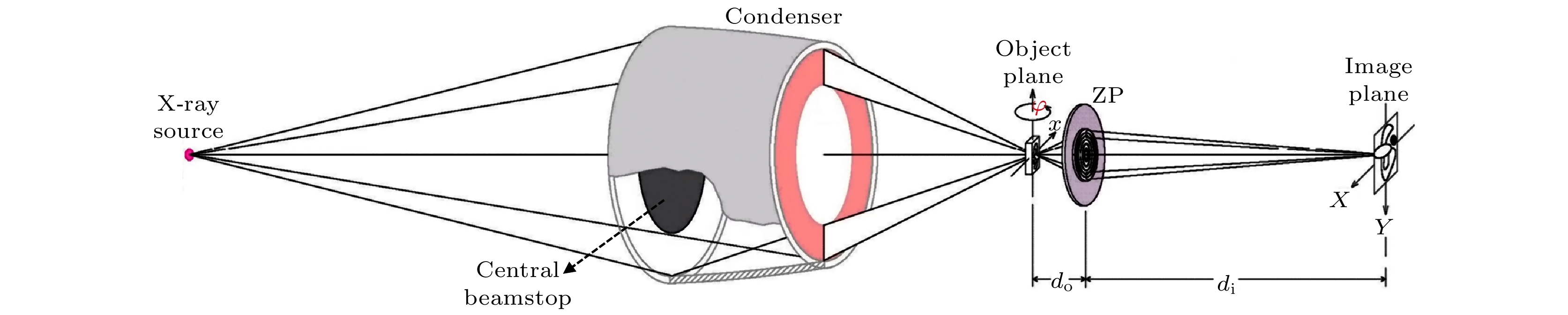
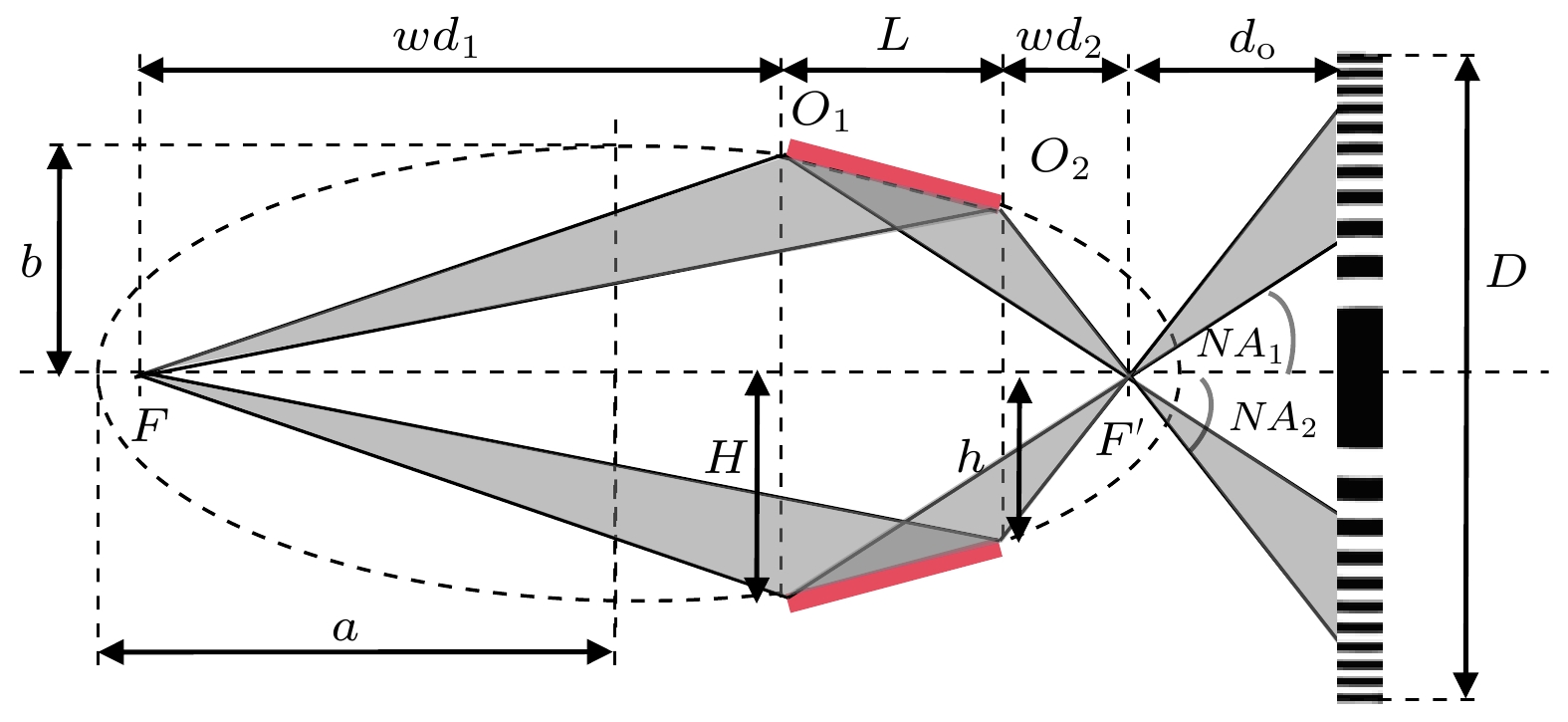
图 1 基于实验室X射线源的TXM仪器光路布局图
Fig. 1. Optical layout of TXM based on laboratory X-ray source.
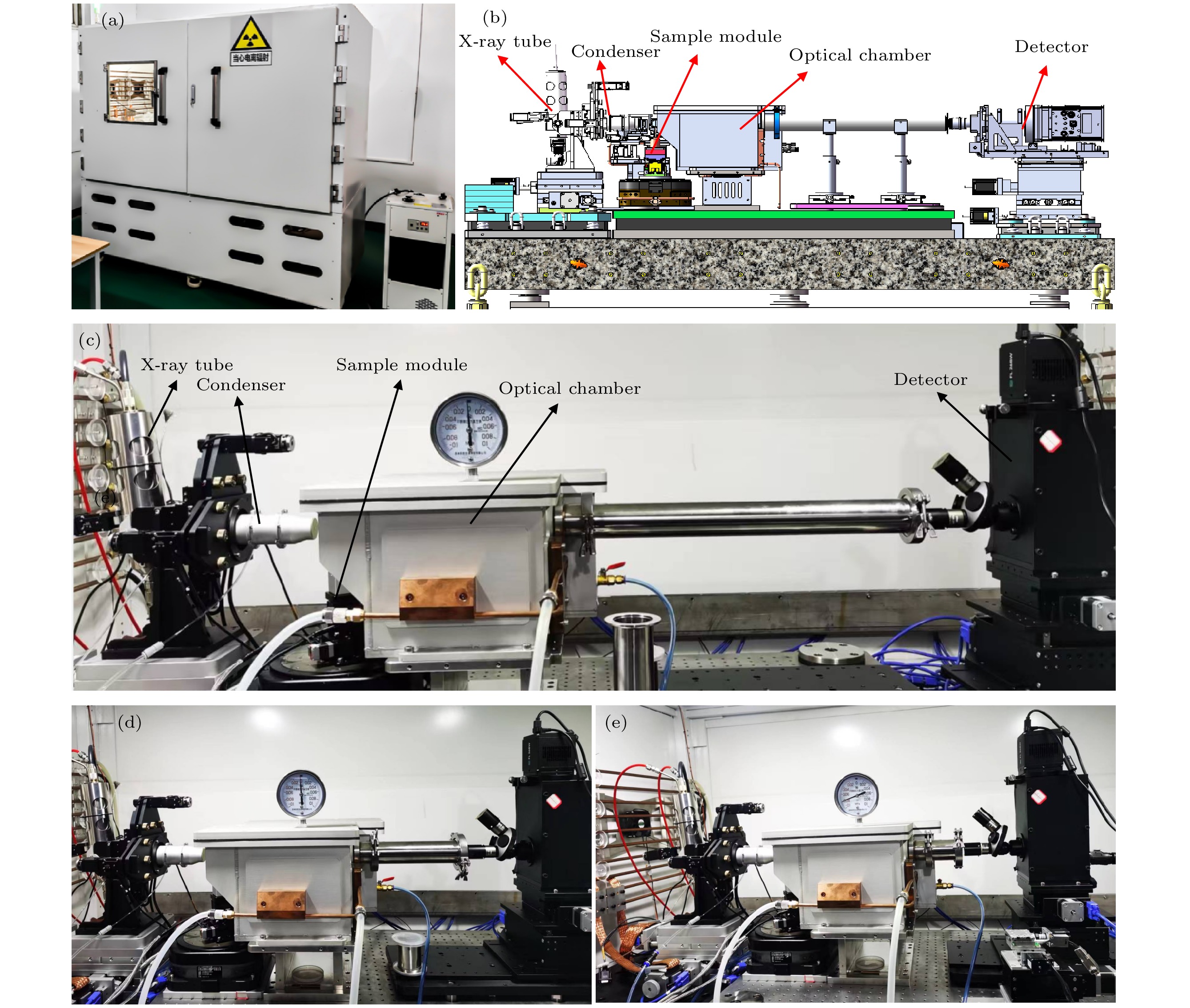
图 2 基于TXM的纳米CT仪器设计与仪器实物图 (a) TXM仪器实物图; (b) TXM仪器的主体结构设计图; (c) 100×放大倍率; (d) 75×放大倍率; (e) 50×放大倍率
Fig. 2. Schematic and photograph of the TXM instrument: (a) Photograph of the TXM instrument; (b) schematic of the main structure of the TXM; photograph of the main structure of the TXM under (c) 100×, (d) 75×, (e) 50× magnification ratio.
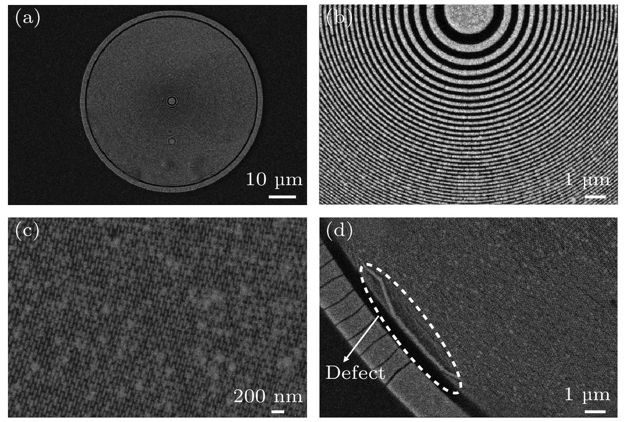
图 4 波带片的SEM图像 (a) 波带片整体结构的SEM图像; (b), (c)波带片中心两处局部区域SEM图像; (d)波带片最外环结构的SEM图像
Fig. 4. SEM graphs of the zone plate: (a) The global SEM graph of the ZP; (b), (c) SEM graphs of two local regions of the ZP; (d) SEM graph of the outmost region of the ZP.
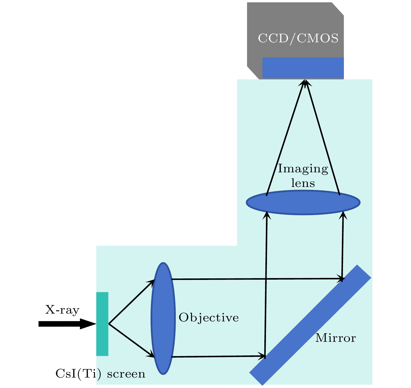
图 5 探测器光学耦合系统示意图
Fig. 5. Schematic of the optical coupling system of the X-ray detector.
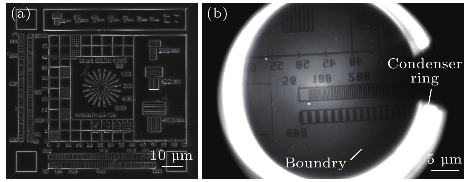
图 6 分辨率测试卡的SEM图像和TXM图像 (a)分辨率测试卡整体图案的SEM图像; (b)分辨率测试卡局部图案的TXM图像
Fig. 6. SEM and TXM graph of the resolution test card: (a) SEM graph of the global pattern; (b) TXM graph of a local pattern.
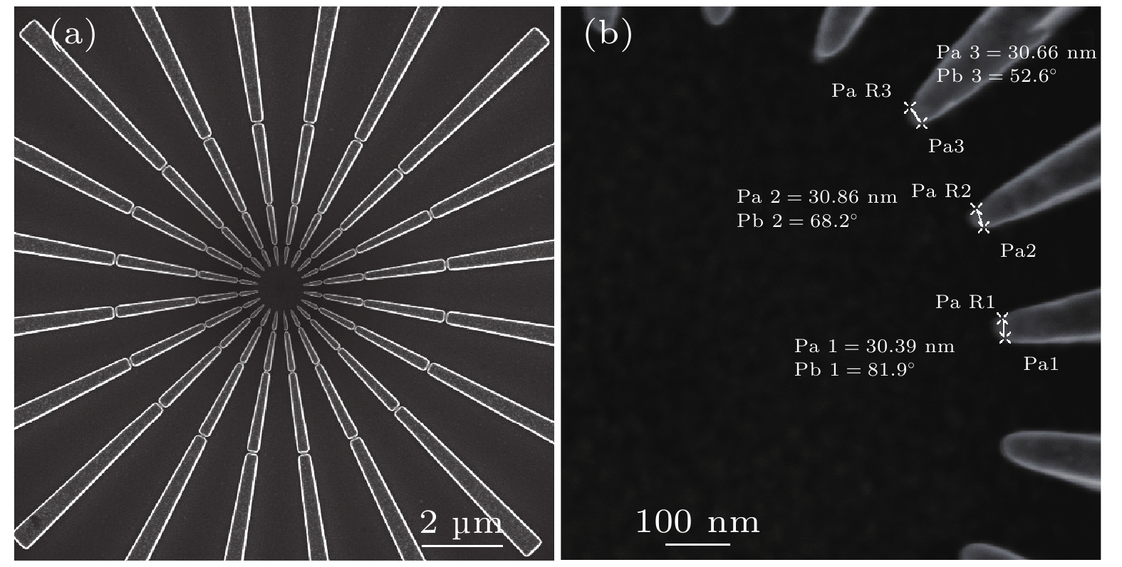
图 7 (a)分辨率测试卡中央的西门子星结构的SEM图像; (b)西门子星最内环局部结构的SEM图像
Fig. 7. (a) SEM graph of the Siemens star on the resolution test card; (b) SEM graph of the inner local structure.

图 8 分辨率测试卡TXM测试结果 (a) 西门子星结构的TXM图像; (b)扣除背底后的西门子星结构的透过率图像; (c)西门子星结构的内环结构局部放大图; (d) 30 nm线宽的栅条结构的透过率分布曲线; (e)径向功率谱强度曲线
Fig. 8. TXM results of the resolution test card: (a) TXM graph of the Siemens star; (b) partial enlarged view of the inner ring structure of the Siemens star; (c) the local structure of the Siemens star; (d) the transmission curve of 30 nm line width grating structure; (e) the radial power spectrum density curve.
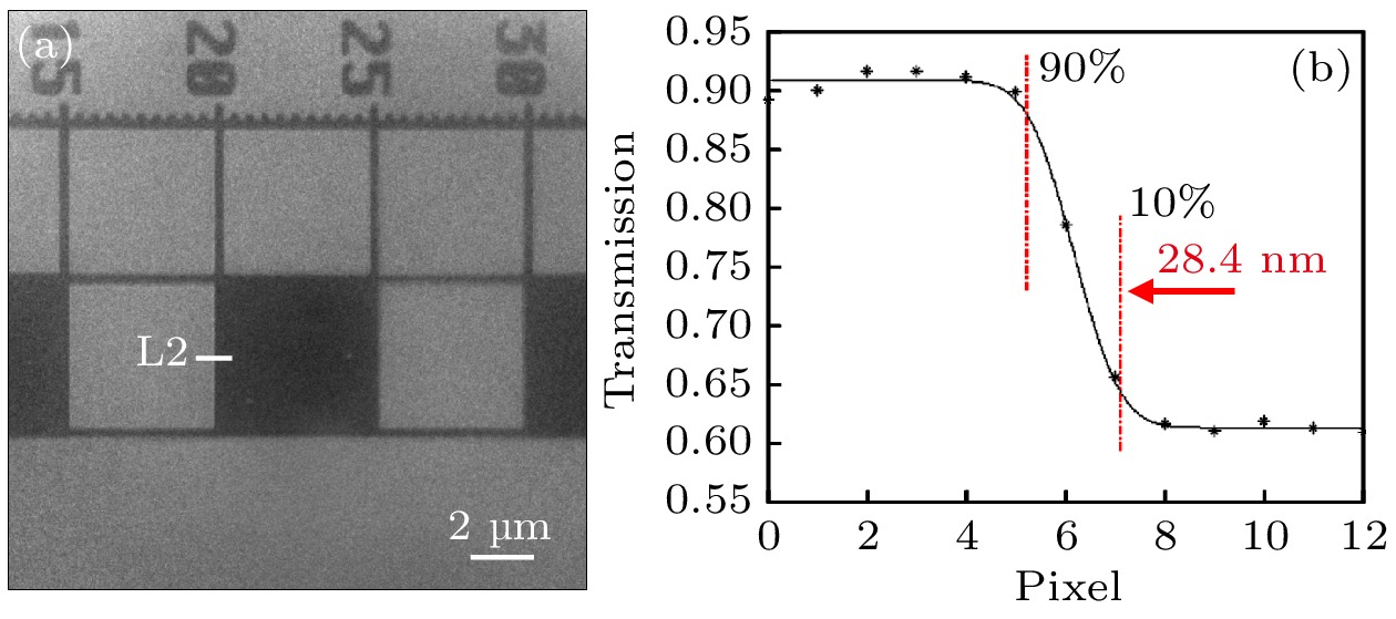
图 9 刀口测试结果 (a) 刀口的TXM图像; (b) L2处的刀口响应曲线
Fig. 9. Knife edge test results: (a) TXM graph of the knife edge; (b) the response curve of the knife edge at L2.
表 1 不同放大倍率M下的光路参数
Table 1. Optical parameters under different magnification M.
放大倍数M 物距 do/mm 像距 di/mm 光路总长/mm 50 8.874 443.7 752.574 75 8.816 661.2 970.016 100 8.787 878.7 1187.487  下载: 导出CSV
表 2 波带片参数
Table 2. Parameter of zone plate.
能量
E/keV材
料直径
D/μm最外环宽度
$ {{\Delta r}}$/nm波带数
N高度/
nm焦距
f/mm5.41 Au 70 28.5 616 ≥800 8.7
下载: 导出CSV
-
[1] Zhao L M, Wang T X, Ma R K, Gu Y, Luo M S, Chen H, Wang Z L, Ge X 2023 Chin. Phys. B 32 028701
Google Scholar
[2] Wang Z L, Chen Z H, Gu Y, Chen H, Ge X 2023 Chinese Phys. B 32 038704
Google Scholar
[3] Winarski R P, Holt M V, Rose V, Fuesz P, Carbaugh D, Benson C, Shu D, Kline D, Stephenson G B, McNulty I, Maser J 2012 J. Synchrotron Rad. 19 1056
Google Scholar
[4] 周光照, 胡哲, 杨树敏, 廖可梁, 周平, 刘科, 滑文强, 王玉柱, 边风刚, 王劼 2020 物理学报 69 034102
Google Scholar
Zhou G Z, Hu Z, Yang S M, Liao K L, Zhou P, Liu K, Hua W Q, Wang Y Z, Bian F G, Wang J 2020 Acta Phys. Sin. 69 034102
Google Scholar
[5] 聂勇敢, 高梓宸, 佟亚军, 范家东, 刘功发, 江怀东 2024 物理学报 73 120701
Google Scholar
Nie Y G, Gao Z C, Tong Y J, Fan J D, Liu G F, Jiang H D 2024 Acta Phys. Sin. 73 120701
Google Scholar
[6] von Hofsten O, Bertilson M, Reinspach J, Holmberg A, Hertz H M, Vogt U 2009 Opt. Lett. 34 2631
Google Scholar
[7] Mao W L, Lin Y, Liu Y, Liu J 2019 Engineering 5 479
Google Scholar
[8] Flenner S, Hagemann J, Wittwer F, et al. 2023 J. Synchrotron Rad. 30 390
Google Scholar
[9] De Andrade V, Nikitin V, Wojcik M, et al. 2021 Adv. Mater. 33 2008653
Google Scholar
[10] Yuan Q, Zhang K, Hong Y, et al. 2012 J. Synchrotron Rad. 19 1021
Google Scholar
[11] Chen J, Li W, Liu Y, et al. 2009 J. Phys.: Conf. Ser. 186 012005
Google Scholar
[12] Tao F, Wang J, Du G, Su B, Zhang L, Hou C, Deng B, Xiao T 2023 J. Synchrotron Rad. 30 815
Google Scholar
[13] Zeiss, https://www.zeiss.com/microscopy/en/products/X-ray-microscopy.html [2024-6-11]
[14] Sigray, https://www.sigray.com/trilambda-30/#resolution [2024-6-11]
[15] 周腊珍, 夏文静, 许倩倩, 陈赞, 李坊佐, 刘志国, 孙天希 2022 物理学报 71 090701
Google Scholar
Zhou L Z, Xia W J, Xu Q Q, Chen Z, Li F Z, Liu Z G, Sun T X 2022 Acta Phys. Sin. 71 090701
Google Scholar
[16] Yun W, Lai B, Cai Z, Maser J, Legnini D, Gluskin E, Chen Z, Krasnoperova A A, Vladimirsky Y, Cerrina F, Di Fabrizio E, Gentili M 1999 Review of Scientific Instruments 70 2238
Google Scholar
[17] Chao W L, Harteneck B D, Liddle J A, Anderson E H, Attwood D T 2005 Nature 435 1210
Google Scholar
[18] 李乾利, 胡亚华, 马逸凡, 孙志祥, 王敏, 刘小林, 赵景泰, 张志军 2020 物理学报 69 102902
Google Scholar
Li Q L, Hu Y H, Ma Y F, Sun Z X, Wang M, Liu X L, Zhao J T, Zhang Z J 2020 Acta Phys. Sin. 69 102902
Google Scholar
[19] Tong X, Chen Y, Xu Z, Li Y, Xing Z, Mu C, Zhao J, Zhen X, Mao C, Tai R 2023 J. Synchrotron Rad. 30 319
Google Scholar
[20] Zhu J, Chen Y, Xie S, Zhang L, Wang C, Tai R 2020 Microelectron. Eng. 225 111254
Google Scholar
[21] Liu J, Li X, Chen S, Zhang S, Xie S, Xu C, Chen Y, Deng B, Mao C 2017 J. Synchrotron Rad. 24 595
Google Scholar
[22] Wang S, Zhang K, Huang W, Gao L, Yang F, Li M, Zhu P, Yuan Q 2021 Nucl. Instrum. Meth. A 993 165089
Google Scholar
[23] Liao K L, He Q L, Li P Y, Song M H, Zhao H F, Zhu P P 2024 Proc. SPIE 13155 1315518
Google Scholar
[24] 陶芬, 王玉丹, 任玉琦, 丰丙刚, 佟亚军, 杜国浩, 邓彪, 孙天希, 谢红兰, 肖体乔 2017 光学学报 37 1034002
Google Scholar
Tao F, Wang Y D, Ren Y Q, Feng B G, Tong Y J, Du G H, Deng B, Sun T X, Xie H L, Xiao T Q 2017 Acta Opt. Sin. 37 1034002
Google Scholar


 下载:
下载:
计量
- 文章访问数: 5912
- PDF下载量: 672
- 被引次数: 0
