


-
Coherent diffractive imaging (CDI) using ultra-short wavelength light source has become an three-dimensional(3D) nanoimaging technique. In CDI, a target sample is first illuminated by a coherent EUV and soft X-ray light, then the diffraction pattern is recorded by using a charge coupled device (CCD), and finally the image of the sample is obtained based on the pattern by using a phase retrieval algorithm. Of the many currently available coherent EUV and soft X-ray light sources, the high-order harmonic generation (HHG) is the simplest in structure, the lowest in cost, and most compact in size. Therefore, it has become the most promising light source for CDI. Through years of development, HHG based CDI technique(HHG-CDI) has become an outstanding 3D nano-imaging technique with the advantages of no aberration, no damage, and no contact either, and it also possesses the extra-capabilities of probing the dynamics, chemical composition and quantum information in various semiconductor and quantum devices. We believe that the HHG-CDI will soon become a generic nano-imaging tool that can complement or even replace the matured nanoimaging techniques, such as atomic force, near field, X-ray, electron, or scanning tunneling microscopes.
-
Keywords:
- high order harmonic generation /
- phase retrieval /
- coherent diffraction imaging /
- reflection mode imaging /
- X-ray laser /
- 3D imaging
[1] Shadfan A, Pawlowski M, Wang Y, Subramanian K, Gabay I, Ben-Yakar A, Tkaczyk T 2016 Opt. Eng. 55 025107
 Google Scholar
[2] Parimi P V, Lu W T T, Vodo P, Sridhar S 2003 Nature 426 404
Google Scholar
[3] Hell S W, Wichmann J 1994 Opt. Lett. 19 780
Google Scholar
[4] Betzig E, Lewis A, Harootunian A, Isaacson M, Kratschmer E 1986 Biophys. J. 49 269
Google Scholar
[5] Wokosin D L, Centonze V E, Crittenden S, White J 2015 Bioimaging 4 208
[6] Denk W, Strickler J H, Webb W W 1990 Science 248 73
Google Scholar
[7] Rust M J, Bates M, Zhuang X 2006 Nat. Methods 3 793
Google Scholar
[8] Hunt B R, Overman T L, Gough P 1998 Opt. Lett. 23 1123
Google Scholar
[9] Miao J, Charalambous P, Kirz J, Sayre D 1999 Nature 400 342
[10] Seibert M M, Ekeberg T, Maia F R, Svenda M, Andreasson J, Jonsson O, Odic D, Iwan B, Rocker A, Westphal D, Hantke M, DePonte D P, Barty A, Schulz J, Gumprecht L, Coppola N, Aquila A, Liang M, White T A, Martin A, Caleman C, Stern S, Abergel C, Seltzer V, Claverie J M, Bostedt C, Bozek J D, Boutet S, Miahnahri A A, Messerschmidt M, Krzywinski J, Williams G, Hodgson K O, Bogan M J, Hampton C Y, Sierra R G, Starodub D, Andersson I, Bajt S, Barthelmess M, Spence J C, Fromme P, Weierstall U, Kirian R, Hunter M, Doak R B, Marchesini S, Hau-Riege S P, Frank M, Shoeman R L, Lomb L, Epp S W, Hartmann R, Rolles D, Rudenko A, Schmidt C, Foucar L, Kimmel N, Holl P, Rudek B, Erk B, Homke A, Reich C, Pietschner D, Weidenspointner G, Struder L, Hauser G, Gorke H, Ullrich J, Schlichting I, Herrmann S, Schaller G, Schopper F, Soltau H, Kuhnel K U, Andritschke R, Schroter C D, Krasniqi F, Bott M, Schorb S, Rupp D, Adolph M, Gorkhover T, Hirsemann H, Potdevin G, Graafsma H, Nilsson B, Chapman H N, Hajdu J 2011 Nature 470 78
Google Scholar
[11] Ekeberg T E, Svenda M, Abergel C, Maia F R N C, Seltzer V, Claverie J-M, Hantke M, Joensson O, Nettelblad C, van der Schot G, Liang M, DePonte D P, Barty A, Seibert M M, Iwan B, Andersson I, Loh N D, Martin A V, Chapman H, Bostedt C, Bozek J D, Ferguson K R, Krzywinski J, Epp S W, Rolles D, Rudenko A, Hartmann R, Kimmel N, Hajdu J 2015 Phys. Rev. Lett. 114 098102
Google Scholar
[12] Sandberg R L, Paul A, Raymondson D A, Haedrich S, Gaudiosi D M, Holtsnider J, Tobey R a I, Cohen O, Murnane M M, Kapteyn H C, Song C, Miao J, Liu Y, Salmassi F 2007 Phys. Rev. Lett. 99 098103
Google Scholar
[13] Iii C D, Rundquist A R, Murnane M M, Kapteyn H C 1998 Science 280 1412
Google Scholar
[14] Gardner D F, Zhang B, Seaberg M D, Martin L S, Adams D E, Salmassi F, Gullikson E, Kapteyn H, Murnane M 2012 Opt. Express 20 19050
Google Scholar
[15] Seaberg M D, Adams D E, Zhang B, Murnane M M, Kapteyn H C 2012 Conference on Lasers and Electro-Optics San Jose, California, USA, May 06 2012 p CF1 L. 8
[16] Seaberg M D, Zhang B, Gardner D F, Shanblatt E R, Murnane M M, Kapteyn H C, Adams D E 2014 Optica 1 39
Google Scholar
[17] Abbey B, Nugent K A, Williams G J, Clark J N, Peele A G, Pfeifer M A, de Jonge M, McNulty I 2008 Nat. Phys. 4 394
Google Scholar
[18] Zhang B, Seaberg M D, Adams D E, Gardner D F, Shanblatt E R, Shaw J M, Chao W, Gullikson E M, Salmassi F, Kapteyn H C, Murnane M M 2013 Opt. Express 21 21970
Google Scholar
[19] Gardner D F, Tanksalvala M, Shanblatt E R, Zhang X, Galloway B R, Porter C L, Karl R, Jr., Bevis C, Adams D E, Kapteyn H C, Murnane M, Mancini G F 2017 Nat. Photonics 11 259
Google Scholar
[20] Mancini G F, Gardner D F, Tanksalvala M, Shanblatt E R, Zhang X, Galloway B R, Porter C R, Karl R, Bevis C, Kapteyn H, Murnane M M, Adams D E 2016 International Conference on Ultrafast Phenomena Santa Fe, New Mexico, USA, July 17 2016 pUTu2 B. 2
[21] Porter C L, Tanksalvala M, Gerrity M, Miley G, Zhang X, Bevis C, Shanblatt E, Karl R, Jr., Murnane M M, Adams D E, Kapteyn H C 2017 Optica 4 1552
Google Scholar
[22] Whitehead L W, Williams G J, Quiney H M, Vine D J, Dilanian R A, Flewett S, Nugent K A, Peele A G, Balaur E, McNulty I 2009 Phys. Rev. Lett. 103 243902
Google Scholar
[23] Thibault P, Menzel A 2013 Nature 494 68
Google Scholar
[24] Karl R, Mancini G, Gardner D, Knobloch J, Frazer T, Hernandez-Charpak J N, Mayor B A, Shanblatt E, Tanksalvala M, Porter C, Bevis C, Adams D, Kapteyn H, Murnane M M 2017 Imaging and Applied Optics San Francisco, California, USA, June 26, 2017 pCW1 B. 2
[25] Karl R, Mancini G, Gardner D, Shanblatt E, Knobloch J, Frazer T, Hernandez-Charpak J N, Mayor B A, Tanksalvala M, Porter C, Bevis C, Adams D, Kapteyn H, Murnane M 2018 High-Brightness Sources and Light-driven Interactions Strasbourg, France, March 26, 2018 pET2B.6
[26] Pan X, Liu C, Zhu J 2013 Appl. Phys. Lett. 103 171105
Google Scholar
[27] Sidorenko P, Cohen O 2016 Optica 3 9
Google Scholar
[28] Sidorenko P, Lahav O, Cohen O 2017 Opt. Express 25 10997
Google Scholar
[29] Wengrowicz O, Peleg O, Loevsky B, Chen B K, Haham G I, Sainadh U S, Cohen O 2019 Opt. Express 27 24568
Google Scholar
[30] Tanksalvala M, Porter C L, Esashi Y, Wang B, Jenkins N W, Zhang Z, Miley G P, Knobloch J L, McBennett B, Horiguchi N, Yazdi S, Zhou J, Jacobs M N, Bevis C S, Karl R M, Jr., Johnsen P, Ren D, Waller L, Adams D E, Cousin S L, Liao C T, Miao J, Gerrity M, Kapteyn H C, Murnane M M 2021 Sci. Adv. 7 9667
Google Scholar
[31] Le H V, Dinh K B, Hannaford P, Van Dao L 2014 J. Appl. Phys. 116 173104
[32] Karl R M, Mancini G F, Knobloch J L, Frazer T D, Hernandez-Charpak J N, Abad B, Gardner D F, Shanblatt E R, Tanksalvala M, Porter C L, Bevis C S, Adams D E, Kapteyn H C, Murnane M M 2018 Sci. Adv. 4 eaau4295
Google Scholar
[33] Antunez P D, Bishop D M, Luo Y, Haight R 2017 Nat. Energy 2
[34] Frazer T D, Knobloch J L, Hernández-Charpak J N, Hoogeboom-Pot K M, Nardi D, Yazdi S, Chao W, Anderson E H, Tripp M K, King S W, Kapteyn H C, Murnane M M, Abad B 2020 Phys. Rev. Mater. 4 073603
Google Scholar
[35] King S W, Simka H, Herr D, Akinaga H, Garner M 2013 APL Mater. 1 040701
Google Scholar
[36] Mochi I, Fernandez S, Nebling R, Locans U, Helfenstein P, Rajeev R, Dejkameh A, Kazazis D, Tseng L T, Ekinci Y 2019 Amplitude and Phase Defect Inspection on EUV Reticles Using RESCAN p29
[37] Moler K A 2017 Nat. Mater. 16 1049
Google Scholar
[38] Klas R, Kirsche A, Gebhardt M, Buldt J, Stark H, Hädrich S, Rothhardt J, Limpert J 2021 PhotoniX 2 4
Google Scholar
[39] McPherson A, Gibson G, Jara H, Johann U, Luk T S, McIntyre I A, Boyer K, Rhodes C K 1987 J. Opt. Soc. Am. B 4 595
Google Scholar
[40] Krause J L, Schafer K J, Kulander K C 1992 Phys. Rev. Lett. 68 3535
Google Scholar
[41] Corkum P B 1993 Phys. Rev. Lett. 71 1994
Google Scholar
[42] Ammosov M V, Delone N B, Krainov V P 1986 Proceedings of SPIE Quebec, Canada, October 21, 1986 p138
[43] 盛政明编 2003 强场激光物理研究前沿(上海: 上海交通大学出版社) 第5, 57页
Sheng Z M 2014 Advances in High Field Laser Physics (Shanghai: Shanghai Jiao Tong University Press) pp5, 57 (in Chinese)
[44] Zhang X, Libertun A R, Paul A, Gagnon E, Backus S, Christov I P, Murnane M M, Kapteyn H C, Bartels R A, Liu Y, Attwood D T 2004 Opt. Lett. 29 1357
Google Scholar
[45] Rundquist A, Durfee C G, Chang Z H, Herne C, Backus S, Murnane M M, Kapteyn H C 1998 Science 280 1412
Google Scholar
[46] Bartels R A, Paul A, Green H, Kapteyn H C, Murnane M M, Backus S, Christov I P, Liu Y W, Attwood D, Jacobsen C 2002 Science 297 376
[47] Zhang X S, Lytle A, Popmintchev T, Paul A, Wagner N, Murnane M, Kapteyn H, Christov I P 2005 Opt. Lett. 30 1971
Google Scholar
[48] Lytle A L, Zhang X, Arpin P, Cohen O, Murnane M M, Kapteyn H C, Ieee 2008 Conference on Lasers and Electro-Optics/Quantum Electronics and Laser Science Conference San Jose, CA, USA, May 4–9 p1984
[49] Corkum P B, Krausz F 2007 Nat. Phys. 3 381
[50] Martin G, Tobias H, Robert K, Alexander K, Chang L, Ziyao W, Mathias L, Christian G, Cesar J, Jose A L, Axel S, Rodrigo A C, Jan R, Jens L Proc. SPIE
[51] Feehan J S, Price J H V, Butcher T J, Brocklesby W S, Frey J G, Richardson D J 2017 Appl. Phys. B 123 43
[52] Hoppe W 1969 Acta Crystallogr. Sect. A 25 508
Google Scholar
[53] Robinson I K, Vartanyants I A, Williams G J, Pfeifer M A, Pitney J A 2001 Phys. Rev. Lett. 87 195505
Google Scholar
[54] Rodenburg J M, Faulkner H M L 2004 Appl. Phys. Lett. 85 4795
Google Scholar
[55] Williams G J, Quiney H M, Dhal B B, Tran C Q, Nugent K A, Peele A G, Paterson D, de Jonge M D 2006 Phys. Rev. Lett. 97 025506
Google Scholar
[56] Roy S, Parks D, Seu K A, Su R, Turner J J, Chao W, Anderson E H, Cabrini S, Kevan S D 2011 Nat. Photonics 5 243
Google Scholar
[57] Bates R H T 1982 Phys. Rep. 90 203
Google Scholar
[58] Miao J, Sayre D, Chapman H N 1998 J. Opt. Soc. Am. A 15 1662
Google Scholar
[59] Gerchberg R W, Saxton, W. O. 1972 Optik 35 237
[60] Burge R E 1981 Scanning 4 159
[61] Fienup J R 1982 Appl. Opt. 21 2758
Google Scholar
[62] Streibl N 1984 Opt. Commun. 49 6
Google Scholar
[63] Teague M R 1983 J. Opt. Soc. Am. A 73 1434
Google Scholar
[64] Sayre D 1952 Acta Crystallogr. 5 843
[65] Hoppe W 1969 Acta Crystallogr. Sect. A 25 495
Google Scholar
[66] Hoppe W, Strube G 1969 Acta Crystallogr. Sect. A 25 502
Google Scholar
[67] Hegerl R, Hoppe W 1972 Proceedings of the 5th European Congress on Electron Microscopy p628
[68] Marchesini S 2007 Rev. Sci. Instrum. 78 011301
Google Scholar
[69] Bauschke H H, Combettes P L, Luke D R 2002 J. Opt. Soc. Am. A 19 1334
Google Scholar
[70] Maiden A M, Rodenburg J M 2009 Ultramicroscopy 109 1256
Google Scholar
[71] Pan X C, Liu C, Tao H, Liu H G, Zhu J Q 2020 Acta Optica Sinica 40 111010
Google Scholar
[72] Maiden A M, Humphry M J, Rodenburg J M 2012 J. Opt. Soc. Am. A 29 1606
[73] Zhang F, Peterson I, Vila-Comamala J, Berenguer A D F, Bean R, Chen B, Menzel A, Robinson I K, Rodenburg J M 2013 Opt. Express 21 13592
Google Scholar
[74] Zheng G, Horstmeyer R, Yang C 2015 Nat. Photonics 9 621
Google Scholar
[75] Shanblatt E R, Porter C L, Gardner D F, Mancini G F, Karl R M, Tanksalvala M,Bevis C S, Vartanian V H, Kapteyn H C, Adams D E 2016 Computational Optical Sensing and Imaging 2016 CT4C.1
[76] Raines K S, Salha S, Sandberg R L, Jiang H, Rodriguez J A, Fahimian B P,Kapteyn H C, Du J, Miao J 2010 Nature 463 214
[77] Miao J, Ishikawa T, Robinson I K, Murnane M M 2015 Science 348 530
Google Scholar
[78] Spence J C H, Weierstall U, Howells M 2004 Ultramicroscopy 101 149
Google Scholar
[79] Abbey B, Whitehead L W, Quiney H M, Vine D J, Cadenazzi G A, Henderson C A, Nugent K A, Balaur E, Putkunz C T, Peele A G, Williams G J, McNulty I 2011 Nat. Photonics 5 420
Google Scholar
[80] Batey D J, Claus D, Rodenburg J M 2014 Ultramicroscopy 138 13
Google Scholar
[81] Williams G J, Quiney H M, Peele A G, Nugent K A 2007 Phys. Rev. B 75 4102
[82] Chen B, Dilanian R A, Teichmann S, Abbey B, Peele A, Williams G J, Hannaford P, Dao L V, Quiney H M, Nugent K A 2009 Phys. Rev. A 79 023809
Google Scholar
[83] Zhang B, Gardner D F, Seaberg M H, Shanblatt E R, Porter C L, Karl R, Mancuso C A, Kapteyn H C, Murnane M M, Adams D E 2016 Opt. Express 24 18745
Google Scholar
[84] Bevis C, Karl R, Reichanadter J, Gardner D F, Porter C, Shanblatt E, Tanksalvala M, Mancini G F, Kapteyn H, Murnane M, Adams D 2018 Ultramicroscopy 184 164
Google Scholar
[85] Karl R, Bevis C, Lopez-Rios R, Reichanadter J, Gardner D, Porter C, Shanblatt E, Tanksalvala M, Mancini G F, Murnane M, Kapteyn H, Adams D 2015 Opt. Express 23 30250
Google Scholar
[86] Rönsch-Schulenburg J, Faatz B, Honkavaara K, Kuhlmann M, Schreiber S, Treusch R, Vogt M 2017 J. Phys. Conf. Ser. 874 012023
Google Scholar
[87] Ellis J L, Dorney K M, Hickstein D D, Brooks N J, Gentry C, Hernández-García C, Zusin D, Shaw J M, Nguyen Q L, Mancuso C A, Matthijs Jansen G S, Witte S, Kapteyn H C, Murnane M M 2018 Optica 5 479
Google Scholar
[88] Hirose M, Higashino T, Ishiguro N, Takahashi Y 2020 Opt. Express 28 1216
Google Scholar
[89] Yao Y, Jiang Y, Klug J A, Wojcik M, Maxey E R, Sirica N S, Roehrig C, Cai Z, Vogt S, Lai B, Deng J 2020 Sci. Rep. 10 19550
Google Scholar
[90] Rokitski R, Sun P C, Fainman Y 2001 Opt. Lett. 26 1125
Google Scholar
[91] Beck A, Teboulle M 2009 IEEE Trans. Image Process. 18 2419
Google Scholar
[92] Cho A 2012 Science 338 1136
Google Scholar
-
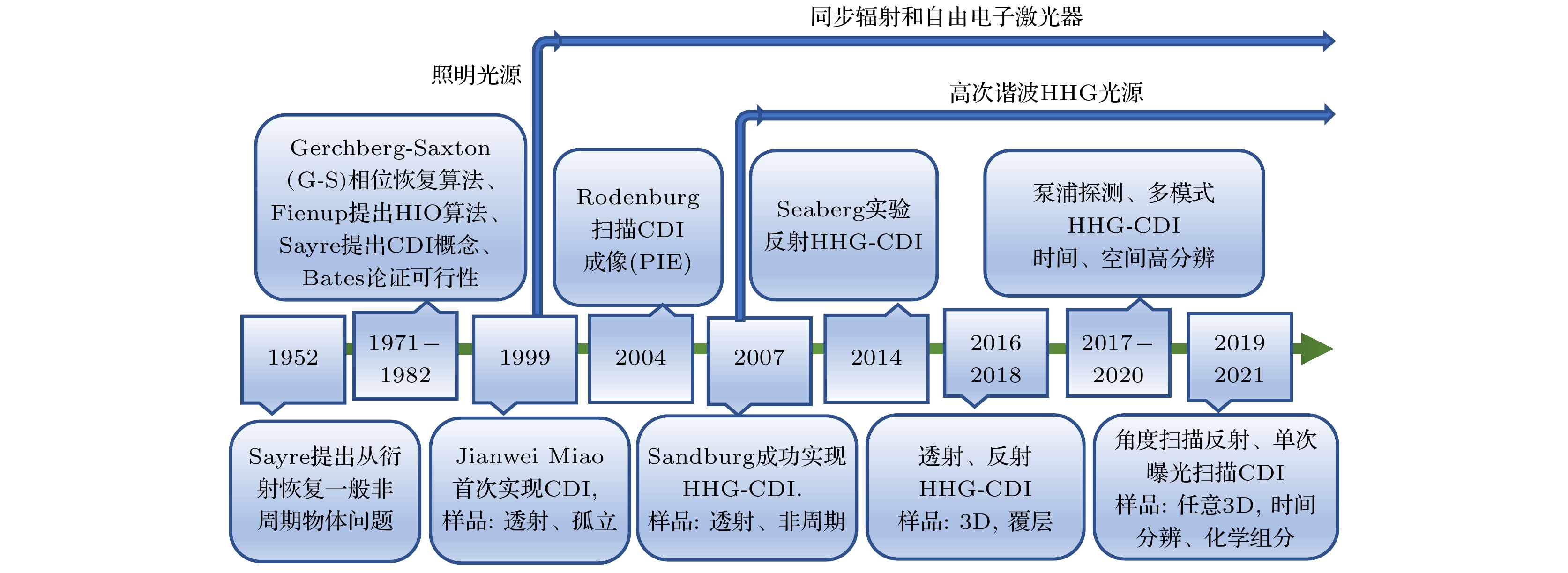
图 1 HHG相干衍射成像(HHG-CDI)的发展史
Figure 1. The evolution of HHG-based coherent diffraction imaging (HHG-CDI).

图 2 (a)上海光源主加速器; (b)台面HHG-EUV/SXR射线光源
Figure 2. (a)Shanghai synchrotron radiation facility(SSRF); (b) a HHG-EUV/SXR source.

图 3 HHG产生的“三步模型”. 原子势垒会被激光场调制, 电子发生隧穿电离; 然后在激光电场加速; 随着电场反向, 电离电子与母核复合, 把获得能量以HHG光子辐射 (制作本图参考了文献 [49] )
Figure 3. The illustration of the three-step Model. The tunneling ionization can occur as the atomic barrier is modulated by the laser field. Then the electron is accelerated in the electric field; As the electric field is reversed, the ionized electron recombines with the parent nucleus and radiates its energy as HHG photons,Figure reproduced from Ref. [49]
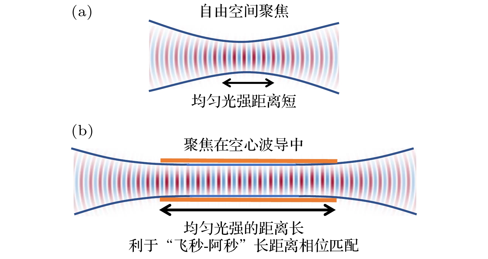
图 4 自由空间聚焦与空心波导HHG对比图
Figure 4. The comparison of HHG in free space focusing and hollow waveguide.
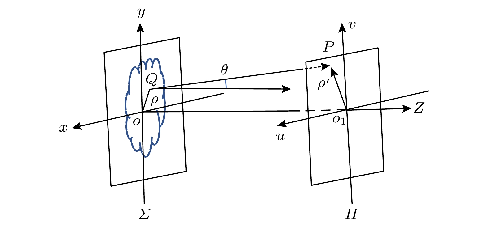
图 5 平面屏衍射示意图
Figure 5. The schematic chart of plane diffraction.

图 6 CDI相位恢复算法原理
Figure 6. The technical schematic and algorithm flow chart of CDI.
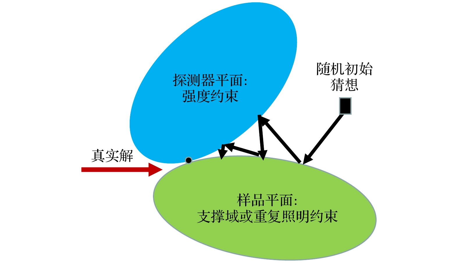
图 7 凸集映射示意图, 一个随机猜测投影到检测器平面约束集, 然后投影到样本平面约束集, 完成一个更新周期. 多次迭代后, 找到两个约束集的交点: 真解
Figure 7. Diagram of convex-set mapping, a random guess is first projected to the detector plane constraint set, then to the sample plane constraint set to finish a full updating cycle. After many iterations, the solution is found at the intersection of the two constraint sets.
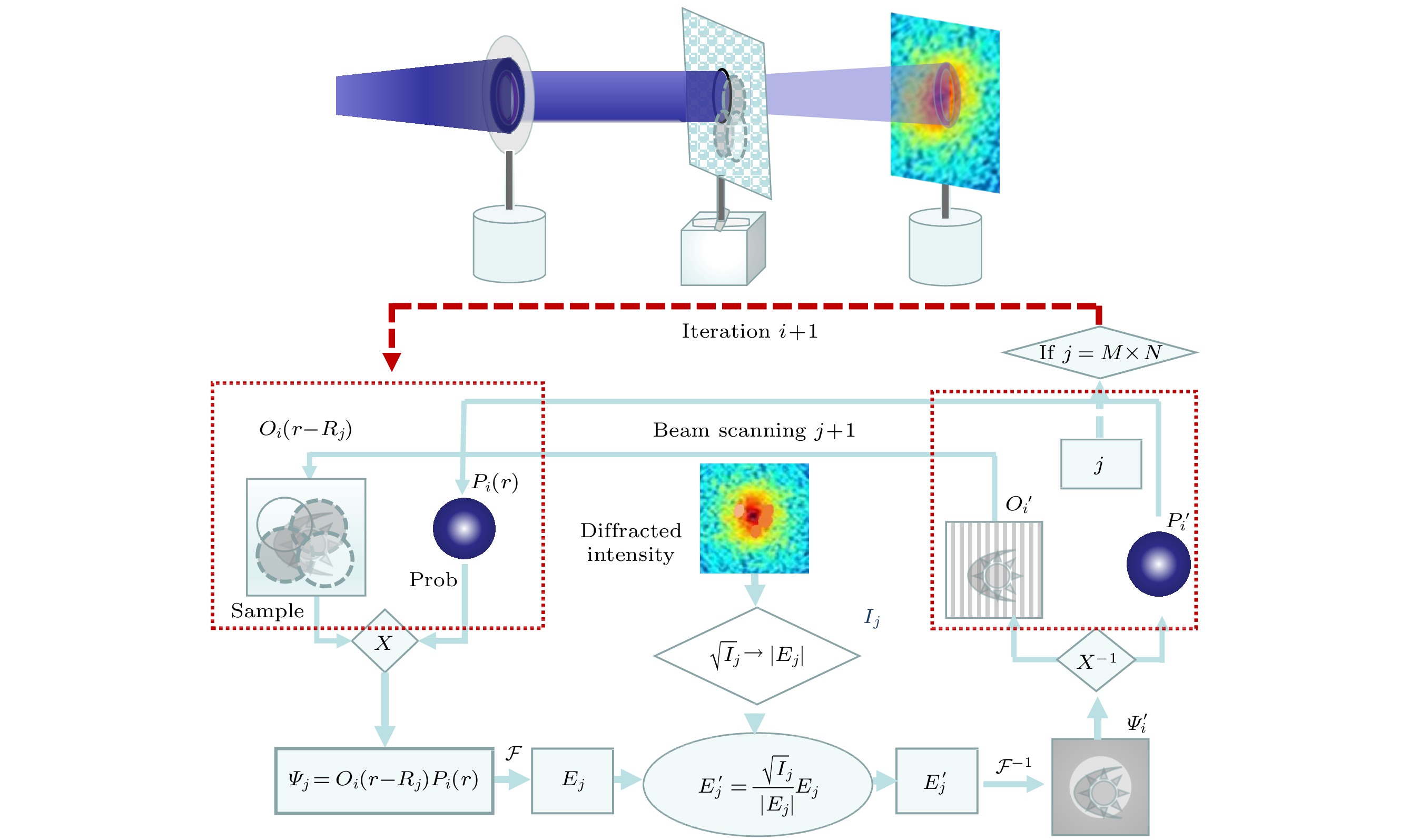
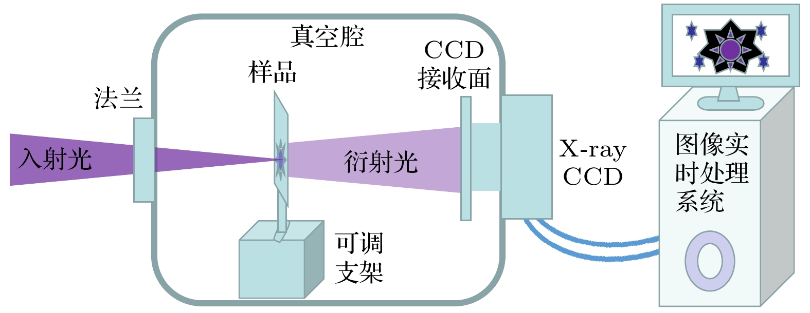
图 9 HHG-CDI纳米成像系统
Figure 9. The coherent diffraction imaging system for a HHG extreme ultraviolet laser source.

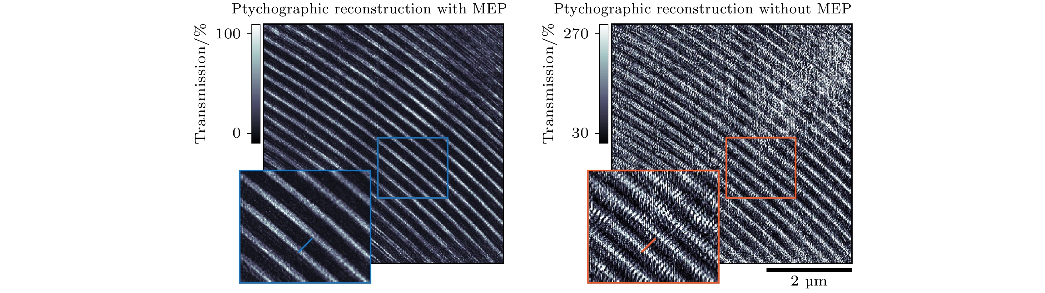
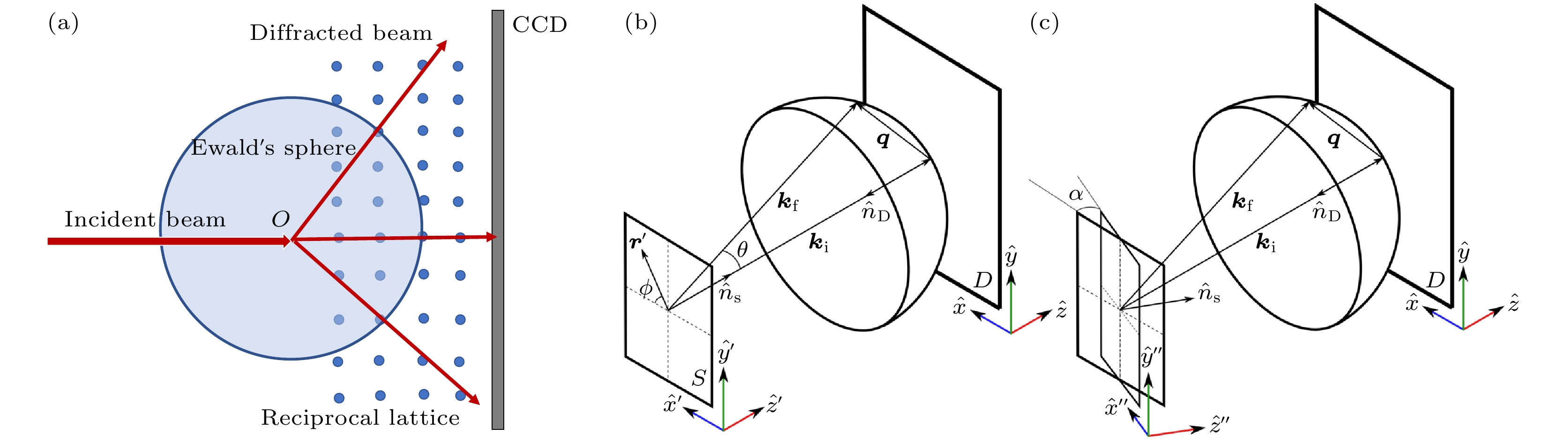
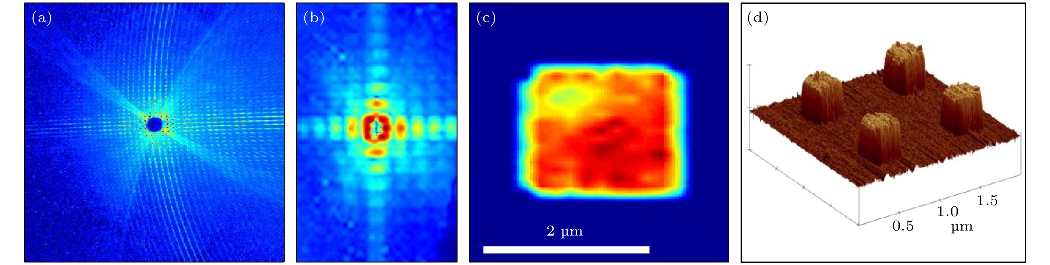
图 13 反射模式相干衍射成像 (a) CCD上的实测衍射图; (b)采用校正算法, 提取图(a)中每个衍射峰的值, 重采样衍射图; (c)重建显示所有照明柱的平均值; (d)类似柱状结构的原子力显微镜图像[15]
Figure 13. Reflection-mode coherent diffraction imaging: (a) measured diffraction pattern on CCD; (b) resampled diffraction pattern in panel (a); (c) reconstruction showing the average of all illuminated pillars; (d) atomic force microscope image of similar pillar structures[15].
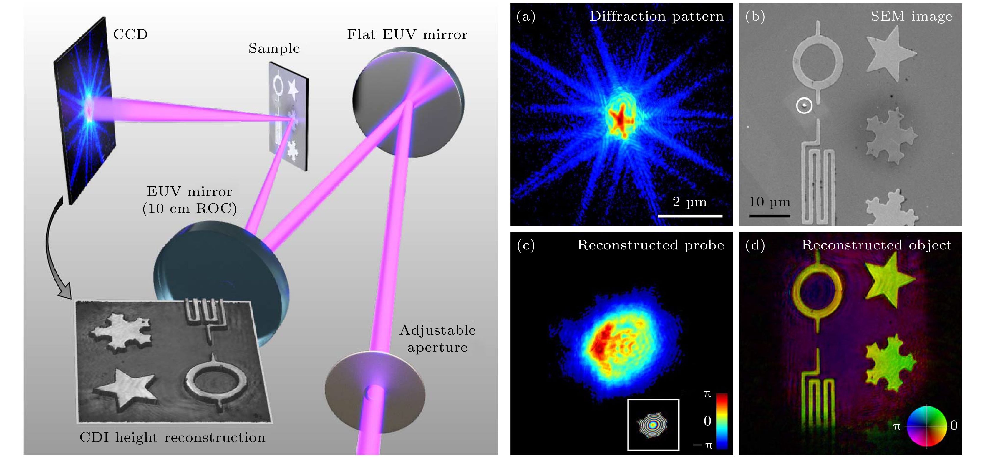
图 14 实验装置、衍射数据和Ptychography重建结果 (a) 90次扫描数据集的代表性衍射图样; (b) SEM像; (c)探针重建; (d)样品重建[16]
Figure 14. Experimental setup for reflection-mode ptychography, diffraction data and ptychographic reconstruction: (a) Representative diffraction pattern taken from the 90-scan dataset; (b) SEM image of the sample; (c) reconstructed amplitude of the HHG beam; (d) Ptychographic reconstruction of the object[16].
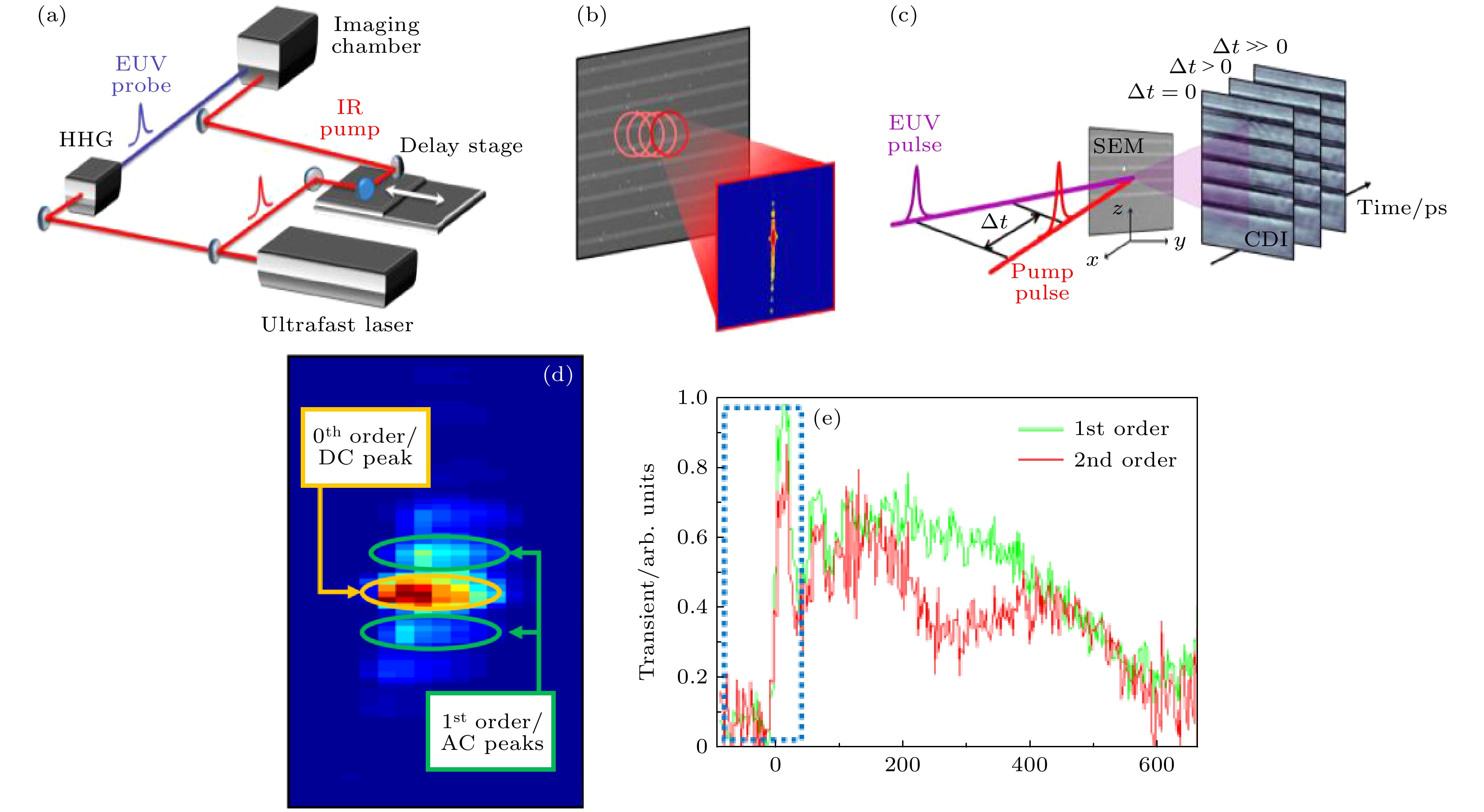
图 15 (a)频闪CDI动态成像实验布局示意图; (b)在每个时间延迟时, 用Ptychography获得样本的图像; (c)不同时间延迟下动态成像实验; (d)硅基镍纳米线的衍射图; (e)衍射效率作为泵浦探测延迟时间的函数的瞬态信号图[24]
Figure 15. (a) Schematic of the experimental layout for dynamic imaging on a tabletop; (b) tt every time delay, the image of the sample is obtained with Ptychographic CDI; (c) general concept of dynamic imaging experiment; (d) diffraction pattern of the Nickel lines on Silicon; (e) plot of the transient signal from diffraction efficiency as a function of pump-probe delay time[24].
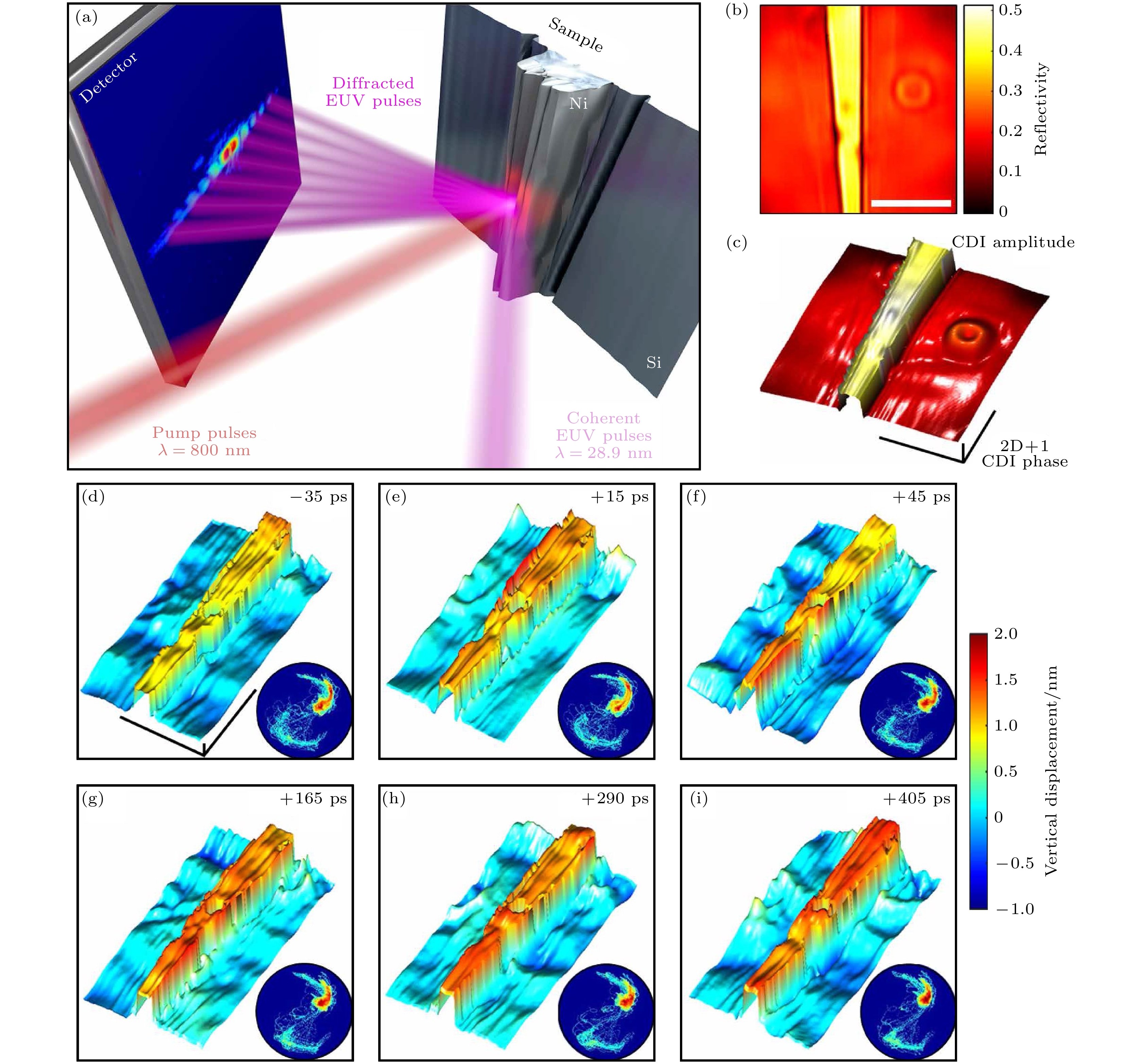
图 16 单个纳米结构中声波的动态成像. (a)频闪CDI显微镜动态成像实验装置; (b)重建样品振幅图像; (c)重建样品相位得到的高度图; (d)—(i) 重建镍纳米结构热膨胀和随后声波在基板中传播的快照[32]
Figure 16. Dynamic imaging of acoustic waves in an individual nanostructure: (a) Stroboscopic CDI microscope for dynamic imaging; (b) reconstructed quantitative amplitude image; (c) height map of the sample obtained from the reconstructed phase image; (d)–(i) ieconstructed snapshots of the nickel nanostructure thermal expansion and subsequent propagation of acoustic waves in the substrate[32]
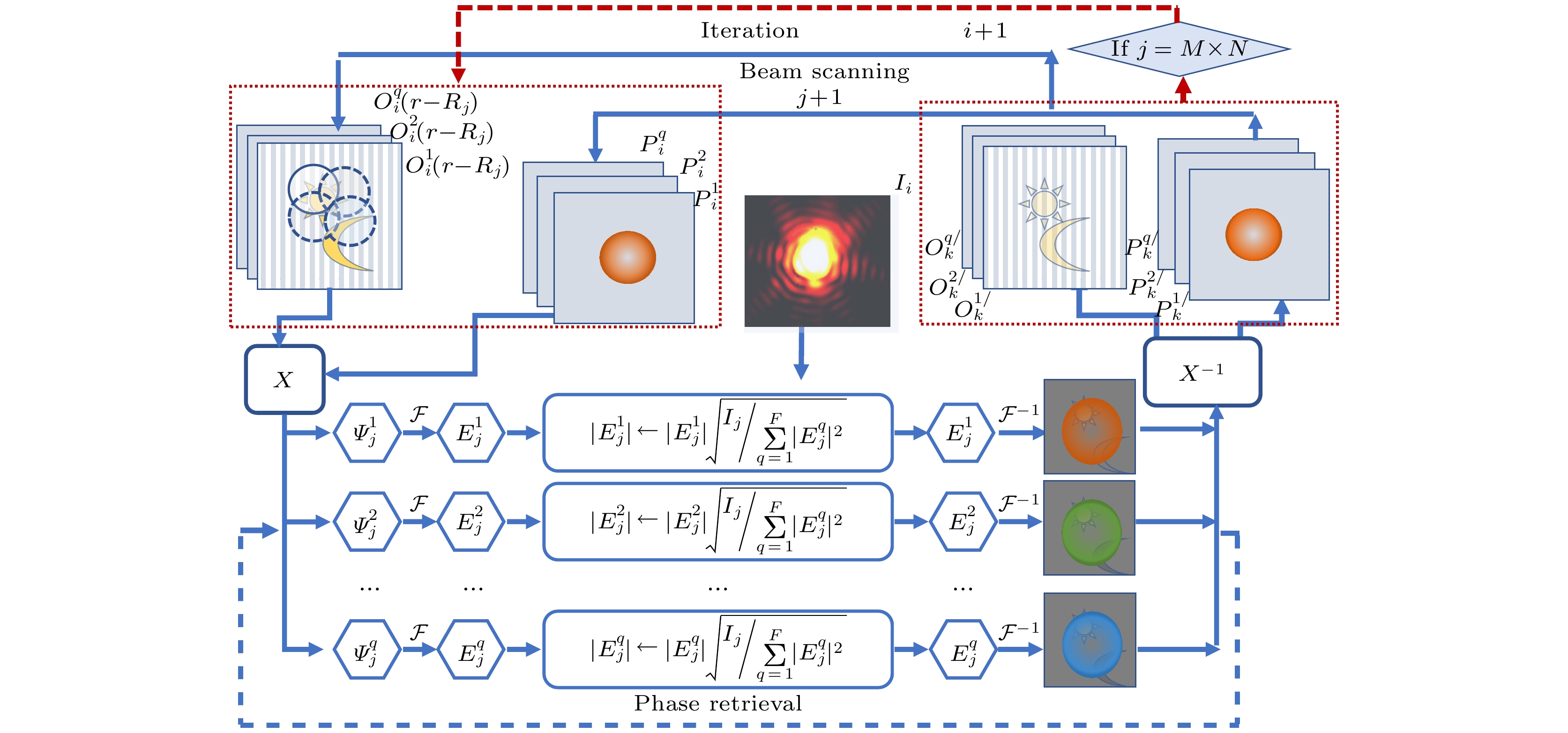
图 17 部分相干光(多色光)的 ePIE 迭代原理
Figure 17. The ePIE system for partially coherent light.
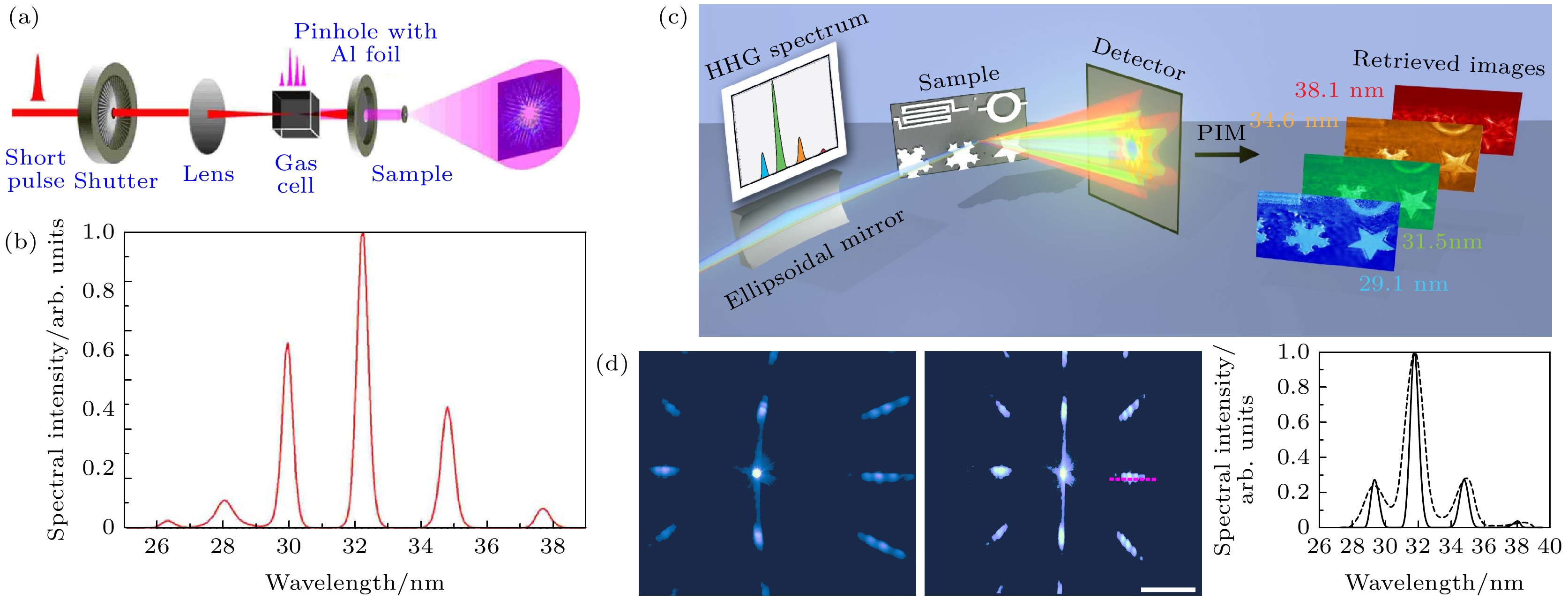
图 18 结合HHG多次极紫外谐波的多光谱衍射成像 (a), (b) 6波长非扫描透射成像模式[82]; (c), (d) 4波长的叠层扫描反射成像模式[83]
Figure 18. Hyperspectral imaging by combining multiple EUV harmonics and PIM: (a), (b) a 6-wavelength non-scanning transmission mode CDI[82]; (c), (d) a ptychographic hyperspectral spectromicroscopy with a 4-wavelength comb[83].
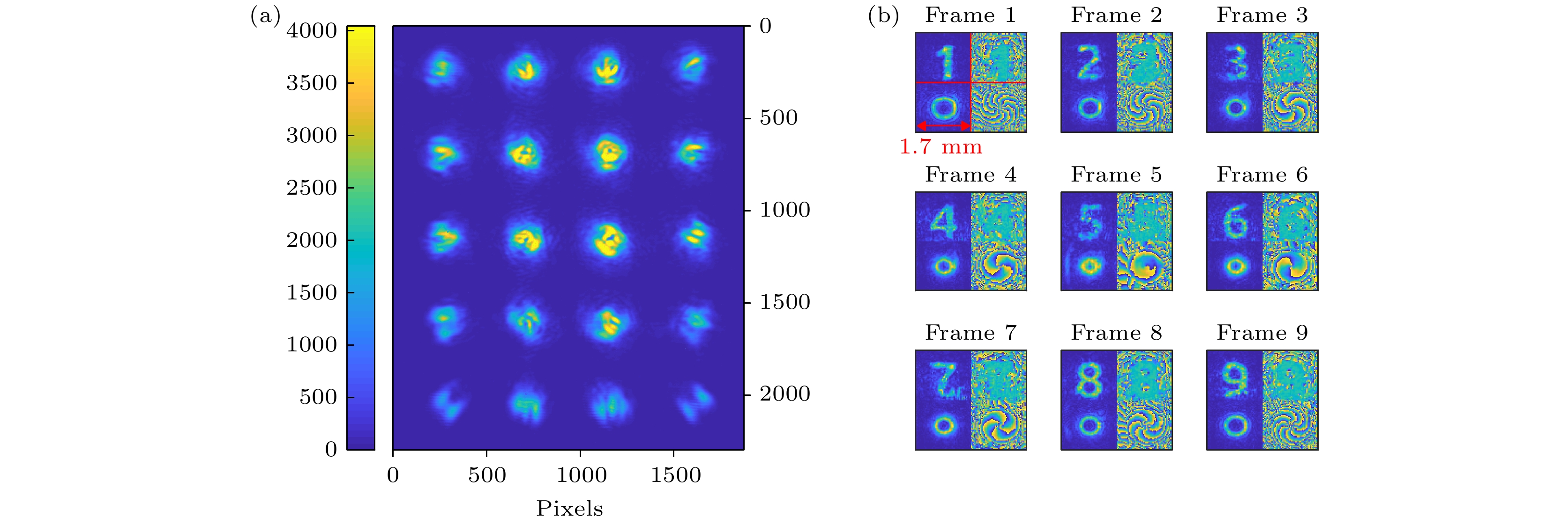
图 21 使用OAME重建9个复值对象和探针 (a)单架照相机抓拍所记录的强度图样;(b)重建帧复值对象和探头, 每帧分为4个区域(如第一帧):左上为物体振幅, 右上为物体相位, 左下为探头振幅, 右下为探头相位[27]
Figure 21. Reconstruction of 9 complex-valued objects and probes using OAME: (a) The intensity pattern recorded in a single camera snapshot; (b) reconstructed frames - complex-valued objects and probes. Each frame is divided to 4 quarters (as marked on the first frame): top-left is object amplitude, top-right is object phase, bottom left is probe amplitude and bottom-right is probe phase[27].

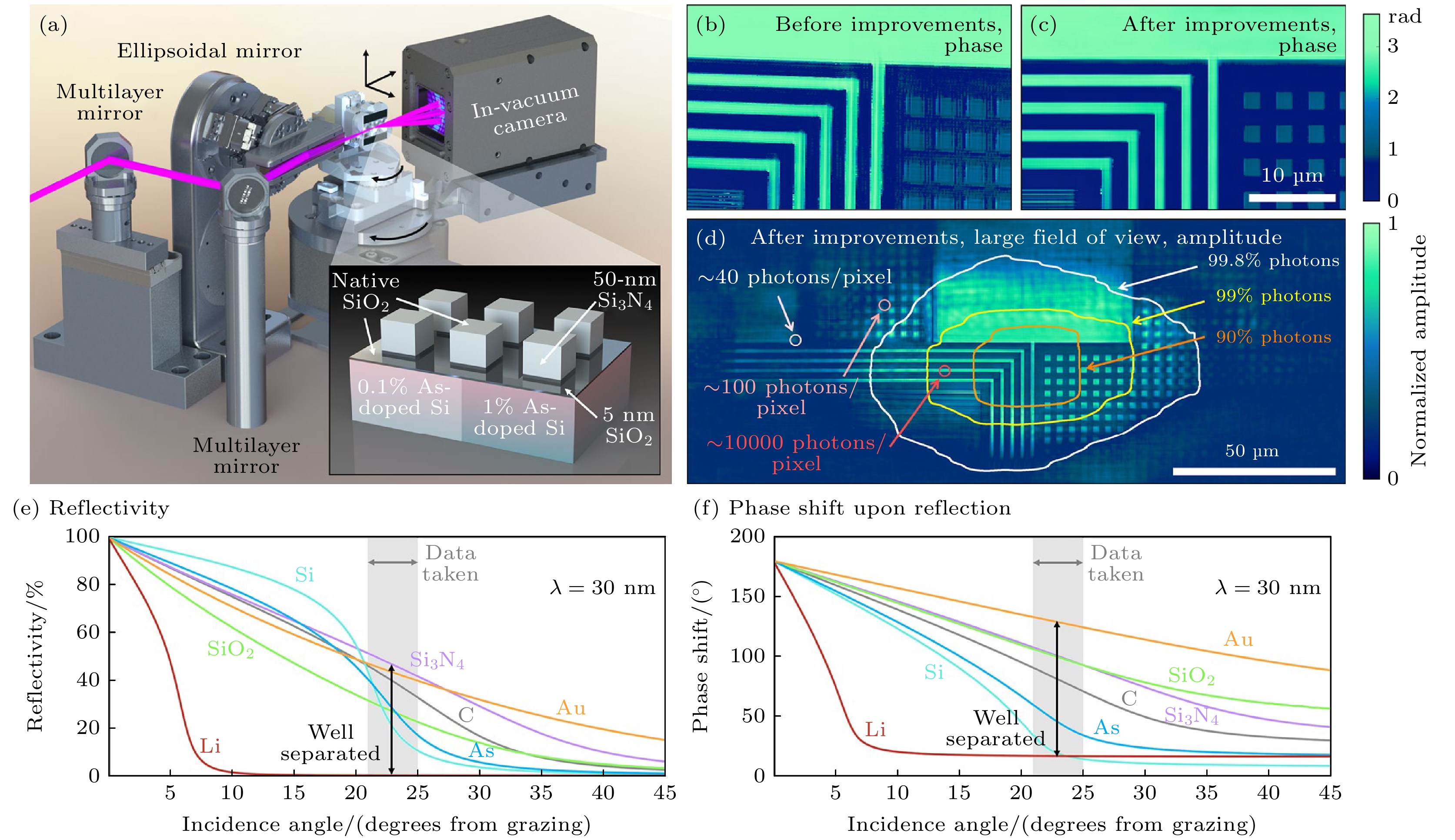
图 23 纳米结构成像 (a)幅值和相位敏感成像反射仪的原理图; (b), (c)实施3D倾斜平面校正和全变分正则化处理和未作相应处理的相位重建; (d)宽视场振幅重建; (e) (f)材料的特征反射率与角度曲线—EUV光对材料成分的敏感性[30]
Figure 23. Experiment overview and nanostructure imaging: (a) Schematic of the amplitude- and phase-sensitive imaging reflectometer. Zoom-in of EUV ptychographic phase reconstructions of the sample, (b) before and (c) after precise implementation of 3D tilted-plane correction and total variation (TV) regularization. (d) Entire, wide field-of-view amplitude reconstruction. (e), (f) Characteristic reflectivity versus angle curves for several materials, showing the sensitivity of EUV light to material composition[30].

图 24 空间分辨、组成敏感和三维纳米结构表征 (a)高掺杂结构, (b)低掺杂衬底和(c)高掺杂衬底中的成分与深度重建; (d)全重构样品的放大(插图); (e) Ptychography相位图像与遗传算法结果相结合得到的结果; (f)同一区域的AFM图像[30]
Figure 24. Spatially resolved, composition-sensitive, 3D nanostructure characterization: Composition versus depth reconstruction in the (a) higher-doped structures, (b) lower-doped substrate, and (c) higher-doped substrate; (D) zoom-out and zoom-in (inset) of fully reconstructed sample; (e) topography map obtained by combining the ptychographic phase image with the results of the genetic algorithm; (f) AFM image of the same region[30].
表 1 半导体器件技术领域常用的几种纳米成像技术和相干衍射成像技术的对比
Table 1. Comparison of several nano-imaging techniques commonly used in semiconductor technology.
纳米成像
技术分辨率/nm 光源 镜头 样品损伤/
预处理表面3D
形貌镀层下结构/
层厚度检测化学成分/浓度 成像
速度光学显微镜 ~100 红外, 可见光 透镜 无损伤无需处理 可探测 部分可探测/
透明料可以可探测/半
导体不可快 X射线显微镜 ~20 SRS, XFEL 波带片 有损伤
无需处理不能探测 均可实现 可探测 慢 扫描电子显微镜 ~0.1 电子束 磁透镜 有损伤
需处理可探测 金属层不可/厚度不可 无法探测 较慢 原子力显微镜 ~0.1 nm 无 纳米探针 无损伤
无需处理可探测 无法探测 无法探测 最慢 HHG-CDI <10 nm SRS, FEL.HHG 无透镜 无损伤无需处理 可探测 均可探测 可探测 快  DownLoad: CSV
-
[1] Shadfan A, Pawlowski M, Wang Y, Subramanian K, Gabay I, Ben-Yakar A, Tkaczyk T 2016 Opt. Eng. 55 025107
Google Scholar
[2] Parimi P V, Lu W T T, Vodo P, Sridhar S 2003 Nature 426 404
Google Scholar
[3] Hell S W, Wichmann J 1994 Opt. Lett. 19 780
Google Scholar
[4] Betzig E, Lewis A, Harootunian A, Isaacson M, Kratschmer E 1986 Biophys. J. 49 269
Google Scholar
[5] Wokosin D L, Centonze V E, Crittenden S, White J 2015 Bioimaging 4 208
[6] Denk W, Strickler J H, Webb W W 1990 Science 248 73
Google Scholar
[7] Rust M J, Bates M, Zhuang X 2006 Nat. Methods 3 793
Google Scholar
[8] Hunt B R, Overman T L, Gough P 1998 Opt. Lett. 23 1123
Google Scholar
[9] Miao J, Charalambous P, Kirz J, Sayre D 1999 Nature 400 342
[10] Seibert M M, Ekeberg T, Maia F R, Svenda M, Andreasson J, Jonsson O, Odic D, Iwan B, Rocker A, Westphal D, Hantke M, DePonte D P, Barty A, Schulz J, Gumprecht L, Coppola N, Aquila A, Liang M, White T A, Martin A, Caleman C, Stern S, Abergel C, Seltzer V, Claverie J M, Bostedt C, Bozek J D, Boutet S, Miahnahri A A, Messerschmidt M, Krzywinski J, Williams G, Hodgson K O, Bogan M J, Hampton C Y, Sierra R G, Starodub D, Andersson I, Bajt S, Barthelmess M, Spence J C, Fromme P, Weierstall U, Kirian R, Hunter M, Doak R B, Marchesini S, Hau-Riege S P, Frank M, Shoeman R L, Lomb L, Epp S W, Hartmann R, Rolles D, Rudenko A, Schmidt C, Foucar L, Kimmel N, Holl P, Rudek B, Erk B, Homke A, Reich C, Pietschner D, Weidenspointner G, Struder L, Hauser G, Gorke H, Ullrich J, Schlichting I, Herrmann S, Schaller G, Schopper F, Soltau H, Kuhnel K U, Andritschke R, Schroter C D, Krasniqi F, Bott M, Schorb S, Rupp D, Adolph M, Gorkhover T, Hirsemann H, Potdevin G, Graafsma H, Nilsson B, Chapman H N, Hajdu J 2011 Nature 470 78
Google Scholar
[11] Ekeberg T E, Svenda M, Abergel C, Maia F R N C, Seltzer V, Claverie J-M, Hantke M, Joensson O, Nettelblad C, van der Schot G, Liang M, DePonte D P, Barty A, Seibert M M, Iwan B, Andersson I, Loh N D, Martin A V, Chapman H, Bostedt C, Bozek J D, Ferguson K R, Krzywinski J, Epp S W, Rolles D, Rudenko A, Hartmann R, Kimmel N, Hajdu J 2015 Phys. Rev. Lett. 114 098102
Google Scholar
[12] Sandberg R L, Paul A, Raymondson D A, Haedrich S, Gaudiosi D M, Holtsnider J, Tobey R a I, Cohen O, Murnane M M, Kapteyn H C, Song C, Miao J, Liu Y, Salmassi F 2007 Phys. Rev. Lett. 99 098103
Google Scholar
[13] Iii C D, Rundquist A R, Murnane M M, Kapteyn H C 1998 Science 280 1412
Google Scholar
[14] Gardner D F, Zhang B, Seaberg M D, Martin L S, Adams D E, Salmassi F, Gullikson E, Kapteyn H, Murnane M 2012 Opt. Express 20 19050
Google Scholar
[15] Seaberg M D, Adams D E, Zhang B, Murnane M M, Kapteyn H C 2012 Conference on Lasers and Electro-Optics San Jose, California, USA, May 06 2012 p CF1 L. 8
[16] Seaberg M D, Zhang B, Gardner D F, Shanblatt E R, Murnane M M, Kapteyn H C, Adams D E 2014 Optica 1 39
Google Scholar
[17] Abbey B, Nugent K A, Williams G J, Clark J N, Peele A G, Pfeifer M A, de Jonge M, McNulty I 2008 Nat. Phys. 4 394
Google Scholar
[18] Zhang B, Seaberg M D, Adams D E, Gardner D F, Shanblatt E R, Shaw J M, Chao W, Gullikson E M, Salmassi F, Kapteyn H C, Murnane M M 2013 Opt. Express 21 21970
Google Scholar
[19] Gardner D F, Tanksalvala M, Shanblatt E R, Zhang X, Galloway B R, Porter C L, Karl R, Jr., Bevis C, Adams D E, Kapteyn H C, Murnane M, Mancini G F 2017 Nat. Photonics 11 259
Google Scholar
[20] Mancini G F, Gardner D F, Tanksalvala M, Shanblatt E R, Zhang X, Galloway B R, Porter C R, Karl R, Bevis C, Kapteyn H, Murnane M M, Adams D E 2016 International Conference on Ultrafast Phenomena Santa Fe, New Mexico, USA, July 17 2016 pUTu2 B. 2
[21] Porter C L, Tanksalvala M, Gerrity M, Miley G, Zhang X, Bevis C, Shanblatt E, Karl R, Jr., Murnane M M, Adams D E, Kapteyn H C 2017 Optica 4 1552
Google Scholar
[22] Whitehead L W, Williams G J, Quiney H M, Vine D J, Dilanian R A, Flewett S, Nugent K A, Peele A G, Balaur E, McNulty I 2009 Phys. Rev. Lett. 103 243902
Google Scholar
[23] Thibault P, Menzel A 2013 Nature 494 68
Google Scholar
[24] Karl R, Mancini G, Gardner D, Knobloch J, Frazer T, Hernandez-Charpak J N, Mayor B A, Shanblatt E, Tanksalvala M, Porter C, Bevis C, Adams D, Kapteyn H, Murnane M M 2017 Imaging and Applied Optics San Francisco, California, USA, June 26, 2017 pCW1 B. 2
[25] Karl R, Mancini G, Gardner D, Shanblatt E, Knobloch J, Frazer T, Hernandez-Charpak J N, Mayor B A, Tanksalvala M, Porter C, Bevis C, Adams D, Kapteyn H, Murnane M 2018 High-Brightness Sources and Light-driven Interactions Strasbourg, France, March 26, 2018 pET2B.6
[26] Pan X, Liu C, Zhu J 2013 Appl. Phys. Lett. 103 171105
Google Scholar
[27] Sidorenko P, Cohen O 2016 Optica 3 9
Google Scholar
[28] Sidorenko P, Lahav O, Cohen O 2017 Opt. Express 25 10997
Google Scholar
[29] Wengrowicz O, Peleg O, Loevsky B, Chen B K, Haham G I, Sainadh U S, Cohen O 2019 Opt. Express 27 24568
Google Scholar
[30] Tanksalvala M, Porter C L, Esashi Y, Wang B, Jenkins N W, Zhang Z, Miley G P, Knobloch J L, McBennett B, Horiguchi N, Yazdi S, Zhou J, Jacobs M N, Bevis C S, Karl R M, Jr., Johnsen P, Ren D, Waller L, Adams D E, Cousin S L, Liao C T, Miao J, Gerrity M, Kapteyn H C, Murnane M M 2021 Sci. Adv. 7 9667
Google Scholar
[31] Le H V, Dinh K B, Hannaford P, Van Dao L 2014 J. Appl. Phys. 116 173104
[32] Karl R M, Mancini G F, Knobloch J L, Frazer T D, Hernandez-Charpak J N, Abad B, Gardner D F, Shanblatt E R, Tanksalvala M, Porter C L, Bevis C S, Adams D E, Kapteyn H C, Murnane M M 2018 Sci. Adv. 4 eaau4295
Google Scholar
[33] Antunez P D, Bishop D M, Luo Y, Haight R 2017 Nat. Energy 2
[34] Frazer T D, Knobloch J L, Hernández-Charpak J N, Hoogeboom-Pot K M, Nardi D, Yazdi S, Chao W, Anderson E H, Tripp M K, King S W, Kapteyn H C, Murnane M M, Abad B 2020 Phys. Rev. Mater. 4 073603
Google Scholar
[35] King S W, Simka H, Herr D, Akinaga H, Garner M 2013 APL Mater. 1 040701
Google Scholar
[36] Mochi I, Fernandez S, Nebling R, Locans U, Helfenstein P, Rajeev R, Dejkameh A, Kazazis D, Tseng L T, Ekinci Y 2019 Amplitude and Phase Defect Inspection on EUV Reticles Using RESCAN p29
[37] Moler K A 2017 Nat. Mater. 16 1049
Google Scholar
[38] Klas R, Kirsche A, Gebhardt M, Buldt J, Stark H, Hädrich S, Rothhardt J, Limpert J 2021 PhotoniX 2 4
Google Scholar
[39] McPherson A, Gibson G, Jara H, Johann U, Luk T S, McIntyre I A, Boyer K, Rhodes C K 1987 J. Opt. Soc. Am. B 4 595
Google Scholar
[40] Krause J L, Schafer K J, Kulander K C 1992 Phys. Rev. Lett. 68 3535
Google Scholar
[41] Corkum P B 1993 Phys. Rev. Lett. 71 1994
Google Scholar
[42] Ammosov M V, Delone N B, Krainov V P 1986 Proceedings of SPIE Quebec, Canada, October 21, 1986 p138
[43] 盛政明编 2003 强场激光物理研究前沿(上海: 上海交通大学出版社) 第5, 57页
Sheng Z M 2014 Advances in High Field Laser Physics (Shanghai: Shanghai Jiao Tong University Press) pp5, 57 (in Chinese)
[44] Zhang X, Libertun A R, Paul A, Gagnon E, Backus S, Christov I P, Murnane M M, Kapteyn H C, Bartels R A, Liu Y, Attwood D T 2004 Opt. Lett. 29 1357
Google Scholar
[45] Rundquist A, Durfee C G, Chang Z H, Herne C, Backus S, Murnane M M, Kapteyn H C 1998 Science 280 1412
Google Scholar
[46] Bartels R A, Paul A, Green H, Kapteyn H C, Murnane M M, Backus S, Christov I P, Liu Y W, Attwood D, Jacobsen C 2002 Science 297 376
[47] Zhang X S, Lytle A, Popmintchev T, Paul A, Wagner N, Murnane M, Kapteyn H, Christov I P 2005 Opt. Lett. 30 1971
Google Scholar
[48] Lytle A L, Zhang X, Arpin P, Cohen O, Murnane M M, Kapteyn H C, Ieee 2008 Conference on Lasers and Electro-Optics/Quantum Electronics and Laser Science Conference San Jose, CA, USA, May 4–9 p1984
[49] Corkum P B, Krausz F 2007 Nat. Phys. 3 381
[50] Martin G, Tobias H, Robert K, Alexander K, Chang L, Ziyao W, Mathias L, Christian G, Cesar J, Jose A L, Axel S, Rodrigo A C, Jan R, Jens L Proc. SPIE
[51] Feehan J S, Price J H V, Butcher T J, Brocklesby W S, Frey J G, Richardson D J 2017 Appl. Phys. B 123 43
[52] Hoppe W 1969 Acta Crystallogr. Sect. A 25 508
Google Scholar
[53] Robinson I K, Vartanyants I A, Williams G J, Pfeifer M A, Pitney J A 2001 Phys. Rev. Lett. 87 195505
Google Scholar
[54] Rodenburg J M, Faulkner H M L 2004 Appl. Phys. Lett. 85 4795
Google Scholar
[55] Williams G J, Quiney H M, Dhal B B, Tran C Q, Nugent K A, Peele A G, Paterson D, de Jonge M D 2006 Phys. Rev. Lett. 97 025506
Google Scholar
[56] Roy S, Parks D, Seu K A, Su R, Turner J J, Chao W, Anderson E H, Cabrini S, Kevan S D 2011 Nat. Photonics 5 243
Google Scholar
[57] Bates R H T 1982 Phys. Rep. 90 203
Google Scholar
[58] Miao J, Sayre D, Chapman H N 1998 J. Opt. Soc. Am. A 15 1662
Google Scholar
[59] Gerchberg R W, Saxton, W. O. 1972 Optik 35 237
[60] Burge R E 1981 Scanning 4 159
[61] Fienup J R 1982 Appl. Opt. 21 2758
Google Scholar
[62] Streibl N 1984 Opt. Commun. 49 6
Google Scholar
[63] Teague M R 1983 J. Opt. Soc. Am. A 73 1434
Google Scholar
[64] Sayre D 1952 Acta Crystallogr. 5 843
[65] Hoppe W 1969 Acta Crystallogr. Sect. A 25 495
Google Scholar
[66] Hoppe W, Strube G 1969 Acta Crystallogr. Sect. A 25 502
Google Scholar
[67] Hegerl R, Hoppe W 1972 Proceedings of the 5th European Congress on Electron Microscopy p628
[68] Marchesini S 2007 Rev. Sci. Instrum. 78 011301
Google Scholar
[69] Bauschke H H, Combettes P L, Luke D R 2002 J. Opt. Soc. Am. A 19 1334
Google Scholar
[70] Maiden A M, Rodenburg J M 2009 Ultramicroscopy 109 1256
Google Scholar
[71] Pan X C, Liu C, Tao H, Liu H G, Zhu J Q 2020 Acta Optica Sinica 40 111010
Google Scholar
[72] Maiden A M, Humphry M J, Rodenburg J M 2012 J. Opt. Soc. Am. A 29 1606
[73] Zhang F, Peterson I, Vila-Comamala J, Berenguer A D F, Bean R, Chen B, Menzel A, Robinson I K, Rodenburg J M 2013 Opt. Express 21 13592
Google Scholar
[74] Zheng G, Horstmeyer R, Yang C 2015 Nat. Photonics 9 621
Google Scholar
[75] Shanblatt E R, Porter C L, Gardner D F, Mancini G F, Karl R M, Tanksalvala M,Bevis C S, Vartanian V H, Kapteyn H C, Adams D E 2016 Computational Optical Sensing and Imaging 2016 CT4C.1
[76] Raines K S, Salha S, Sandberg R L, Jiang H, Rodriguez J A, Fahimian B P,Kapteyn H C, Du J, Miao J 2010 Nature 463 214
[77] Miao J, Ishikawa T, Robinson I K, Murnane M M 2015 Science 348 530
Google Scholar
[78] Spence J C H, Weierstall U, Howells M 2004 Ultramicroscopy 101 149
Google Scholar
[79] Abbey B, Whitehead L W, Quiney H M, Vine D J, Cadenazzi G A, Henderson C A, Nugent K A, Balaur E, Putkunz C T, Peele A G, Williams G J, McNulty I 2011 Nat. Photonics 5 420
Google Scholar
[80] Batey D J, Claus D, Rodenburg J M 2014 Ultramicroscopy 138 13
Google Scholar
[81] Williams G J, Quiney H M, Peele A G, Nugent K A 2007 Phys. Rev. B 75 4102
[82] Chen B, Dilanian R A, Teichmann S, Abbey B, Peele A, Williams G J, Hannaford P, Dao L V, Quiney H M, Nugent K A 2009 Phys. Rev. A 79 023809
Google Scholar
[83] Zhang B, Gardner D F, Seaberg M H, Shanblatt E R, Porter C L, Karl R, Mancuso C A, Kapteyn H C, Murnane M M, Adams D E 2016 Opt. Express 24 18745
Google Scholar
[84] Bevis C, Karl R, Reichanadter J, Gardner D F, Porter C, Shanblatt E, Tanksalvala M, Mancini G F, Kapteyn H, Murnane M, Adams D 2018 Ultramicroscopy 184 164
Google Scholar
[85] Karl R, Bevis C, Lopez-Rios R, Reichanadter J, Gardner D, Porter C, Shanblatt E, Tanksalvala M, Mancini G F, Murnane M, Kapteyn H, Adams D 2015 Opt. Express 23 30250
Google Scholar
[86] Rönsch-Schulenburg J, Faatz B, Honkavaara K, Kuhlmann M, Schreiber S, Treusch R, Vogt M 2017 J. Phys. Conf. Ser. 874 012023
Google Scholar
[87] Ellis J L, Dorney K M, Hickstein D D, Brooks N J, Gentry C, Hernández-García C, Zusin D, Shaw J M, Nguyen Q L, Mancuso C A, Matthijs Jansen G S, Witte S, Kapteyn H C, Murnane M M 2018 Optica 5 479
Google Scholar
[88] Hirose M, Higashino T, Ishiguro N, Takahashi Y 2020 Opt. Express 28 1216
Google Scholar
[89] Yao Y, Jiang Y, Klug J A, Wojcik M, Maxey E R, Sirica N S, Roehrig C, Cai Z, Vogt S, Lai B, Deng J 2020 Sci. Rep. 10 19550
Google Scholar
[90] Rokitski R, Sun P C, Fainman Y 2001 Opt. Lett. 26 1125
Google Scholar
[91] Beck A, Teboulle M 2009 IEEE Trans. Image Process. 18 2419
Google Scholar
[92] Cho A 2012 Science 338 1136
Google Scholar

 DownLoad:
Catalog
Metrics
- Abstract views: 21867
- PDF Downloads: 504
- Cited By: 0
