


-
Up-conversion nanoparticle (UCNP) can collect near-infrared (NIR) light and convert it into visible light. Therefore, UCNP has potential applications in fields such as biomedicine, anti-counterfeiting, and solar cells. However, the efficiency of traditional UCNP in the above-mentioned fields is relatively low, greatly limiting its use in related fields. Therefore, enhancing the up-conversion luminescence intensity of up-conversion nanoparticles is particularly important and urgently needed. In this work, anodic alumina templates are used to enhance the luminescence intensity of up-conversion nanocrystals. NaYF4:Yb3+, Er3+with a diameter of 35 nm is prepared by using co-precipitation method. Single pass AAO templates with pore size and pore spacing of 88 nm and 100 nm are prepared by using two-step anodization method. Finally, NaYF4:Yb3+, Er3+/AAO composite structures are formed by using spin coating method. The red green light emission intensity of NaYF4:Yb3+, Er3+/AAO sample can increase 4.4 and 9.0 times that of NaYF4:Yb3+, Er3+/Al reference sample, respectively. The enhancement mechanism is explored by using the finite difference time domain method, and the results show that the primary source of enhancement is the localized surface plasmon resonance effect of the pores in the anodic alumina template. At the same time, the relationship between the up-conversion luminescence intensity of NaYF4:Yb3+, Er3+/AAO sample and the incident angle is investigated. The experimental results show that as the incident angle increases, the luminescence intensity of the red and green light of NaYF4:Yb3+, Er3+/AAO samples first decrease and then increase. Due to the coupling of the local surface plasmon resonance with the excitation wavelength and emission wavelength, the up-conversion luminescence intensity of the sample can be affected. The relationship of AAO channel enhancement factor with incident angle at excitation wavelength and emission wavelength is studied by using the finite difference time domain method. The results indicate that as the incident angle increases, the enhancement factor at the excitation wavelength decreases, while the enhancement factor at the emission wavelength increases after being illuminated at an incident angle of 15°. Therefore, when the incident angle is less than 20°, the electric field intensity at 980 nm dominates, but when it is greater than 20°, the electric field intensity at 540 nm and 650 nm takes precedence. The above results provide a reference for putting them into practical applications in the fields of anti-counterfeiting and solar cells.
-
Keywords:
- up-conversion luminescence /
- anodized aluminum /
- local surface plasmon resonance /
- finite-difference time-domain solutions
[1] Haase M, Schäfer H 2011 Angew. Chem. Int. Ed. 50 5808
 Google Scholar
[2] Shao B, Yang Z W, Li J, Yang J Z, Wang Y D, Qiu J B, Song Z G 2017 Opt. Mater. Express 7 1188
Google Scholar
[3] Zhang J, Shen H O, Guo W, Wang S H, Zhu C T, Xue F, Hou J F, Su H Q, Yuan Z B 2013 J. Power Sources 226 47
Google Scholar
[4] He L, Dragavon J, Cho S, Mao C, Yildirim A, Ma K, Chattaraj R, Goodwin A P, Park W, Cha J N 2016 J. Mater. Chem. B 4 4455
Google Scholar
[5] Kumar A, Tiwari S P, Esteves Da Silva J C G, Kumar K 2018 Laser Phys. Lett. 15 075901
Google Scholar
[6] Janjua R A, Iqbal O, Ahmed M A, Al-Kahtani A A, Saeed S, Imran M, Wattoo A G 2021 RSC Adv. 11 20746
Google Scholar
[7] Chen D, Huang P 2014 Dalton Trans. 43 11299
Google Scholar
[8] Zhu W, Wu Q, Zhao S, Liang Z, Yang Y, Zhang J, Xu Z 2016 Opt. Mater. Express 6 3001
Google Scholar
[9] 高伟, 董军, 王瑞博, 王朝晋, 郑海荣 2016 物理学报 65 084205
Google Scholar
Gao W, Dong J, Wang R B, Wang Z J, Zheng H R 2016 Acta Phys. Sin. 65 084205
Google Scholar
[10] Niu W, Su L T, Chen R, Chen H, Wang Y, Palaniappan A, Sun H, Yoong Tok A I 2014 Nanoscale 6 817
Google Scholar
[11] Xu W, Zhu Y, Chen X, Wang J, Tao L, Xu S, Liu T, Song H 2013 Nano Res. 6 795
Google Scholar
[12] Wang H, Zhan S, Wu X, Wu L, Liu Y 2021 RSC Adv. 11 565
Google Scholar
[13] 高伟, 王博扬, 韩庆艳, 韩珊珊, 程小同, 张晨雪, 孙泽煜, 刘琳, 严学文, 王勇凯, 董军 2020 物理学报 69 184213
Google Scholar
Gao W, Wang B Y, Han Q Y, Han S S, Cheng X T, Zhang C X, Sun Z Y, Liu L, Yan X W, Wang Y K, Dong J 2020 Acta Phys. Sin. 69 184213
Google Scholar
[14] Verhagen E, Kuipers L, Polman A 2009 Opt. Express 17 14586
Google Scholar
[15] Saboktakin M, Ye X, Chettiar U K, Engheta N, Murray C B, Kagan C R 2013 ACS Nano 7 7186
Google Scholar
[16] Chu A, He H, Yin Z, Peng R, Yang H, Gao X, Luo D, Chen R, Xing G, Liu Y J 2020 ACS Appl. Mater. Interfaces 12 1292
Google Scholar
[17] 薛映仙, 戎有英, 马强, 潘诚达, 陈凌霄, 武愕, 吴伯涛 2017 光学学报 37 0724002
Google Scholar
Xue Y X, Rong Y Y, Ma Q, Pan C D, Chen L X, Wu E, Wu B T 2017 Acta Opt. Sin. 37 0724002
Google Scholar
[18] Yin D, Wang C, Ouyang J, Zhang X, Jiao Z, Feng Y, Song K, Liu B, Cao X, Zhang L, Han Y, Wu M 2014 ACS Appl. Mater. Interfaces 6 18480
Google Scholar
[19] Yuan P, Lee Y H, Gnanasammandhan M K, Guan Z, Zhang Y, Xu Q H 2012 Nanoscale 4 5132
Google Scholar
[20] 刘忆森 2012 博士学位论文 (广州: 华南理工大学)
Sen L Y 2012 Ph. D. Dissertation (Guangzhou: South China University of Technology of China
[21] Smith D Y, Shiles E, Inokuti M, Palik E D 1997 Handbook of Optical Constants of Solids (Burlington: Academic Press) pp369–406
[22] Li C, Quan Z, Yang P, Huang S, Lian H, Lin J 2008 J. Phys. Chem. C 112 13395
Google Scholar
[23] Tang H, Xu Y, Cheng X 2020 J. Solid State Chem. 285 121229
Google Scholar
[24] Bhiri N M, Dammak M, Carvajal J J, Aguiló M, Díaz F, Pujol M C 2022 Mater. Res. Bull. 151 111801
Google Scholar
[25] Zhan S, Wu X, Tan C, Xiong J, Hu S, Hu J, Wu S, Gao Y, Liu Y 2018 J. Alloys Compd. 735 372
Google Scholar
-
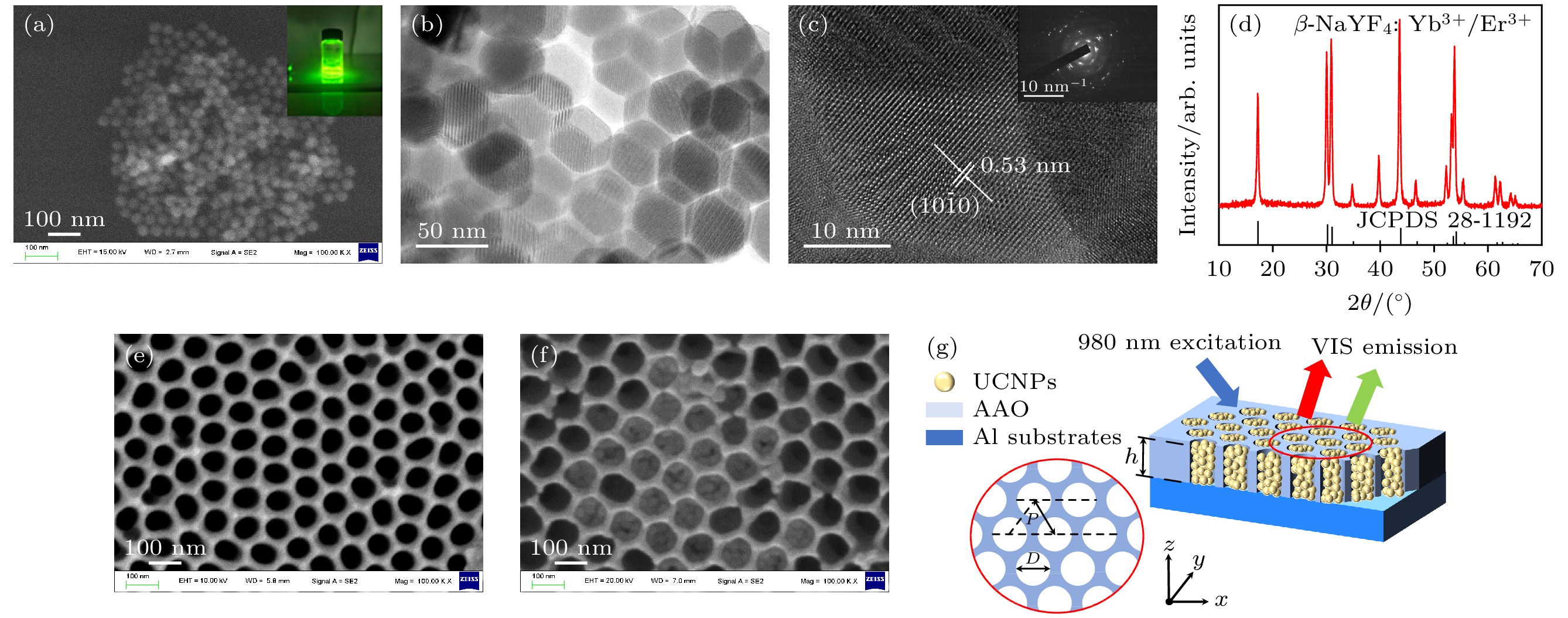
图 1 (a) β-NaYF4:Yb3+, Er3+ SEM图, 插图显示980 nm激光激发下纳米颗粒溶液的上转换发光照片; (b) NaYF4:Yb3+, Er3+的TEM图; (c) NaYF4:Yb3+, Er3+的HRTEM图, 插图为NaYF4:Yb3+, Er3+的SAED图; (d) β-NaYF4:Yb3+, Er3+ XRD谱图; (e)氧化电压40 V二次氧化时间 30 min 得D = 88 nm, P = 100 nm AAO的SEM图; (f) NaYF4:Yb3+, Er3+填充AAO孔道结构SEM图; (g)结构示意图
Figure 1. (a) Scanning electron microscope image of β-NaYF4:Yb3+, Er3+, where the inset is up-conversion luminescence photo of nanoparticle solution under 980 nm laser excitation; (b) TEM diagram of NaYF4:Yb3+, Er3+; (c) HRTEM diagram of NaYF4:Yb3+, Er3+, illustration showing SAED diagram of NaYF4:Yb3+, Er3+; (d) β-NaYF4:Yb3+, Er3+ X-ray diffraction pattern; (e) the SEM images of D = 88 nm, P = 100 nm AAO obtained by oxidation voltage of 40 V and secondary oxidation time of 30 min; (f) SEM image of NaYF4:Yb3+, Er3+ filled AAO pore structure; (g) structural diagram.
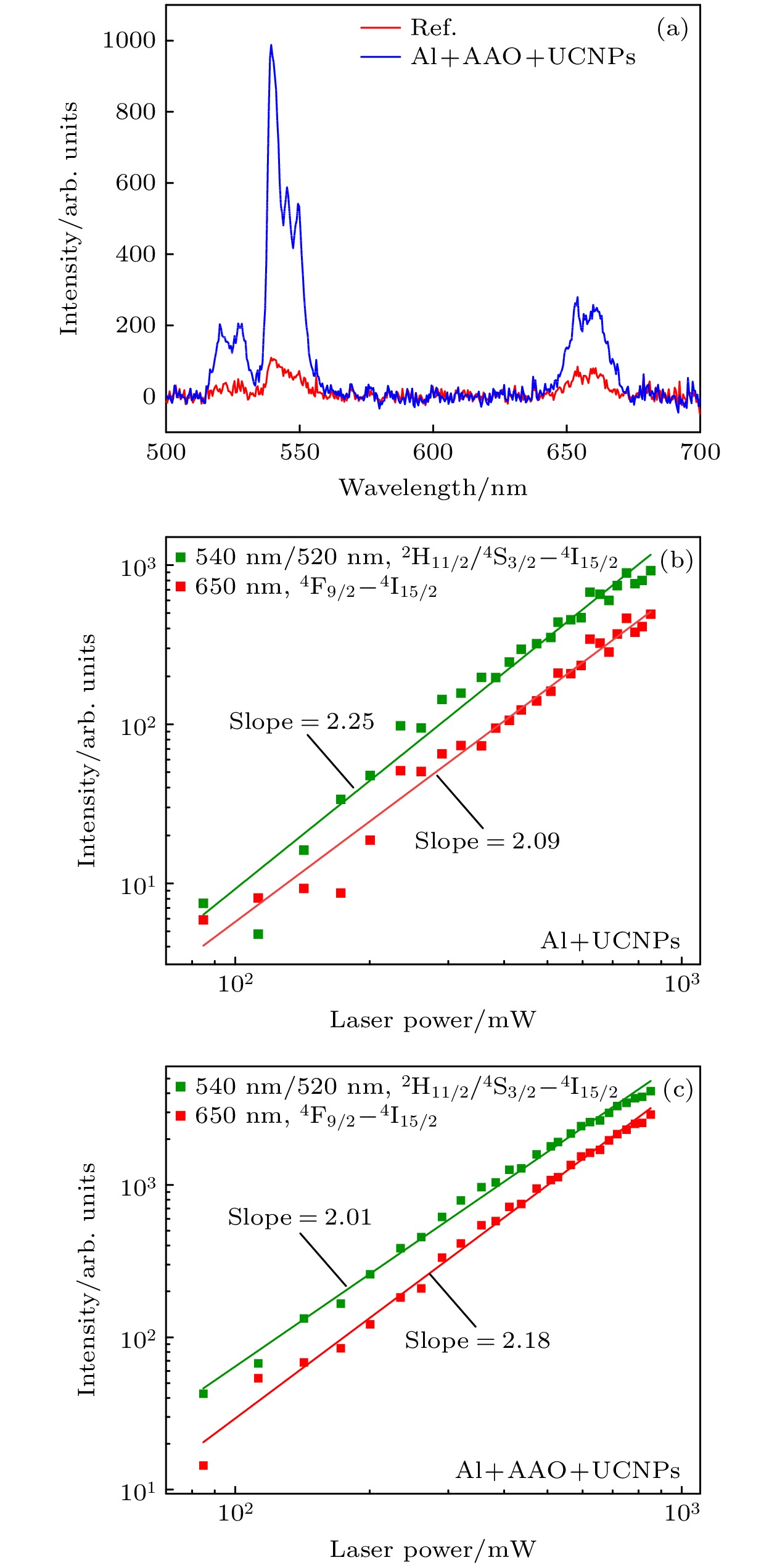
图 2 (a)增强光谱图; (b) NaYF4:Yb3+, Er3+/Al功率密度曲线; (c) NaYF4:Yb3+, Er3+/AAO功率密度曲线
Figure 2. (a) Enhanced spectral diagram; (b) NaYF4:Yb3+, Er3+/Al power density curve; (c) NaYF4:Yb3+, Er3+/AAO power density curve.
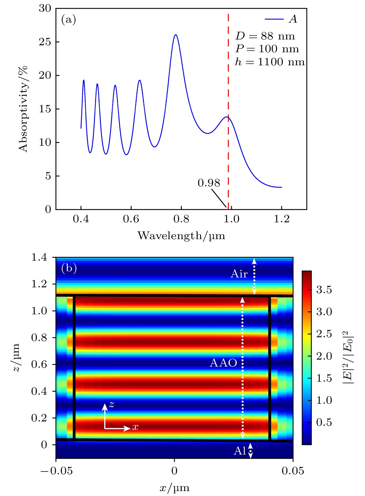
图 3 (a)吸收光谱图; (b)波长980 nm近场增强图像
Figure 3. (a) Absorption spectrogram; (b) near-field enhancement image with wavelength 980 nm.
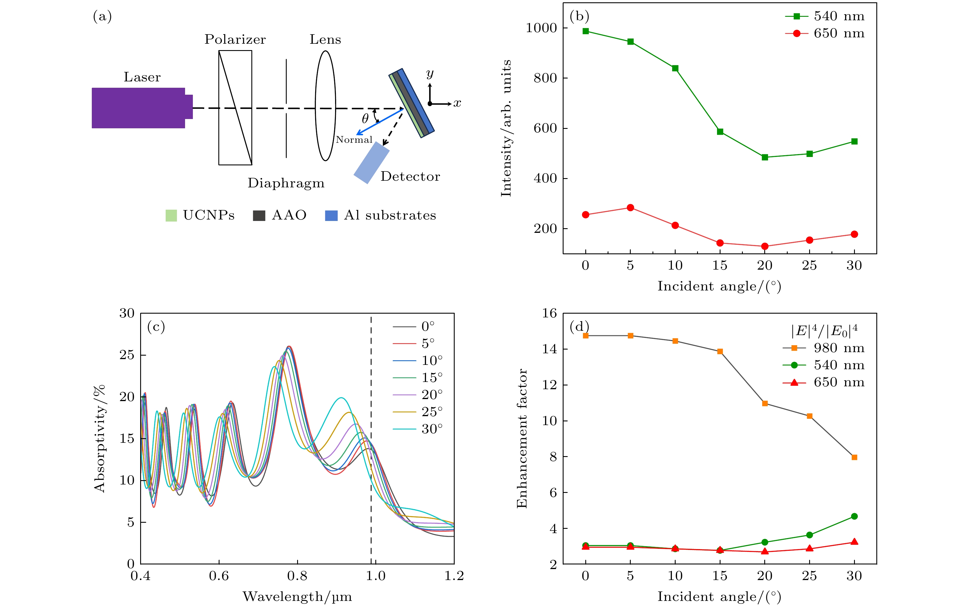
图 4 (a)测试光路示意图; (b) AAO增强NaYF4:Yb3+, Er3+的红绿上转换发光对入射角度的依赖关系; (c) FDTD仿真模拟计算不同入射角下NaYF4:Yb3+, Er3+/AAO吸收曲线; (d) NaYF4:Yb3+, Er3+/AAO的980 nm, 650 nm, 540 nm电场增强因子与入射角度的关系
Figure 4. (a) Test optical path schematic; (b) the dependence of the red-green up-conversion luminescence of NaYF4:Yb3+, Er3+/AAO on the angle of incidence; (c) the absorption curves of NaYF4:Yb3+, Er3+/AAO at different incidence angles calculated by FDTD simulation; (d) 980 nm, 650 nm, 540 nm electric field enhancement factor vs. incidence angle curve of NaYF4:Yb3+, Er3+/AAO.
-
[1] Haase M, Schäfer H 2011 Angew. Chem. Int. Ed. 50 5808
Google Scholar
[2] Shao B, Yang Z W, Li J, Yang J Z, Wang Y D, Qiu J B, Song Z G 2017 Opt. Mater. Express 7 1188
Google Scholar
[3] Zhang J, Shen H O, Guo W, Wang S H, Zhu C T, Xue F, Hou J F, Su H Q, Yuan Z B 2013 J. Power Sources 226 47
Google Scholar
[4] He L, Dragavon J, Cho S, Mao C, Yildirim A, Ma K, Chattaraj R, Goodwin A P, Park W, Cha J N 2016 J. Mater. Chem. B 4 4455
Google Scholar
[5] Kumar A, Tiwari S P, Esteves Da Silva J C G, Kumar K 2018 Laser Phys. Lett. 15 075901
Google Scholar
[6] Janjua R A, Iqbal O, Ahmed M A, Al-Kahtani A A, Saeed S, Imran M, Wattoo A G 2021 RSC Adv. 11 20746
Google Scholar
[7] Chen D, Huang P 2014 Dalton Trans. 43 11299
Google Scholar
[8] Zhu W, Wu Q, Zhao S, Liang Z, Yang Y, Zhang J, Xu Z 2016 Opt. Mater. Express 6 3001
Google Scholar
[9] 高伟, 董军, 王瑞博, 王朝晋, 郑海荣 2016 物理学报 65 084205
Google Scholar
Gao W, Dong J, Wang R B, Wang Z J, Zheng H R 2016 Acta Phys. Sin. 65 084205
Google Scholar
[10] Niu W, Su L T, Chen R, Chen H, Wang Y, Palaniappan A, Sun H, Yoong Tok A I 2014 Nanoscale 6 817
Google Scholar
[11] Xu W, Zhu Y, Chen X, Wang J, Tao L, Xu S, Liu T, Song H 2013 Nano Res. 6 795
Google Scholar
[12] Wang H, Zhan S, Wu X, Wu L, Liu Y 2021 RSC Adv. 11 565
Google Scholar
[13] 高伟, 王博扬, 韩庆艳, 韩珊珊, 程小同, 张晨雪, 孙泽煜, 刘琳, 严学文, 王勇凯, 董军 2020 物理学报 69 184213
Google Scholar
Gao W, Wang B Y, Han Q Y, Han S S, Cheng X T, Zhang C X, Sun Z Y, Liu L, Yan X W, Wang Y K, Dong J 2020 Acta Phys. Sin. 69 184213
Google Scholar
[14] Verhagen E, Kuipers L, Polman A 2009 Opt. Express 17 14586
Google Scholar
[15] Saboktakin M, Ye X, Chettiar U K, Engheta N, Murray C B, Kagan C R 2013 ACS Nano 7 7186
Google Scholar
[16] Chu A, He H, Yin Z, Peng R, Yang H, Gao X, Luo D, Chen R, Xing G, Liu Y J 2020 ACS Appl. Mater. Interfaces 12 1292
Google Scholar
[17] 薛映仙, 戎有英, 马强, 潘诚达, 陈凌霄, 武愕, 吴伯涛 2017 光学学报 37 0724002
Google Scholar
Xue Y X, Rong Y Y, Ma Q, Pan C D, Chen L X, Wu E, Wu B T 2017 Acta Opt. Sin. 37 0724002
Google Scholar
[18] Yin D, Wang C, Ouyang J, Zhang X, Jiao Z, Feng Y, Song K, Liu B, Cao X, Zhang L, Han Y, Wu M 2014 ACS Appl. Mater. Interfaces 6 18480
Google Scholar
[19] Yuan P, Lee Y H, Gnanasammandhan M K, Guan Z, Zhang Y, Xu Q H 2012 Nanoscale 4 5132
Google Scholar
[20] 刘忆森 2012 博士学位论文 (广州: 华南理工大学)
Sen L Y 2012 Ph. D. Dissertation (Guangzhou: South China University of Technology of China
[21] Smith D Y, Shiles E, Inokuti M, Palik E D 1997 Handbook of Optical Constants of Solids (Burlington: Academic Press) pp369–406
[22] Li C, Quan Z, Yang P, Huang S, Lian H, Lin J 2008 J. Phys. Chem. C 112 13395
Google Scholar
[23] Tang H, Xu Y, Cheng X 2020 J. Solid State Chem. 285 121229
Google Scholar
[24] Bhiri N M, Dammak M, Carvajal J J, Aguiló M, Díaz F, Pujol M C 2022 Mater. Res. Bull. 151 111801
Google Scholar
[25] Zhan S, Wu X, Tan C, Xiong J, Hu S, Hu J, Wu S, Gao Y, Liu Y 2018 J. Alloys Compd. 735 372
Google Scholar

 DownLoad:
Catalog
Metrics
- Abstract views: 4442
- PDF Downloads: 95
- Cited By: 0
