


-
Nanoparticles have been widely used in many fields such as nanomedicine and cell imaging. Understanding the microscopic mechanism of the interaction between nanoparticles and biomembranes is very vital for the synthesis and applications of nanoparticles. In this paper, using coarse-grained molecular dynamics simulation, we study the interaction between nanoparticles coated with fully or partially charged ligands and phase-separated biomembranes containing charged lipids. The results show that the final positions or states of nanoparticles on/in the biomembranes can be readily modulated by varying the grafting density, ratio, and type of charged ligands as well as the type of charged lipids. For the nanoparticle with a highly hydrophilic surface, the nanoparticle prefers to be adsorbed on the surface of the biomembrane. In this case, the electrostatic interaction determines that the nanoparticle is adsorbed on the surface of liquid-ordered domain or the surface of liquid-disordered domain. For the nanoparticle with a (partially) hydrophobic surface, the nanoparticle tends to penetrate into the lipid bilayer from the liquid-disordered domain. In this case, the hydrophobicity of the nanoparticle plays a crucial role in the penetrating of the nanoparticle. The hydrophilicity or hydrophobicity of the nanoparticle is affected by the ratio between the charged and neutral ligands, the grafting density of the charged ligands, and the ionic concentration in the system. Furthermore, the microscopic mechanism of the interaction between charged nanoparticles and charged biomembranes is revealed by using the potential of mean force between nanoparticles and lipid domains. The potential of mean force shows that none of the (partially) charged nanoparticles can spontaneously penetrate into the liquid-ordered domain due to a high free energy barrier but they can spontaneously penetrate into the liquid-disordered domain with a certain probability. However, due to the limitation of the simulation time and the number of sampling of the simulations, only some of the partially hydrophobic nanoparticles which are not initially adsorbed onto the surface of liquid-ordered domain are found to finally penetrate into the liquid-disordered domain in this work. This work yields some theoretical insights into the application of nanoparticles in nanomedicine, cell imaging, etc.
-
Keywords:
- biomembrane /
- nanoparticle /
- phase separation /
- molecular simulation
[1] Bogart L K, Pourroy G, Murphy C J, Puntes V, Pellegrino T, Rosenblum D, Peer D, Lévy R 2014 ACS Nano 8 3107
 Google Scholar
[2] Giljohann D A, Seferos D S, Daniel W L, Massich M D, Patel P C, Mirkin C A 2010 Angew. Chem. Int. Ed. 49 3280
Google Scholar
[3] Li N, Zhao P X, Astruc D 2014 Angew. Chem. Int. Ed. 53 1756
Google Scholar
[4] Miller K P, Wang L, Benicewicz B C, Decho A W 2015 Chem. Soc. Rev. 44 7787
Google Scholar
[5] Nicolas J, Mura S, Brambilla D, Mackiewicz N, Couvreur P 2013 Chem. Soc. Rev. 42 1147
Google Scholar
[6] Yang X, Yang M X, Pang B, Vara M, Xia Y N 2015 Chem. Rev. 115 10410
Google Scholar
[7] Blanco E, Shen H P, Ferrari M 2015 Nat. Biotechnol. 33 941
Google Scholar
[8] Bobo D, Robinson K J, Islam J, Thurecht K J, Corrie S R 2016 Pharm. Res. 33 2373
Google Scholar
[9] Ding H M, Ma Y Q 2018 Nanoscale Horiz. 3 6
Google Scholar
[10] Lee B K, Yun Y H, Park K 2015 Chem. Eng. Sci. 125 158
Google Scholar
[11] Sharifi S, Behzadi S, Laurent S, Laird Forrest M, Stroeve P, Mahmoudi M 2012 Chem. Soc. Rev. 41 2323
Google Scholar
[12] Soenen S J, Parak W J, Rejman J, Manshian B 2015 Chem. Rev. 115 2109
Google Scholar
[13] Chang Y Z, He L Z, Li Z B, Zeng L L, Song Z H, Li P H, Chan L, You Y Y, Yu X F, Chu P K, Chen T F 2017 ACS Nano 11 4848
Google Scholar
[14] Docter D, Strieth S, Westmeier D, Hayden O, Gao M Y, Knauer S K, Stauber R H 2015 Nanomedicine 10 503
Google Scholar
[15] Kobayashi K, Wei J J, Iida R 2014 Polym. J. 46 460
Google Scholar
[16] Ong Q, Luo Z, Stellacci F 2017 Acc. Chem. Res. 50 1911
Google Scholar
[17] Bhattacharjee S, Rietjens I M C M, Singh M P, Atkins T M, Purkait T K, Xu Z J, Regli S, Shukaliak A, Clark R J, Mitchell B S, Alink G M, Marcelis A T M, Fink M J, Veinot J G C, Kauzlarich S M, Zuilhof H 2013 Nanoscale 5 4870
Google Scholar
[18] Mout R, Moyano D F, Rana S, Rotello V M 2012 Chem. Soc. Rev. 41 2539
Google Scholar
[19] Saei A A, Yazdani M, Lohse S E, Bakhtiary Z, Serpooshan V, Ghavami M, Asadian M, Mashaghi S, Dreaden E C, Mashaghi A, Mahmoudi M 2017 Chem. Mater. 29 6578
Google Scholar
[20] Deserno M, Bickel T 2003 Europhys. Lett. 62 767
Google Scholar
[21] Mayor S, Pagano R E 2007 Nat. Rev. Mol. Cell Biol. 8 603
Google Scholar
[22] Osaka T, Nakanishi T, Shanmugam S, Takahama S, Zhang H 2009 Colloids. Surf. B 71 325
Google Scholar
[23] Cho E C, Xie J W, Wurm P A, Xia Y N 2009 Nano Lett. 9 1080
Google Scholar
[24] Gao Y C, Terazzi E, Seemann R, Fleury J B, Baulin V A 2016 Sci. Adv. 2 e1600261
Google Scholar
[25] Lu F, Wu S H, Hung Y, Mou C Y 2009 Small 5 1408
Google Scholar
[26] Osaki F, Kanamori T, Sando S, Sera T, Aoyama Y 2004 J. Am. Chem. Soc. 126 6520
Google Scholar
[27] Wittenberg N J, Im H, Johnson T W, Xu X H, Warrington A E, Rodriguez M, Oh S H 2011 ACS Nano 5 7555
Google Scholar
[28] Ding H M, Ma Y Q 2012 Biomaterials 33 5798
Google Scholar
[29] Ding H M, Ma Y Q 2015 Small 11 1055
Google Scholar
[30] Dubavik A, Sezgin E, Lesnyak V, Gaponik N, Schwille P, Eychmüller A 2012 ACS Nano 6 2150
Google Scholar
[31] Ji Q J, Yuan B, Lu X M, Yang K, Ma Y Q 2016 Small 12 1140
Google Scholar
[32] Reynwar B J, Illya G, Harmandaris V A, Müller M M, Kremer K, Deserno M 2007 Nature 447 461
Google Scholar
[33] Vácha R, Martinez V F J, Frenkel D 2012 ACS Nano 6 10598
Google Scholar
[34] Yang K, Ma Y Q 2010 Nat. Nanotechnol. 5 579
Google Scholar
[35] Van Lehn R C, Alexander-Katz A 2015 Soft Matter 11 3165
Google Scholar
[36] Van Lehn R C, Atukorale P U, Carney R P, Yang Y S, Stellacci F, Irvine D J, Alexander-Katz A 2013 Nano Lett. 13 4060
Google Scholar
[37] Van Lehn R C, Ricci M, Silva P H J, Andreozzi P, Reguera J, Voïtchovsky K, Stellacci F, Alexander-Katz A 2014 Nat. Commun. 5 4482
Google Scholar
[38] Lin J Q, Zhang H W, Chen Z, Zheng Y G 2010 ACS Nano 4 5421
Google Scholar
[39] Lin J Q, Zheng Y G, Zhang H W, Chen Z 2011 Langmuir 27 8323
Google Scholar
[40] Ding H M, Li J, Chen N, Hu X J, Yang X F, Guo L J, Li Q, Zuo X L, Wang L H, Ma Y Q, Fan C H 2018 ACS Cent. Sci. 4 1344
Google Scholar
[41] Ding H M, Ma Y Q 2012 Nanoscale 4 1116
Google Scholar
[42] Simonelli F, Bochicchio D, Ferrando R, Rossi G 2015 J. Phys. Chem. Lett. 6 3175
Google Scholar
[43] Baumgart T, Hammond A T, Sengupta P, Hess S T, Holowka D A, Baird B A, Webb W W 2007 Proc. Natl. Acad. Sci. USA 104 3165
Google Scholar
[44] Lloyd D R, Kinzer K E, Tseng H S 1990 J. Membr. Sci. 52 239
Google Scholar
[45] van de Witte P, Dijkstra P J, van den Berg J W A, Feijen J 1996 J. Membr. Sci. 117 1
Google Scholar
[46] Chen X J, Tieleman D P, Liang Q 2018 Nanoscale 10 2481
Google Scholar
[47] Marrink S J, de Vries A H, Mark A E 2004 J. Phys. Chem. B 108 750
Google Scholar
[48] Marrink S J, Tieleman D P 2013 Chem. Soc. Rev. 42 6801
Google Scholar
[49] Ingólfsson H I, Melo M N, van Eerden F J, Arnarez C, Lopez C A, Wassenaar T A, Periole X, de Vries A H, Tieleman D P, Marrink S J 2014 J. Am. Chem. Soc. 136 14554
Google Scholar
[50] Li Z L, Ding H M, Ma Y Q 2013 Soft Matter 9 1281
Google Scholar
[51] Rocha E L D, Caramori G F, Rambo C R 2013 Phys. Chem. Chem. Phys. 15 2282
Google Scholar
[52] Nosé S, Klein M L 1983 Mol. Phys. 50 1055
Google Scholar
[53] Parrinello M, Rahman A 1981 J. Appl. Phys. 52 7182
Google Scholar
[54] Bussi G, Donadio D, Parrinello M 2007 J. Chem. Phys. 126 014101
Google Scholar
[55] Jackson A M, Hu Y, Silva P J, Stellacci F 2006 J. Am. Chem. Soc. 128 11135
Google Scholar
[56] Terrill R H, Postlethwaite T A, Chen C H, Poon C D, Terzis A, Chen A, Hutchison J E, Clark M R, Wignall G 1995 J. Am. Chem. Soc. 117 12537
Google Scholar
[57] Wu J Z, Bratko D, Blanch H W, Prausnitz J M 1999 J. Chem. Phys. 111 7084
Google Scholar
-
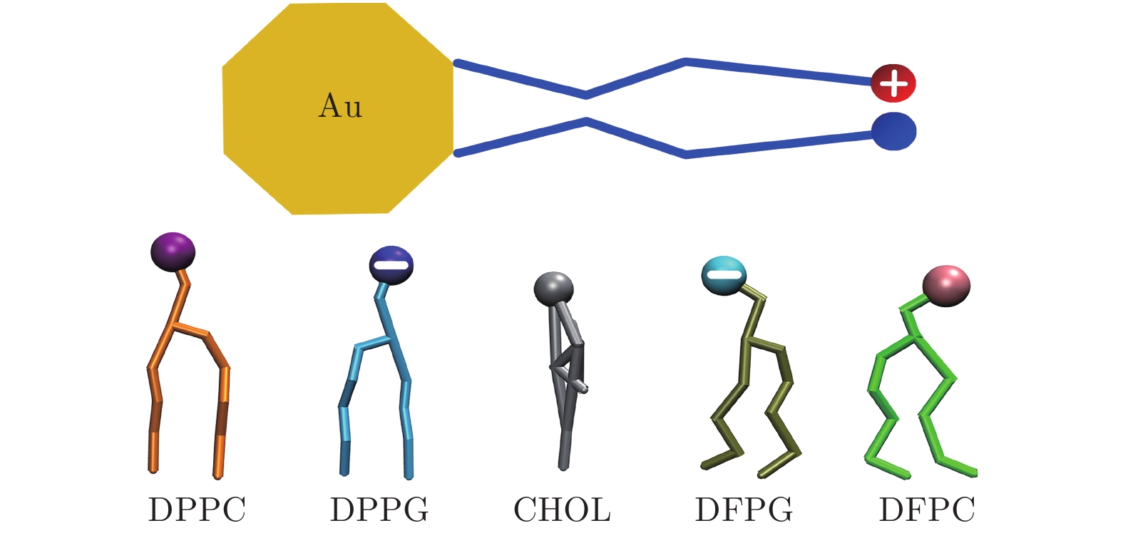
图 1 金纳米颗粒和脂分子的分子结构示意图 其中DPPC, DFPC以及CHOL (胆固醇)为不带电的脂分子, 而DPPG和DFPG为头部带负电的脂分子; 另外DPPC和DPPG为饱和脂分子, 而DFPC和DFPG为不饱和脂分子; 颗粒和各种脂分子的各部分颜色表示通用于全文
Fig. 1. Molecular schematic illustrations of Au nanoparticle and lipids. Here, DPPC, DFPC and cholesterol (CHOL) are electrically neutral, while DPPG or DFPG has a negatively charged headgroup. Additionally, DPPC and DPPG are fully saturated, while DFPC and DFPG are poly-unsaturated. The coloring scheme of the nanoparticle and lipids is used throughout the whole paper.
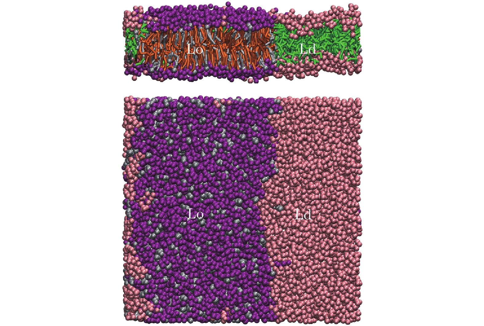
图 2 由DPPC (紫色), DFPC (粉红色)及CHOL (灰色)按4 ∶ 3 ∶ 3摩尔比组成的三组分相分离脂质双层膜的侧视图(上)和俯视图(下). 其中, Lo畴富含DPPC和CHOL, Ld畴富含DFPC
Fig. 2. Phase-separated lipid bilayer composed of DPPC (purple), DFPC (pink) and CHOL (gray) with the molar ratio of 4 ∶ 3 ∶ 3. Here, Lo domain is enriched in DPPC and CHOL, while Ld domain is enriched in DFPC.
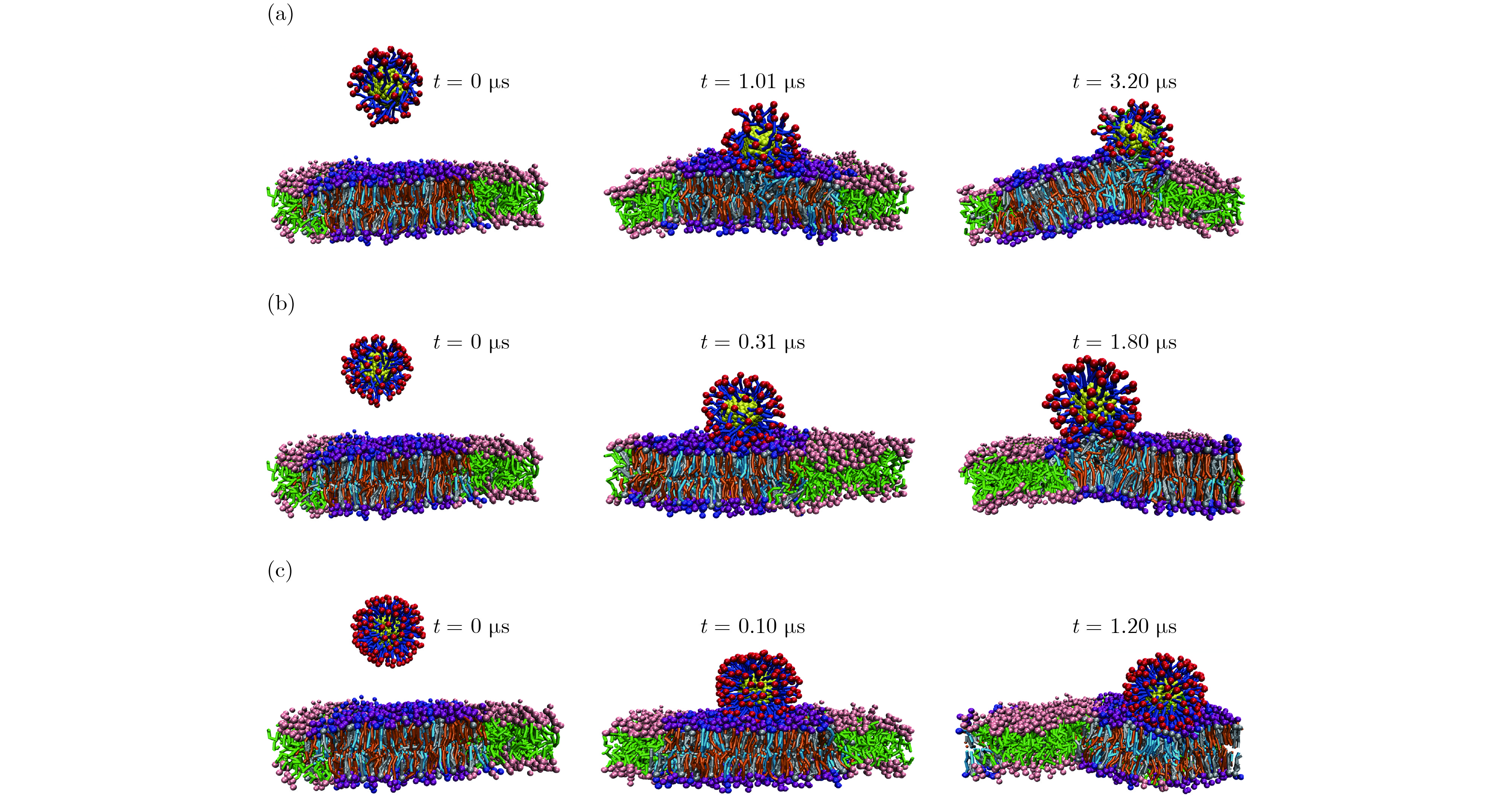
图 3 3种不同带正电的纳米颗粒吸附于由DPPC&DPPG/DFPC/CHOL组成的相分离膜上的动力学过程 (a) Au70/+70; (b) Au104/+104; (c) Au174/+174
Fig. 3. Dynamic processes of adsorption of three different positively charged nanoparticles onto the surface of phase-separated lipid bilayer composed of DPPC & DPPG/DFPC/CHOL: (a) Au70/+70; (b) Au104/+104; (c) Au174/+174.

图 4 3种不同带正电的纳米颗粒吸附于由DPPC/DFPC&DFPG/CHOL组成的相分离膜上的动力学过程 (a) Au70/+70; (b) Au104/+104; (c) Au174/+174
Fig. 4. Dynamic processes of adsorption of three different positively charged nanoparticles into/onto the surface of the phase-separated lipid bilayer composed of DPPC/DFPC&DFPG/CHOL: (a) Au70/+70; (b) Au104/+104; (c) Au174/+174.

图 5 两种带正电的颗粒与带电/中性的Lo/Ld脂质畴之间的PMF曲线 (a) Au70/+70; (b) Au174/+174
Fig. 5. PMF curves of two kinds of positively charged nanoparticles with charged/neutral Lo/Ld lipid domains: (a) Au70/+70; (b) Au174/+174.

图 6 3种不同带负电的纳米颗粒吸附于DPPC&DPPG/DFPC/CHOL (a)—(c) 和DPPC/DFPC&DFPG/CHOL (d)−(f) 组成的相分离的膜表面上在模拟时间为15 μs时的稳定结构
Fig. 6. Final stable structures of adsorption of Au70/−70, Au104/−104, Au174/−174 onto the surface of DPPC&DPPG/DFPC/CHOL (a)−(c) and DPPC/DFPC&DFPG/CHOL (d)−(f) phase-separated lipid bilayers at the simulation time of 15 μs.

图 7 Au70/−70与带电/中性的Lo/Ld脂质畴之间的PMF曲线
Fig. 7. PMF curves of Au70/−70 with charged/neutral Lo/Ld lipid domains.

图 8 两种不同的表面部分带正电的纳米颗粒吸附或嵌入DPPC&DPPG/DFPC/CHOL (a), (c) 和DPPC/DFPC&DFPG/CHOL (b), (d) 组成的相分离的膜表面或内部在模拟时间为15 μs时的稳定结构
Fig. 8. Final stable structures of adsorption/penetration of Au104/+70 and Au174/+70 onto/into the DPPC&DPPG/DFPC/CHOL (a), (c) and DPPC/DFPC&DFPG/CHOL (b), (d) phase-separated lipid bilayers at the simulation time of 15 μs.
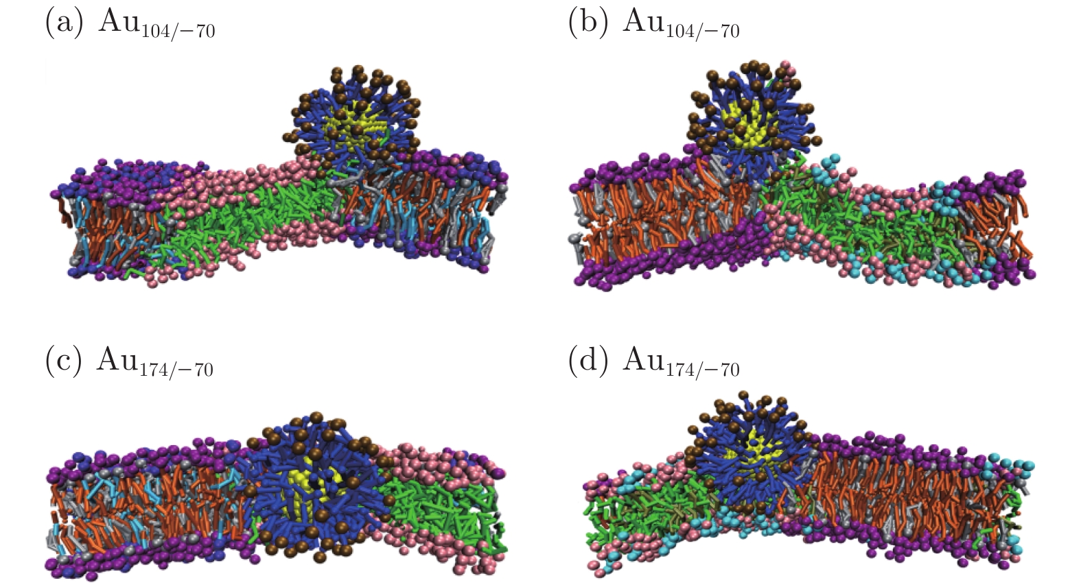
图 9 两种不同的表面部分带负电的纳米颗粒吸附或嵌入DPPC&DPPG/DFPC/CHOL (a), (c) 和DPPC/DFPC&DFPG/CHOL (b), (d) 组成的相分离的膜表面或内部在模拟时间为15 μs时的稳定结构
Fig. 9. Final stable structures of adsorption/penetration of Au104/−70 and Au174/−70 onto/into the DPPC&DPPG/DFPC/CHOL (a), (c) and DPPC/DFPC&DFPG/CHOL (b), (d) phase-separated lipid bilayers at the simulation time of 15 μs.
表 1 本文所研究金纳米颗粒的类型
Table 1. Types of Au nanoparticles in this work.
金纳米颗粒 配体总数 带电配体数 中性配体数 Au70/±70 70 70 0 Au104/±70 104 70 34 Au104/±104 104 104 0 Au174/±70 174 70 104 Au174/±174 174 174 0  下载: 导出CSV
表 2 脂质双层膜及脂质畴的组分
Table 2. Components of lipid bilayers and lipid domains.
脂质双层膜 带电脂 Lo畴组分(带电性) Ld畴组分(带电性) DPPC&DPPG/DFPC/CHOL DPPG DPPC&DPPG/CHOL(带电) DFPC(中性) DPPC/DFPC&DFPG/CHOL DFPG DPPC/CHOL(中性) DFPC&DFPG(带电)
下载: 导出CSV
-
[1] Bogart L K, Pourroy G, Murphy C J, Puntes V, Pellegrino T, Rosenblum D, Peer D, Lévy R 2014 ACS Nano 8 3107
Google Scholar
[2] Giljohann D A, Seferos D S, Daniel W L, Massich M D, Patel P C, Mirkin C A 2010 Angew. Chem. Int. Ed. 49 3280
Google Scholar
[3] Li N, Zhao P X, Astruc D 2014 Angew. Chem. Int. Ed. 53 1756
Google Scholar
[4] Miller K P, Wang L, Benicewicz B C, Decho A W 2015 Chem. Soc. Rev. 44 7787
Google Scholar
[5] Nicolas J, Mura S, Brambilla D, Mackiewicz N, Couvreur P 2013 Chem. Soc. Rev. 42 1147
Google Scholar
[6] Yang X, Yang M X, Pang B, Vara M, Xia Y N 2015 Chem. Rev. 115 10410
Google Scholar
[7] Blanco E, Shen H P, Ferrari M 2015 Nat. Biotechnol. 33 941
Google Scholar
[8] Bobo D, Robinson K J, Islam J, Thurecht K J, Corrie S R 2016 Pharm. Res. 33 2373
Google Scholar
[9] Ding H M, Ma Y Q 2018 Nanoscale Horiz. 3 6
Google Scholar
[10] Lee B K, Yun Y H, Park K 2015 Chem. Eng. Sci. 125 158
Google Scholar
[11] Sharifi S, Behzadi S, Laurent S, Laird Forrest M, Stroeve P, Mahmoudi M 2012 Chem. Soc. Rev. 41 2323
Google Scholar
[12] Soenen S J, Parak W J, Rejman J, Manshian B 2015 Chem. Rev. 115 2109
Google Scholar
[13] Chang Y Z, He L Z, Li Z B, Zeng L L, Song Z H, Li P H, Chan L, You Y Y, Yu X F, Chu P K, Chen T F 2017 ACS Nano 11 4848
Google Scholar
[14] Docter D, Strieth S, Westmeier D, Hayden O, Gao M Y, Knauer S K, Stauber R H 2015 Nanomedicine 10 503
Google Scholar
[15] Kobayashi K, Wei J J, Iida R 2014 Polym. J. 46 460
Google Scholar
[16] Ong Q, Luo Z, Stellacci F 2017 Acc. Chem. Res. 50 1911
Google Scholar
[17] Bhattacharjee S, Rietjens I M C M, Singh M P, Atkins T M, Purkait T K, Xu Z J, Regli S, Shukaliak A, Clark R J, Mitchell B S, Alink G M, Marcelis A T M, Fink M J, Veinot J G C, Kauzlarich S M, Zuilhof H 2013 Nanoscale 5 4870
Google Scholar
[18] Mout R, Moyano D F, Rana S, Rotello V M 2012 Chem. Soc. Rev. 41 2539
Google Scholar
[19] Saei A A, Yazdani M, Lohse S E, Bakhtiary Z, Serpooshan V, Ghavami M, Asadian M, Mashaghi S, Dreaden E C, Mashaghi A, Mahmoudi M 2017 Chem. Mater. 29 6578
Google Scholar
[20] Deserno M, Bickel T 2003 Europhys. Lett. 62 767
Google Scholar
[21] Mayor S, Pagano R E 2007 Nat. Rev. Mol. Cell Biol. 8 603
Google Scholar
[22] Osaka T, Nakanishi T, Shanmugam S, Takahama S, Zhang H 2009 Colloids. Surf. B 71 325
Google Scholar
[23] Cho E C, Xie J W, Wurm P A, Xia Y N 2009 Nano Lett. 9 1080
Google Scholar
[24] Gao Y C, Terazzi E, Seemann R, Fleury J B, Baulin V A 2016 Sci. Adv. 2 e1600261
Google Scholar
[25] Lu F, Wu S H, Hung Y, Mou C Y 2009 Small 5 1408
Google Scholar
[26] Osaki F, Kanamori T, Sando S, Sera T, Aoyama Y 2004 J. Am. Chem. Soc. 126 6520
Google Scholar
[27] Wittenberg N J, Im H, Johnson T W, Xu X H, Warrington A E, Rodriguez M, Oh S H 2011 ACS Nano 5 7555
Google Scholar
[28] Ding H M, Ma Y Q 2012 Biomaterials 33 5798
Google Scholar
[29] Ding H M, Ma Y Q 2015 Small 11 1055
Google Scholar
[30] Dubavik A, Sezgin E, Lesnyak V, Gaponik N, Schwille P, Eychmüller A 2012 ACS Nano 6 2150
Google Scholar
[31] Ji Q J, Yuan B, Lu X M, Yang K, Ma Y Q 2016 Small 12 1140
Google Scholar
[32] Reynwar B J, Illya G, Harmandaris V A, Müller M M, Kremer K, Deserno M 2007 Nature 447 461
Google Scholar
[33] Vácha R, Martinez V F J, Frenkel D 2012 ACS Nano 6 10598
Google Scholar
[34] Yang K, Ma Y Q 2010 Nat. Nanotechnol. 5 579
Google Scholar
[35] Van Lehn R C, Alexander-Katz A 2015 Soft Matter 11 3165
Google Scholar
[36] Van Lehn R C, Atukorale P U, Carney R P, Yang Y S, Stellacci F, Irvine D J, Alexander-Katz A 2013 Nano Lett. 13 4060
Google Scholar
[37] Van Lehn R C, Ricci M, Silva P H J, Andreozzi P, Reguera J, Voïtchovsky K, Stellacci F, Alexander-Katz A 2014 Nat. Commun. 5 4482
Google Scholar
[38] Lin J Q, Zhang H W, Chen Z, Zheng Y G 2010 ACS Nano 4 5421
Google Scholar
[39] Lin J Q, Zheng Y G, Zhang H W, Chen Z 2011 Langmuir 27 8323
Google Scholar
[40] Ding H M, Li J, Chen N, Hu X J, Yang X F, Guo L J, Li Q, Zuo X L, Wang L H, Ma Y Q, Fan C H 2018 ACS Cent. Sci. 4 1344
Google Scholar
[41] Ding H M, Ma Y Q 2012 Nanoscale 4 1116
Google Scholar
[42] Simonelli F, Bochicchio D, Ferrando R, Rossi G 2015 J. Phys. Chem. Lett. 6 3175
Google Scholar
[43] Baumgart T, Hammond A T, Sengupta P, Hess S T, Holowka D A, Baird B A, Webb W W 2007 Proc. Natl. Acad. Sci. USA 104 3165
Google Scholar
[44] Lloyd D R, Kinzer K E, Tseng H S 1990 J. Membr. Sci. 52 239
Google Scholar
[45] van de Witte P, Dijkstra P J, van den Berg J W A, Feijen J 1996 J. Membr. Sci. 117 1
Google Scholar
[46] Chen X J, Tieleman D P, Liang Q 2018 Nanoscale 10 2481
Google Scholar
[47] Marrink S J, de Vries A H, Mark A E 2004 J. Phys. Chem. B 108 750
Google Scholar
[48] Marrink S J, Tieleman D P 2013 Chem. Soc. Rev. 42 6801
Google Scholar
[49] Ingólfsson H I, Melo M N, van Eerden F J, Arnarez C, Lopez C A, Wassenaar T A, Periole X, de Vries A H, Tieleman D P, Marrink S J 2014 J. Am. Chem. Soc. 136 14554
Google Scholar
[50] Li Z L, Ding H M, Ma Y Q 2013 Soft Matter 9 1281
Google Scholar
[51] Rocha E L D, Caramori G F, Rambo C R 2013 Phys. Chem. Chem. Phys. 15 2282
Google Scholar
[52] Nosé S, Klein M L 1983 Mol. Phys. 50 1055
Google Scholar
[53] Parrinello M, Rahman A 1981 J. Appl. Phys. 52 7182
Google Scholar
[54] Bussi G, Donadio D, Parrinello M 2007 J. Chem. Phys. 126 014101
Google Scholar
[55] Jackson A M, Hu Y, Silva P J, Stellacci F 2006 J. Am. Chem. Soc. 128 11135
Google Scholar
[56] Terrill R H, Postlethwaite T A, Chen C H, Poon C D, Terzis A, Chen A, Hutchison J E, Clark M R, Wignall G 1995 J. Am. Chem. Soc. 117 12537
Google Scholar
[57] Wu J Z, Bratko D, Blanch H W, Prausnitz J M 1999 J. Chem. Phys. 111 7084
Google Scholar

 下载:
下载:
计量
- 文章访问数: 18495
- PDF下载量: 132
- 被引次数: 0
