


-
生物阻抗谱是一种非侵入式、免标记、能够定量分析的检测技术, 将其应用于生物细胞及组织的生理、病理分析中具有很大优势. 本文采用数值仿真的方法研究了单细胞电学特性与其结构之间的关系, 并通过实验进行了验证. 根据细胞的生理特征, 依据细胞的双壳模型与单壳模型理论分别建立了不同种类细胞的电学模型, 研究了细胞种类、细胞膜、细胞核对细胞电学特性的影响. 数值分析结果表明: 1)细胞结构尺寸的变化引起细胞电学特性的改变, 因此, 依据细胞电学特性能够准确实现细胞分类; 2)柯尔-柯尔(Cole-Cole)图上高频与低频的两个半圆弧分别是由细胞质或细胞外液的离子极化、细胞膜与细胞外液之间的界面极化引起的; 3)细胞核大小对测量阻抗的影响主要在低频段, 是由细胞核与细胞内液的界面极化引起的, 当存在细胞膜且当细胞核的核质比小于0.25时可忽略其影响. 为验证仿真结果, 对20%不同活性的酵母菌进行了实验. 实验结果表明, 运用本文建立的细胞电学模型, 可以准确检测细胞的不同活性. 该方法对实现细胞的精准电阻抗检测提供了理论依据, 具有重要的应用价值.Bioelectrical impedance spectroscopy is a noninvasive, label-free and quantitative detection technology, which has great advantages in the physiological and pathological analysis of biological cells and tissues. In this paper, the relationship between the electrical properties of a single cell and its structure is studied by numerical simulation. Moreover, experiments are conducted to verify the simulation results. For simulation, three single cell models are used to express its structure. Among of the three models, No Shell Model (NS) is proposed in this paper to study the influence of cell membrane on bioelectrical impedance spectroscopy. In addition, the effects of cell type, cell membrane and cell nucleus on its electrical properties are studied by simulation based on Single Shell Model (SS) and Two Shell Model (TS). The simulation results show that: 1) the electrical characteristics of cells can reflect its structure, therefore, the cell type can be accurately distinguished by its electrical characteristics; 2) the high frequency part of the Cole-Cole Plot is caused by ionic polarization of cytoplasm or extracellular fluid, and the low frequency part of the Cole-Cole Plot is caused by interface polarization between cell membrane and the extracellular fluid; 3) the influence of cell nucleus size on impedance measurement is mainly in the low frequency range, which is caused by the polarization of the interface between cell nucleus and intracellular fluid, and when the nucleocytoplasmic ratio is less than 0.25, the effect of nuclear size on impedance analysis could be ignored. Finally, an experiment was conducted on 20% yeasts suspension with different activity to verify the simulation results. It is known that the cell membranes of dead yeasts are destroyed, however, living yeasts have completed cell structures. The structure difference between living and dead yeast is distinguished by electrical impedance spectroscopy through numerical simulation. The experimental results are consistent with the simulation results, which verifies the fact that the high frequency part of the Cole-Cole Plot is caused by ionic polarization of cytoplasm or extracellular fluid, and the low frequency part of the Cole-Cole Plot is caused by interface polarization between cell membrane and the extracellular fluid.
-
Keywords:
- electrical impedance spectroscopy /
- cell model /
- numerical simulation /
- electrical characteristics
[1] Pierga J Y, Bonneton C, Vincent-Salomon A, Cremoux P D, Magdelénat H 2004 Clin. Cancer Res. 10 1392
 Google Scholar
[2] Haba R, Miki H, Kobayashi S, Ohmori M 1993 Cancer 72 3258
Google Scholar
[3] Kern D H, Drogemuller C R, Kennedy M C, Hildebrand-Zanki S U, Sondak V K 1985 Cancer Res. 45 5436
[4] Bera T K, Nagaraju J, Lubineau G 2016 J. Visualization 19 691
Google Scholar
[5] 郭各朴, 宿慧丹, 丁鹤平, 马青玉 2017 物理学报 66 164301
Google Scholar
Guo G P, Su H D, Ding H P, Ma Q Y 2017 Acta Phys. Sin. 66 164301
Google Scholar
[6] Xu Y, Xie X, Duan Y, Wang L, Cheng Z, Cheng J 2016 Biosens. Bioelectron. 77 824
Google Scholar
[7] Heileman K, Daoud J, Tabrizian M 2013 Biosens. Bioelectron. 49 348
Google Scholar
[8] Hodgkin A L, Huxley A F 1989 Bull. Math. Biol. 52 25
[9] Kanai H, Sakamoto K, Haeno M 1983 J. Microwave Power 18 233
Google Scholar
[10] 方云, 汤治元, 张倩, 赵鑫, 马青 2014 生物医学工程学杂志 5 1070
Google Scholar
Fang Y, Tang Z Y, Zhang Q, Zhao X, Ma Q 2014 J. Biomed. Eng. 5 1070
Google Scholar
[11] 王威, 董秀珍, 付峰, 刘蒙, 杨鹏飞, 史学涛, 刘锐岗 2010 医疗卫生装备 31 20
Google Scholar
Wang W, Dong X Z, Fu F, Liu M, Yang P F, Shi X T 2010 Chin. Med. Equip. J. 31 20
Google Scholar
[12] Amin N, Rayhan S, Anik A A, Jameel R 2016 Proceeding of the Second International Conference on Research in Computational Intelligence and Communication Networks Kolkata, India, September 23–25, 2016 p147
[13] Sun T, Bernabini C, Morgan H 2010 Langmuir 26 3821
Google Scholar
[14] Guo X, Zhu R, Zong X 2015 Analyst 140 156
Google Scholar
[15] Zhu Z, Xu X, Lei F, Pan D, Huang Q A 2016 Sens. Actuators, 235 515
Google Scholar
[16] Asami K, Takahashi Y, Takashima S 1989 Biochim. Biophys. Acta 1010 49
Google Scholar
[17] Irimajiri A, Doida Y, Hanai T, Inouye A 1978 J. Membr. Biol. 38 209
Google Scholar
[18] Hanai T, Asami K, Koizumi N 2005 Phys. Rev. Lett. 57 297
[19] Joshi R P, Hu Q, Schoenbach K H 2004 IEEE Trans. Plasma Sci. 32 1677
Google Scholar
[20] Woolley A T, Hadley D, Landre P, de Mello A J, Mathies R A, Northrup M A 1996 Anal. Chem. 68 4081
Google Scholar
[21] 姚佳烽, 姜祝鹏, 赵桐, 王昊, 陈柏, 吴洪涛 2019 分析化学 47 62
Yao J F, Jiang Z P, Zhao T, Wang H, Chen B, Wu H T 2019 Anal. Chem. 47 62
[22] Asami K 2002 J. Non-Cryst. Solids 305 0
-

图 1 仿真模型 (a)−(c)细胞模型; (d), (e)仿真区域模型
Fig. 1. Simulation model (a)−(c) Model of cell; (d), (e) model of simulation area.
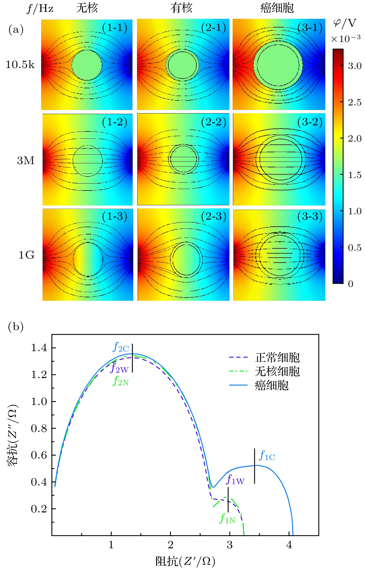
图 2 不同细胞的仿真结果及Cole-Cole Plot (a)不同细胞在不同频率下的仿真结果; (b)不同细胞的Cole-Cole Plot
Fig. 2. Simulation results of different cells and Cole-Cole Plot: (a) Simulation results of different frequencies of different cells; (b) Cole-Cole Plot of different cells.
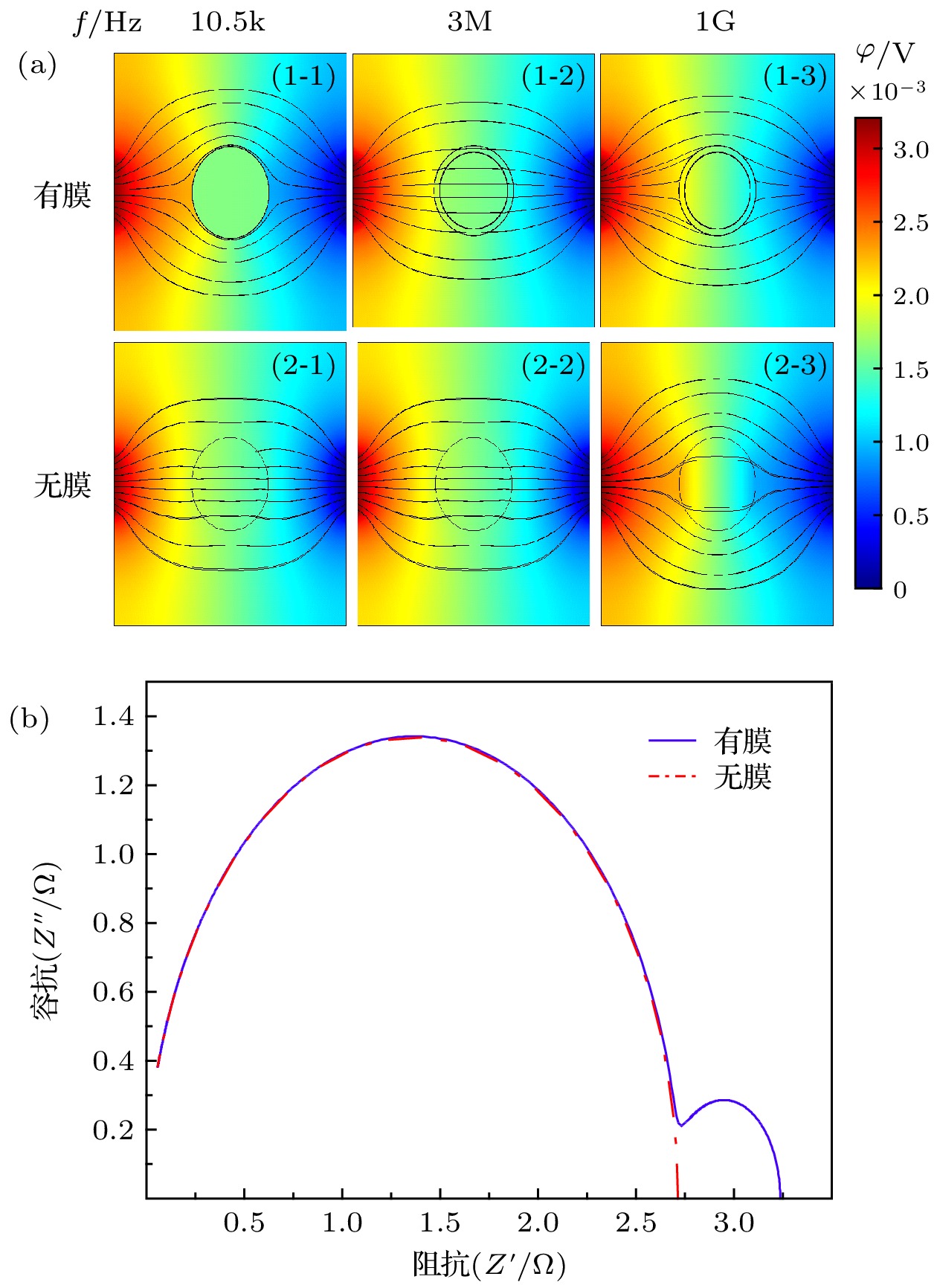
图 3 无核细胞在有无膜下的仿真结果 (a)不同频率下无核细胞的仿真结果; (b)仿真结果对应的Cole-Cole Plot
Fig. 3. Simulation results of no-nuclear cells with or without membrane: (a) Simulation results of non-nucleated cells with different frequencies; (b) the Cole-Cole Plot corresponding to the simulation results.
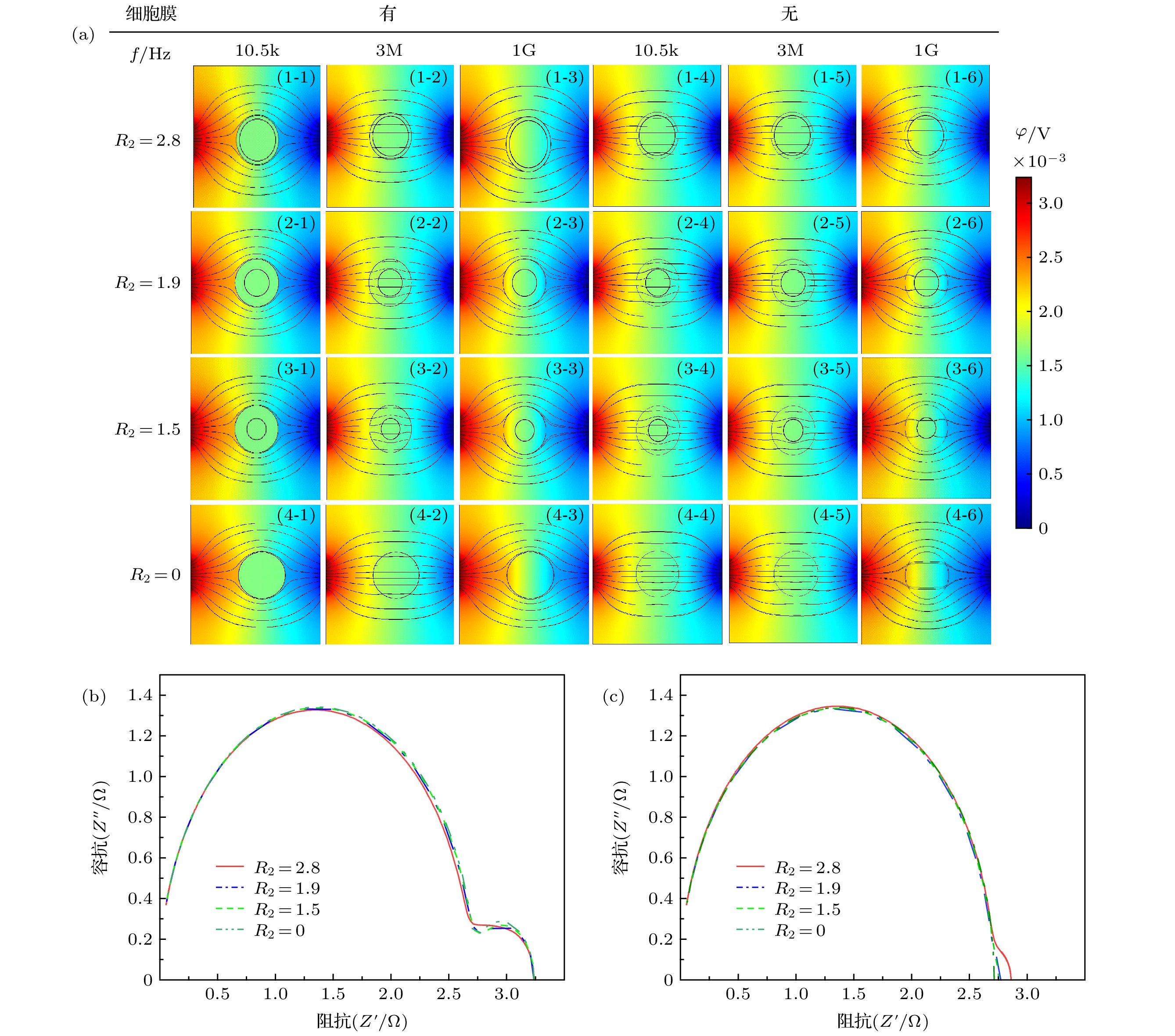
图 4 不同核径细胞的仿真结果 (a)不同频率下的仿真结果; (b), (c)相应的Cole-Cole图
Fig. 4. Simulation results of cells with different nuclear radius: (a) Simulation results of cells at different frequencies; (b), (c) Cole-Cole Plot of different nuclear radius.
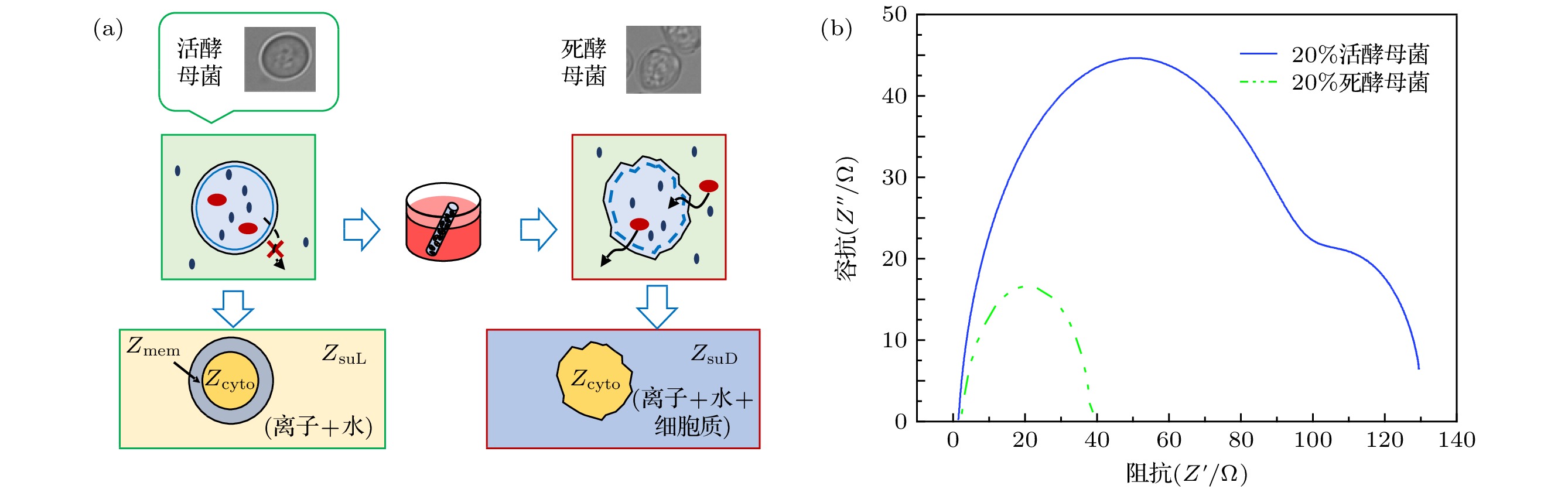
图 6 实验图 (a)死活酵母菌实物与模型图; (b)20%死活酵母菌Cole-Cole图
Fig. 6. Experiment: (a) Object and model of dead and alive yeast; (b) Cole-Cole Plot of 20% dead and alive yeast.
表 1 正常B细胞及病变B细胞的仿真参数
Table 1. Simulation parameters of normal and malignant (farage) onsillar B-cells[19].
参数 正常细胞 癌细胞 导电率σ/S·m–1 环境 0.6 0.6 细胞膜 5.6 × 10⁻⁵ 9.1 × 10⁻⁶ 细胞质 1.31 0.48 核膜 1.11 × 10⁻² 4.4 × 10⁻³ 核质 2.04 1.07 相对介电常数 环境 80 80 细胞膜 12.8 9.8 细胞质 60 60 核膜 106 60.3 核质 120 120 几何参数/μm 仿真区域(L × L) 20 × 20 20 × 20 电极$ (l) $ 2 × 4 2 × 4 细胞半径(R1) 3.3 5.2 细胞核半径(R2) 2.8 4.4 细胞膜厚(d1) 0.007 0.007 核膜厚(d2) 0.04 0.04  下载: 导出CSV
-
[1] Pierga J Y, Bonneton C, Vincent-Salomon A, Cremoux P D, Magdelénat H 2004 Clin. Cancer Res. 10 1392
Google Scholar
[2] Haba R, Miki H, Kobayashi S, Ohmori M 1993 Cancer 72 3258
Google Scholar
[3] Kern D H, Drogemuller C R, Kennedy M C, Hildebrand-Zanki S U, Sondak V K 1985 Cancer Res. 45 5436
[4] Bera T K, Nagaraju J, Lubineau G 2016 J. Visualization 19 691
Google Scholar
[5] 郭各朴, 宿慧丹, 丁鹤平, 马青玉 2017 物理学报 66 164301
Google Scholar
Guo G P, Su H D, Ding H P, Ma Q Y 2017 Acta Phys. Sin. 66 164301
Google Scholar
[6] Xu Y, Xie X, Duan Y, Wang L, Cheng Z, Cheng J 2016 Biosens. Bioelectron. 77 824
Google Scholar
[7] Heileman K, Daoud J, Tabrizian M 2013 Biosens. Bioelectron. 49 348
Google Scholar
[8] Hodgkin A L, Huxley A F 1989 Bull. Math. Biol. 52 25
[9] Kanai H, Sakamoto K, Haeno M 1983 J. Microwave Power 18 233
Google Scholar
[10] 方云, 汤治元, 张倩, 赵鑫, 马青 2014 生物医学工程学杂志 5 1070
Google Scholar
Fang Y, Tang Z Y, Zhang Q, Zhao X, Ma Q 2014 J. Biomed. Eng. 5 1070
Google Scholar
[11] 王威, 董秀珍, 付峰, 刘蒙, 杨鹏飞, 史学涛, 刘锐岗 2010 医疗卫生装备 31 20
Google Scholar
Wang W, Dong X Z, Fu F, Liu M, Yang P F, Shi X T 2010 Chin. Med. Equip. J. 31 20
Google Scholar
[12] Amin N, Rayhan S, Anik A A, Jameel R 2016 Proceeding of the Second International Conference on Research in Computational Intelligence and Communication Networks Kolkata, India, September 23–25, 2016 p147
[13] Sun T, Bernabini C, Morgan H 2010 Langmuir 26 3821
Google Scholar
[14] Guo X, Zhu R, Zong X 2015 Analyst 140 156
Google Scholar
[15] Zhu Z, Xu X, Lei F, Pan D, Huang Q A 2016 Sens. Actuators, 235 515
Google Scholar
[16] Asami K, Takahashi Y, Takashima S 1989 Biochim. Biophys. Acta 1010 49
Google Scholar
[17] Irimajiri A, Doida Y, Hanai T, Inouye A 1978 J. Membr. Biol. 38 209
Google Scholar
[18] Hanai T, Asami K, Koizumi N 2005 Phys. Rev. Lett. 57 297
[19] Joshi R P, Hu Q, Schoenbach K H 2004 IEEE Trans. Plasma Sci. 32 1677
Google Scholar
[20] Woolley A T, Hadley D, Landre P, de Mello A J, Mathies R A, Northrup M A 1996 Anal. Chem. 68 4081
Google Scholar
[21] 姚佳烽, 姜祝鹏, 赵桐, 王昊, 陈柏, 吴洪涛 2019 分析化学 47 62
Yao J F, Jiang Z P, Zhao T, Wang H, Chen B, Wu H T 2019 Anal. Chem. 47 62
[22] Asami K 2002 J. Non-Cryst. Solids 305 0

 下载:
下载:

计量
- 文章访问数: 17272
- PDF下载量: 355
- 被引次数: 0
