


-
超声/光声双模态成像技术因其同时兼具超声的高分辨率结构成像和光声的高对比度功能成像优势, 极大地推动了光声成像技术的临床应用推广. 传统超声/光声双模态成像技术多基于超声成像所用阵列探头同时收集光声信号, 系统结构紧凑且无需图像配准, 操作便捷. 但该类设备使用阵列探头和多通道数据采集, 使得其成本较高; 且成像结果易受通道一致性差异影响. 本文提出了一种基于声学扫描振镜的超声/光声双模态成像技术, 该技术采用单个超声换能器结合一维声学扫描振镜进行快速声束扫描, 实现超声/光声双模态成像, 是一种小型化、低成本的双模态快速成像技术. 本文开展了系列仿体和活体成像研究, 实验结果表明: 系统有效成像范围为15.6 mm, 超声和光声成像B扫描速度分别为1.0 s–1和0.1 s–1 (光声成像速度主要受制于脉冲激光器重复频率). 基于本文所提技术研究, 有助于进一步推动超声/光声双模态成像技术的临床转化和普及; 也为基于超声信号检测的多模态成像技术提供了一种低成本、小型化和快速声信号检测的参考方案.
-
关键词:
- 超声/光声双模态成像 /
- 声学扫描振镜 /
- 低成本 /
- 小型化
Ultrasound/photoacoustic dual-modality imaging technology has greatly promoted the clinical application and photoacoustic imaging technology because it integrates the advantages of high-resolution structural imaging of ultrasound and high-contrast functional imaging of photoacoustic imaging. Traditional ultrasound/photoacoustic dual-modality imaging is based mainly on the array probe used in ultrasound imaging to collect photoacoustic signals at the same time. The system has a compact structure and easy operation. However, this kind of equipment utilizes array probes and multi-channel data acquisition system, which makes it expensive. And the imaging quality can be affected by the difference in channel consistency. In this paper, an ultrasound/photoacoustic dual-modality imaging method based on an acoustic scanning galvanometer is proposed. In this system, a single ultrasonic transducer combined with a one-dimensional acoustic scanning galvanometer is used for fast acoustic beam scanning to realize ultrasound/photoacoustic dual-modality imaging. It is a compact, low-cost and fast dual-modality imaging technology. The experimental results show that the effective imaging range of the system is 15.6 mm, and the temporal resolution of ultrasound and photoacoustic imaging are 1.0 and 0.1 s–1 (B scan), respectively (the temporal resolution of photoacoustic imaging is limited mainly by the laser repetition rate). Based on the proposed technology research, it is helpful to further promote the clinical transformation and popularization of ultrasound/photoacoustic dual-modality imaging. It also provides a low-cost, miniaturized and fast scheme for multimodal imaging technology which is based on ultrasound signal detection.-
Keywords:
- ultrasound/photoacoustic dual-modality imaging /
- acoustic scanning galvanometer /
- low cost /
- miniaturization
[1] 林日强, 冷吉, 陈敬钦, 刘成波, 龚小竞, 宋亮 2018 中国医疗设备 33 1
 Google Scholar
Lin R Q, Leng J, Chen J Q, Liu C B, Gong L J, Song L 2018 Chin. Med. Dev. 33 1
Google Scholar
[2] Beard P 2011 Interface Focus 1 602
Google Scholar
[3] 黄豆豆, 邱棋, 林文珍, 刘基嫣, 黄雅丽, 赵庆亮 2019 光散射学报 31 1
Google Scholar
Huang D D, Qiu Q, Lin W Z, Liu J Y, Huang Y L, Zhao Q L 2019 Chin. J. Light. Scatt. 31 1
Google Scholar
[4] Xu G, Rajian J R, Girish G, Kaplan M J, Fowlkes J B, Carson P L, Wang X D 2012 J. Biomed. Opt. 18 10502
Google Scholar
[5] Berg P J, Bansal R, Daoudi K, Steenbergen W, Prakash J 2016 Biomed. Opt. Express 7 5081
Google Scholar
[6] Garcia-Uribe A, Erpelding T N, Krumholz A, Ke H, Maslov K, Appleton C, Margenthaler J A, Wang L V 2015 Sci. Rep. 5 15748
Google Scholar
[7] Florian R, Julien S, Marilyne L M, Stéphanie R, Sharuja N, Stéphanie L, Alain L P 2016 Plos. One 11 4
Google Scholar
[8] Yang M, Zhao L, He X, Su N, Zhao C, Tang H, Hong T, Li W, Yang F, Lin L, Zhang B, Zhang R, Jiang Y, Li C 2017 Biomed. Opt. Express 8 3449
Google Scholar
[9] Li X, Wang D, Ran H, Hao L, Cao Y, Ao M, Zhang N, Song J, Zhang L, Yi H, Wang Z, Li P 2018 Biochem. Biophys. Res. Commun. 502 255
Google Scholar
[10] Mallidi S, Watanabe K, Timerman D, Schoenfeld D, Hasan T 2015 Theranostics 5 289
Google Scholar
[11] Qian M, Du Y, Wang S, Li C, Jiang H, Shi W, Chen J, Wang Y, Wagner E, Huang R 2018 ACS Appl. Mater. Inter. 10 4031
Google Scholar
[12] Diot G, Metz S, Noske A, Liapis E, Schroeder B, Ovsepian S V, Meier R, Rummeny E, Ntziachristos V 2017 Clin. Cancer Res. 23 6912
Google Scholar
[13] Toi M, Asao Y, Matsumoto Y, Sekiguchi H, Yoshikawa A, Takada M, Kataoka M, Endo T, Kawaguchi-Sakita N, Kawashima M, Fakhrejahani E, Kanao S, Yamaga I, Nakayama Y, Tokiwa M, Torii M, Yagi T, Sakurai T, Togashi K, Shiina T 2017 Sci. Rep. 7 41970
Google Scholar
[14] Yang J-M, Favazza C, Chen R, Yao J, Cai X, Maslov K, Zhou Q, Shung K K, Wang L V 2012 Nat. Med. 18 1297
Google Scholar
[15] Li Y, Lin R, Liu C, Chen J, Liu H, Zheng R, Gong X, Song L 2018 J. Biophotonics 11 e201800034
Google Scholar
[16] Oeri M, Bost W, Senegond N, Tretbar S, Fournelle M 2017 Ultrasound Med. Biol. 43 2200
Google Scholar
[17] Daoudi K, van den Berg P J, Rabot O, Kohl A, Tisserand S, Brands P, Steenbergen W 2014 Opt. Express 22 26365
Google Scholar
[18] 谢实梦, 黄林, 王雪, 迟子惠, 汤永辉, 郑铸, 蒋华北 2021 物理学报 70 100701
Google Scholar
Xie S M, Huang L, Wang L, Chi Z H, Tang Y H, Zheng Z, Jiang H B 2021 Acta Phys. Sin. 70 100701
Google Scholar
[19] Choi S, Kim J Y, Lim H G, Baik J W, Kim H H, Kim C 2020 Sci. Rep. 10 6544
Google Scholar
[20] 齐伟智 2018 博士学位论文 (成都: 电子科技大学)
Qi W Z 2018 Ph. D. Dissertation (Chengdu: University of Electronic Science and Technology of China) (in Chinese)
[21] Xi L, Sun J, Zhu Y, Wu L, Xie H, Jiang H 2010 Biomed. Opt. Express 1 1278
Google Scholar
[22] Lee C, Kim J Y, Kim C 2018 Micromachines 9 584
Google Scholar
[23] Yao J, Huang C H, Wang L, Yang J M, Gao L, Maslov K I, Zou J, Wang L V 2012 J. Biomed. Opt. 17 080505
Google Scholar
[24] Kim J Y, Lee C, Park K, Lim G, Kim C 2015 Sci. Rep. 5 7932
Google Scholar
[25] Kim S, Lee C, Kim J Y, Kim J, Lim G, Kim C 2016 J. Biomed. Opt. 21 106001
Google Scholar
[26] Kim J Y, Lee C, Lim G, Kim C 2016 Photons Plus Ultrasound: Imaging and Sensing San Francisco, 15 March 2016, p970835
[27] Pu Y, Bi R, Ahn J, Kim J Y, Kim C, Olivo M, Zhao X J 2019 Photons Plus Ultrasound: Imaging and Sensing San Francisco, 27 February 2019, p1087843
[28] Park K, Kim J Y, Lee C, Jeon S, Lim G, Kim C 2017 Sci. Rep. 7 13359
Google Scholar
[29] American Laser Institute. American National Standards for the Safe Use of Lasers ANSIZ136.1. Orlando, FL: American Laser Institute, 2014
[30] Wang L V, Hu S 2012 Science 335 1458
Google Scholar
[31] 汤永辉, 郑铸, 谢实梦, 黄林, 蒋华北 2020 物理学报 69 240701
Google Scholar
Tang Y H, Zheng Z, Xie S M, Huang L, Jiang H B 2020 Acta Phys. Sin. 69 240701
Google Scholar
-
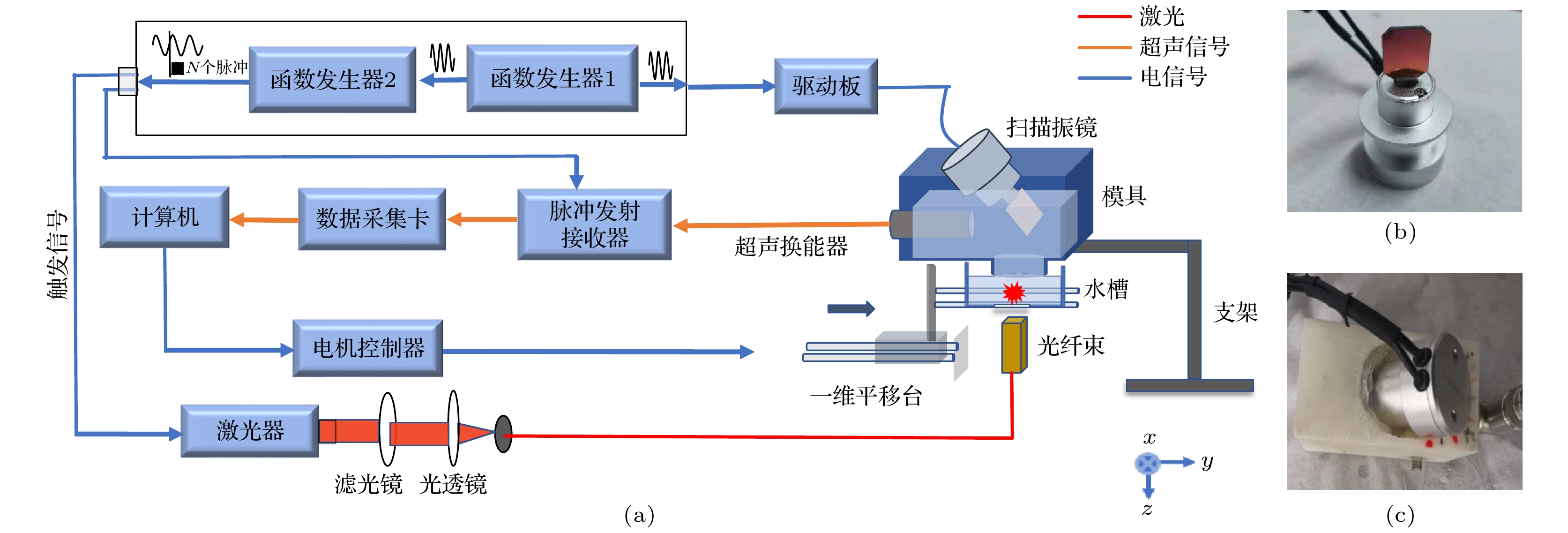
图 1 (a) 超声/光声双模态成像系统框图; (b) 一维声学扫描振镜实物图; (c) 模具实物图
Fig. 1. (a) Schematic of the ultrasound/photoacoustic dual-modality imaging system; (b) photograph of the acoustic scanning galvanometer; (c) picture of the mould.
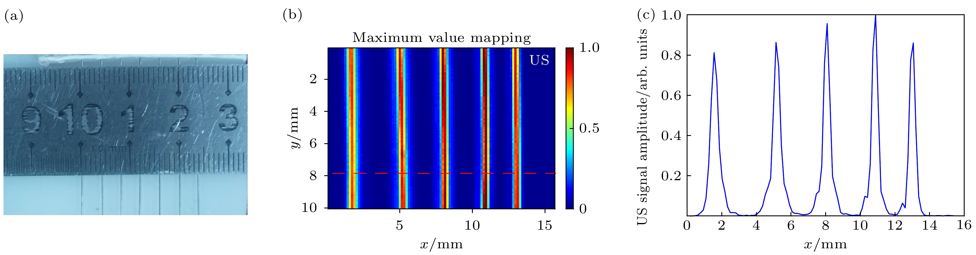
图 2 分辨率和成像区域实验 (a)银针实物图; (b)超声实验结果; (c)沿图(b)红色虚线的一维信号轮廓图像
Fig. 2. Resolution and imaging region experiments: (a) Photograph of the silver needles; (b) ultrasound (US) results; (c) one dimensional signal profile along the red dashed line shown in (b).
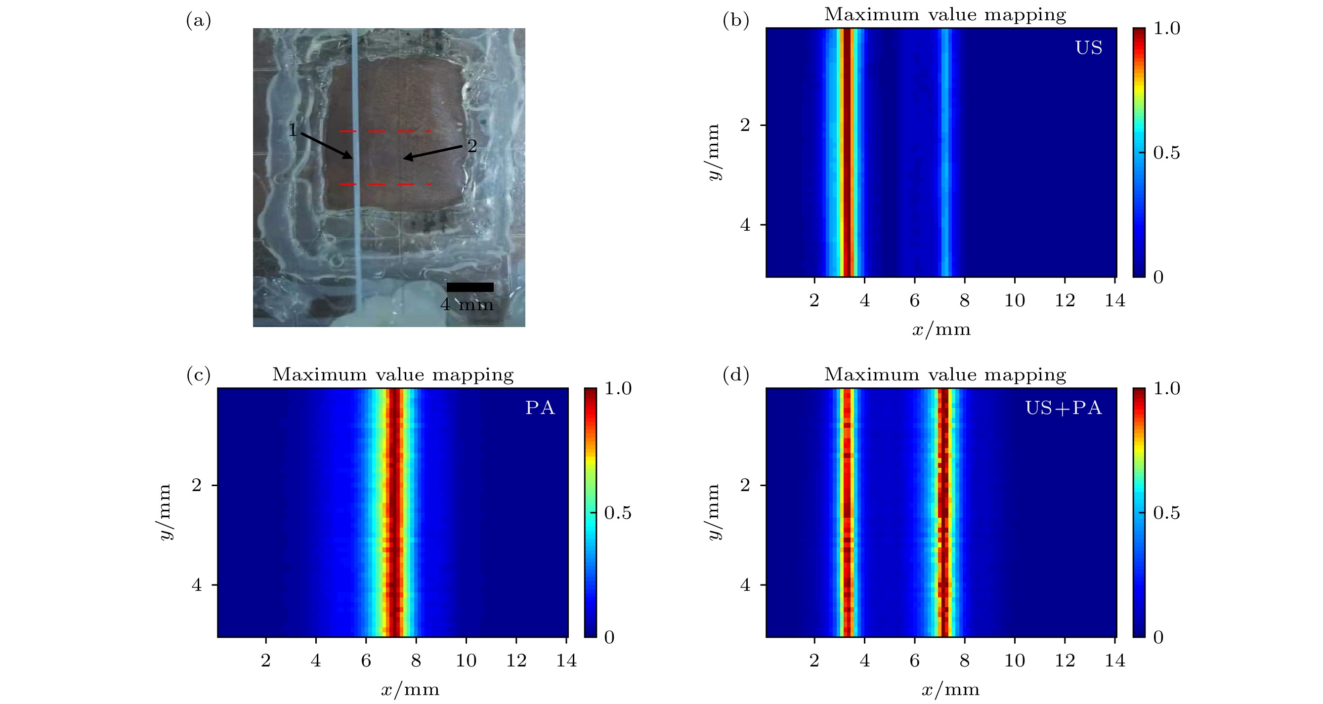
图 3 超声/光声双模态实验结果展示 (a)实验对象实物图; (b)超声图像; (c)光声图像; (d)超声和光声的双模态图像
Fig. 3. Results of ultrasound/photoacoustic dual-modality experiment: (a) Photograph of experimental subject; (b) image of ultrasound (US); (c) image of photoacoustic (PA); (d) the fused US/PA image.
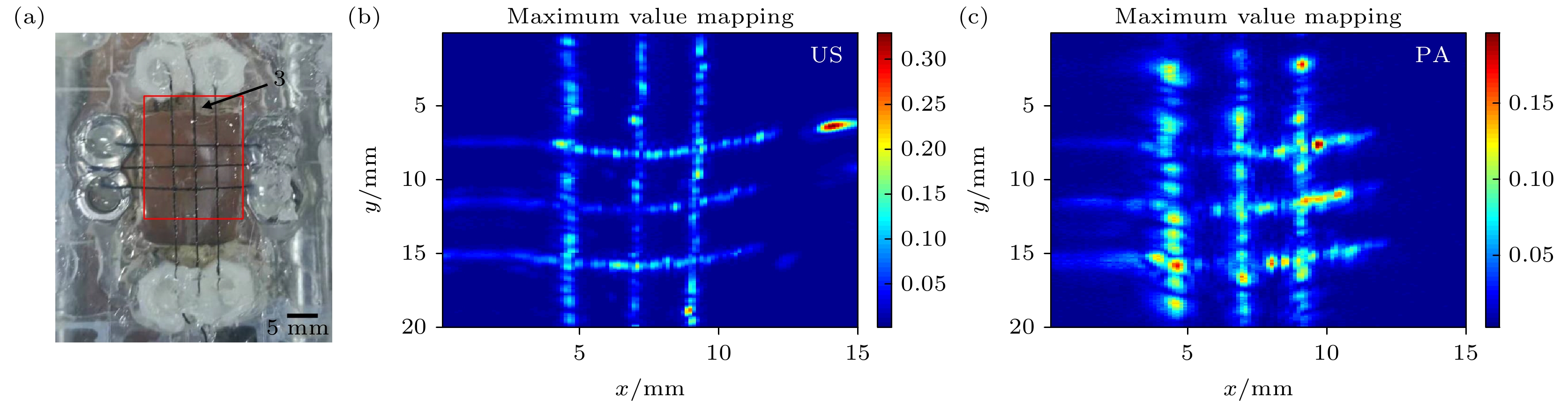
图 4 缝纫线实验结果展示 (a)实物图; (b)超声图像; (c)光声图像
Fig. 4. Experimental results of sewing thread: (a) Photograph of sewing thread; (b) image of ultrasound; (c) image of photoacoustic.
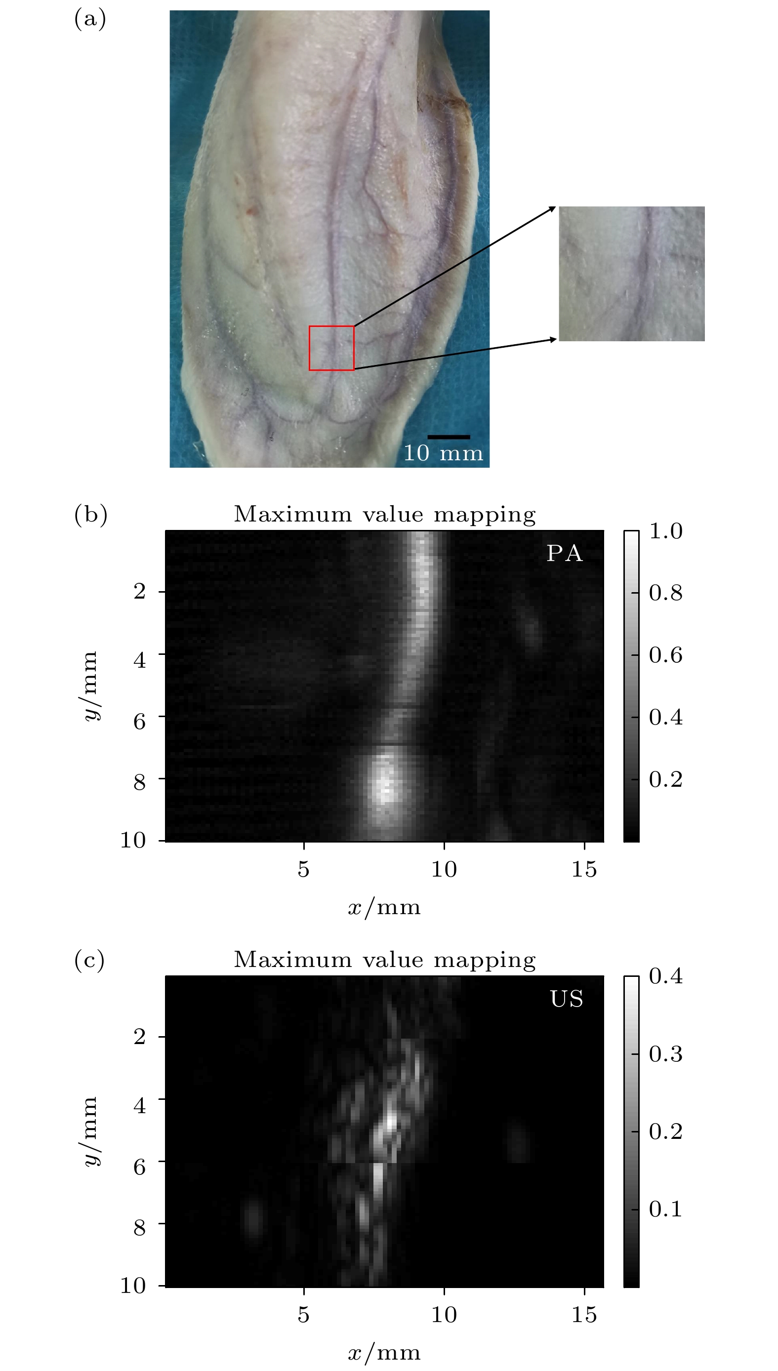
图 5 兔子耳朵活体实验成像结果展示 (a)兔子耳朵实物图; (b)光声图像; (c)超声图像
Fig. 5. Results of rabbit ear in vivo: (a) Photograph of rabbit ear; (b) image of photoacoustic; (c) image of ultrasound.
表 1 光声显微成像中扫描机制对比
Table 1. Comparison of scanning methods in photoacoustic microscopy imaging.
扫描方式 优点 缺点 应用 传统机械扫描 结构简单 体积大、笨重、成像速度慢 多 扇形扫描 体积小 需要特殊的小重量传感器 少 音圈扫描 体积小、速度快 负载能力弱、扫描范围小 少 扫描振镜扫描 体积小、速度快 需要高精度器件和配准 较多  下载: 导出CSV
-
[1] 林日强, 冷吉, 陈敬钦, 刘成波, 龚小竞, 宋亮 2018 中国医疗设备 33 1
Google Scholar
Lin R Q, Leng J, Chen J Q, Liu C B, Gong L J, Song L 2018 Chin. Med. Dev. 33 1
Google Scholar
[2] Beard P 2011 Interface Focus 1 602
Google Scholar
[3] 黄豆豆, 邱棋, 林文珍, 刘基嫣, 黄雅丽, 赵庆亮 2019 光散射学报 31 1
Google Scholar
Huang D D, Qiu Q, Lin W Z, Liu J Y, Huang Y L, Zhao Q L 2019 Chin. J. Light. Scatt. 31 1
Google Scholar
[4] Xu G, Rajian J R, Girish G, Kaplan M J, Fowlkes J B, Carson P L, Wang X D 2012 J. Biomed. Opt. 18 10502
Google Scholar
[5] Berg P J, Bansal R, Daoudi K, Steenbergen W, Prakash J 2016 Biomed. Opt. Express 7 5081
Google Scholar
[6] Garcia-Uribe A, Erpelding T N, Krumholz A, Ke H, Maslov K, Appleton C, Margenthaler J A, Wang L V 2015 Sci. Rep. 5 15748
Google Scholar
[7] Florian R, Julien S, Marilyne L M, Stéphanie R, Sharuja N, Stéphanie L, Alain L P 2016 Plos. One 11 4
Google Scholar
[8] Yang M, Zhao L, He X, Su N, Zhao C, Tang H, Hong T, Li W, Yang F, Lin L, Zhang B, Zhang R, Jiang Y, Li C 2017 Biomed. Opt. Express 8 3449
Google Scholar
[9] Li X, Wang D, Ran H, Hao L, Cao Y, Ao M, Zhang N, Song J, Zhang L, Yi H, Wang Z, Li P 2018 Biochem. Biophys. Res. Commun. 502 255
Google Scholar
[10] Mallidi S, Watanabe K, Timerman D, Schoenfeld D, Hasan T 2015 Theranostics 5 289
Google Scholar
[11] Qian M, Du Y, Wang S, Li C, Jiang H, Shi W, Chen J, Wang Y, Wagner E, Huang R 2018 ACS Appl. Mater. Inter. 10 4031
Google Scholar
[12] Diot G, Metz S, Noske A, Liapis E, Schroeder B, Ovsepian S V, Meier R, Rummeny E, Ntziachristos V 2017 Clin. Cancer Res. 23 6912
Google Scholar
[13] Toi M, Asao Y, Matsumoto Y, Sekiguchi H, Yoshikawa A, Takada M, Kataoka M, Endo T, Kawaguchi-Sakita N, Kawashima M, Fakhrejahani E, Kanao S, Yamaga I, Nakayama Y, Tokiwa M, Torii M, Yagi T, Sakurai T, Togashi K, Shiina T 2017 Sci. Rep. 7 41970
Google Scholar
[14] Yang J-M, Favazza C, Chen R, Yao J, Cai X, Maslov K, Zhou Q, Shung K K, Wang L V 2012 Nat. Med. 18 1297
Google Scholar
[15] Li Y, Lin R, Liu C, Chen J, Liu H, Zheng R, Gong X, Song L 2018 J. Biophotonics 11 e201800034
Google Scholar
[16] Oeri M, Bost W, Senegond N, Tretbar S, Fournelle M 2017 Ultrasound Med. Biol. 43 2200
Google Scholar
[17] Daoudi K, van den Berg P J, Rabot O, Kohl A, Tisserand S, Brands P, Steenbergen W 2014 Opt. Express 22 26365
Google Scholar
[18] 谢实梦, 黄林, 王雪, 迟子惠, 汤永辉, 郑铸, 蒋华北 2021 物理学报 70 100701
Google Scholar
Xie S M, Huang L, Wang L, Chi Z H, Tang Y H, Zheng Z, Jiang H B 2021 Acta Phys. Sin. 70 100701
Google Scholar
[19] Choi S, Kim J Y, Lim H G, Baik J W, Kim H H, Kim C 2020 Sci. Rep. 10 6544
Google Scholar
[20] 齐伟智 2018 博士学位论文 (成都: 电子科技大学)
Qi W Z 2018 Ph. D. Dissertation (Chengdu: University of Electronic Science and Technology of China) (in Chinese)
[21] Xi L, Sun J, Zhu Y, Wu L, Xie H, Jiang H 2010 Biomed. Opt. Express 1 1278
Google Scholar
[22] Lee C, Kim J Y, Kim C 2018 Micromachines 9 584
Google Scholar
[23] Yao J, Huang C H, Wang L, Yang J M, Gao L, Maslov K I, Zou J, Wang L V 2012 J. Biomed. Opt. 17 080505
Google Scholar
[24] Kim J Y, Lee C, Park K, Lim G, Kim C 2015 Sci. Rep. 5 7932
Google Scholar
[25] Kim S, Lee C, Kim J Y, Kim J, Lim G, Kim C 2016 J. Biomed. Opt. 21 106001
Google Scholar
[26] Kim J Y, Lee C, Lim G, Kim C 2016 Photons Plus Ultrasound: Imaging and Sensing San Francisco, 15 March 2016, p970835
[27] Pu Y, Bi R, Ahn J, Kim J Y, Kim C, Olivo M, Zhao X J 2019 Photons Plus Ultrasound: Imaging and Sensing San Francisco, 27 February 2019, p1087843
[28] Park K, Kim J Y, Lee C, Jeon S, Lim G, Kim C 2017 Sci. Rep. 7 13359
Google Scholar
[29] American Laser Institute. American National Standards for the Safe Use of Lasers ANSIZ136.1. Orlando, FL: American Laser Institute, 2014
[30] Wang L V, Hu S 2012 Science 335 1458
Google Scholar
[31] 汤永辉, 郑铸, 谢实梦, 黄林, 蒋华北 2020 物理学报 69 240701
Google Scholar
Tang Y H, Zheng Z, Xie S M, Huang L, Jiang H B 2020 Acta Phys. Sin. 69 240701
Google Scholar

 下载:
下载:
计量
- 文章访问数: 11583
- PDF下载量: 201
- 被引次数: 0
