


-
电子束衍射辐射源具有体积小、波长可调和极化可控等优点, 在纳米光子电路中显示出巨大的应用潜力. 这里展示了等腰直角三角形光阱纳米结构中电子束衍射辐射的光学手性现象, 可以获得手性度高于40%的衍射辐射信号, 光学手性的状态能被有效调控, 甚至可以实现手性的反转. 通过详细分析体系中的真空电磁模式和电荷分布的动态变化, 为这种手性光学效应的产生提供合理的理论解释. 电子束激发源所具有的纳米尺度下聚焦和移动的优势使电子束衍射辐射的光学手性具有灵活的调节潜力. 本文提出的物理机制和独特的实验平台对于未来二进制纳米光子电路和手性纳米光源的发展具有重要意义.
Electron beam diffraction radiation source has the advantages of small size, wavelength tunability, and polarization controllability, showing great potential applications in nanophotonic circuits. However, studies of optical chirality of electron beam diffraction radiation are relatively scarce. The optical chirality of electron beam diffraction radiation in isosceles right triangle light-well is realized and demonstrated in this work. The light-well consists of twelve alternating layers of gold and silicon dioxide, and the electron beam diffraction radiation originates from oscillating dipole sources within the light-well. In the case of the electron beam injected at the geometrically asymmetric position within the structure, the diffraction radiation signal with a maximum chirality of more than 40% can be obtained. By changing the injection position of the electron beam, the state of the optical chirality can be effectively controlled, and even the inversion of the chirality can be realized. By analyzing the vacuum electromagnetic mode in the system and the dynamic evolution of charge distribution in the gold layer, a reasonable theoretical explanation is provided for the generation of the chiral optical effect. A semi-analytical model for explaining the optical chirality of electron beam diffraction radiation is given by solving the incident electromagnetic field and the waveguide modes in the light-well. The observed difference between left-handed circularly polarized and right-handed circularly polarized signals originates from the asymmetry between left-handed and right-handed electromagnetic modes in the light-well caused by the geometrically asymmetric positional excitation, which ultimately leads to far-field chiral radiation determined by the electromagnetic field within the light-well. In addition, the simulation results of the surface charge distribution of the top gold film of the light-well reveal the correlation between the dynamic evolution of the surface charge distribution and the radiation chirality. The advantages of nanoscale focusing and moving of electron beam excitation source make the optical chirality of electron beam diffraction radiation have more flexible adjustment potential. The proposed physical mechanism and unique experimental platform not only provide new ideas for manipulating optical chirality on a nanoscale, but also lay the foundation for binary information processing and integration in nanophotonic circuits and chiral nano-light-sources in the future. -
Keywords:
- electron beam diffraction radiation /
- optical chirality /
- light-well
[1] Zhang Y J, Oka T, Suzuki R, Ye J T, Iwasa Y 2014 Science 344 725
 Google Scholar
[2] Takahashi S, Tajiri T, Ota Y, Tatebayashi J, Iwamoto S, Arakawa Y 2014 Appl. Phys. Lett. 105 051107
Google Scholar
[3] Kim J W, Carbone M, Dil J H, Tallarida M, Flammini R, Casaletto M P, Horn K, Piancastelli M N 2005 Phys. Rev. Lett. 95 107601
Google Scholar
[4] Mannini M, Pineider F, Sainctavit P, Joly L, Fraile-Rodríguez A, Arrio M A, Cartier dit Moulin C, Wernsdorfer W, Cornia A, Gatteschi D, Sessoli R 2009 Adv. Mater. 21 167
Google Scholar
[5] Lodahl P, Mahmoodian S, Stobbe S, Rauschenbeutel A, Schneeweiss P, Volz J, Pichler H, Zoller P 2017 Nature 541 473
Google Scholar
[6] Neufeld O, Cohen O 2018 Phys. Rev. Lett. 120 133206
Google Scholar
[7] Han T Y, Zu S, Li Z W, Jiang M L, Zhu X, Fang Z Y 2018 Nano Lett. 18 567
Google Scholar
[8] Zu S, Han T Y, Jiang M L, Lin F, Zhu X, Fang Z Y 2018 ACS Nano 12 3908
Google Scholar
[9] Potylitsyn A P 1998 Nucl. Instrum. Meth. B 145 169
[10] De Abajo F J G 2010 Rev. Mod. Phys. 82 209
Google Scholar
[11] Helliwell J R 1998 Nat. Struct. Biol. 5 614
Google Scholar
[12] Drummond W E, Rosenbluth M N 1960 Phys. Fluids 3 45
Google Scholar
[13] Ginis V, Danckaert J, Veretennicoff I, Tassin P 2014 Phys. Rev. Lett. 113 167402
Google Scholar
[14] Lin X, Easo S, Shen Y C, Chen H S, Zhang B L, Joannopoulos J D, Soljačić M, Kaminer I 2018 Nat. Phys. 14 816
Google Scholar
[15] Čerenkov P A 1937 Phys. Rev. 52 378
Google Scholar
[16] Urata J, Goldstein M, Kimmitt M F, Naumov A, Platt C, Walsh J E 1998 Phys. Rev. Lett. 80 516
Google Scholar
[17] Gardelle J, Modin P, Donohue J T 2010 Phys. Rev. Lett. 105 224801
Google Scholar
[18] So J K, García de Abajo F J, MacDonald K F, Zheludev N I 2015 ACS Photonics 2 1236
Google Scholar
[19] Kaminer I, Kooi S E, Shiloh R, Zhen B, Shen Y, López J J, Remez R, Skirlo S A, Yang Y, Joannopoulos J D, Arie A, Soljačić M 2017 Phys. Rev. X 7 011003
[20] Wang Z J, Yao K, Chen M, Chen H S, Liu Y M 2016 Phys. Rev. Lett. 117 157401
Google Scholar
[21] Remez R, Shapira N, Roques-Carmes C, Tirole R, Yang Y, Lereah Y, Soljačić M, Kaminer I, Arie A 2017 Phys. Rev. A 96 061801
Google Scholar
[22] Fang Y R, Sun M T 2015 Light Sci. Appl. 4 e294
Google Scholar
[23] Shen Y C, Harris N C, Skirlo S, et al. 2017 Nat. Photonics 11 441
Google Scholar
[24] Adamo G, MacDonald K F, Fu Y H, Wang C M, Tsai D P, De Abajo F J G, Zheludev N I 2009 Phys. Rev. Lett. 103 113901
Google Scholar
[25] Konishi K, Nomura M, Kumagai N, Iwamoto S, Arakawa Y, Kuwata-Gonokami M 2011 Phys. Rev. Lett. 106 057402
Google Scholar
-

图 1 (a) 周期性堆叠的SiO2-Au层结构及等腰直角三角形光阱结构示意图; (b) 等腰直角三角形光阱结构电子显微镜图; (c) 周期性堆叠层截面图; (d) SEM-CL联合系统收集光路示意图
Fig. 1. (a) Schematic diagram of periodically stacked SiO2-Au layers and isosceles right triangle light-well; (b) electron microscopy of isosceles right triangle light-well; (c) section diagram of periodically stacked layers; (d) schematic diagram of SEM-CL excitation and collection light path.
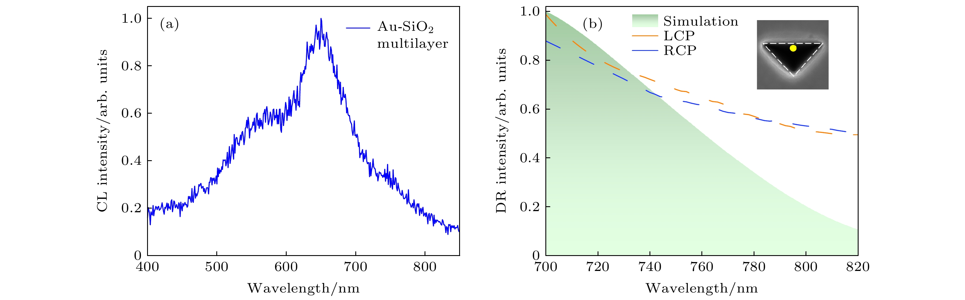
图 2 (a) 30 keV电子束激发SiO2-Au多层得到的归一化CL强度光谱; (b) 30 keV电子束激发三角形光阱斜边中心点获得的归一化LCP和RCP光谱, 虚线和阴影分别对应实验结果和模拟结果. 插图为三角形光阱SEM图, 黄色圆点代表电子束注入位置为斜边中心点
Fig. 2. (a) Normalized total CL spectra obtained by 30 kV electron beam impinging on the top surface of SiO2-Au multilayer; (b) normalized LCP and RCP emission spectra acquired by 30 kV electron beam impinging at the center point of the hypotenuse of the triangle light-well. Dashed lines and shadows correspond to experimental and simulated results, respectively. Inset is SEM image of the triangle light-well with yellow dot representing the center point of the hypotenuse where the electron beam is injected.
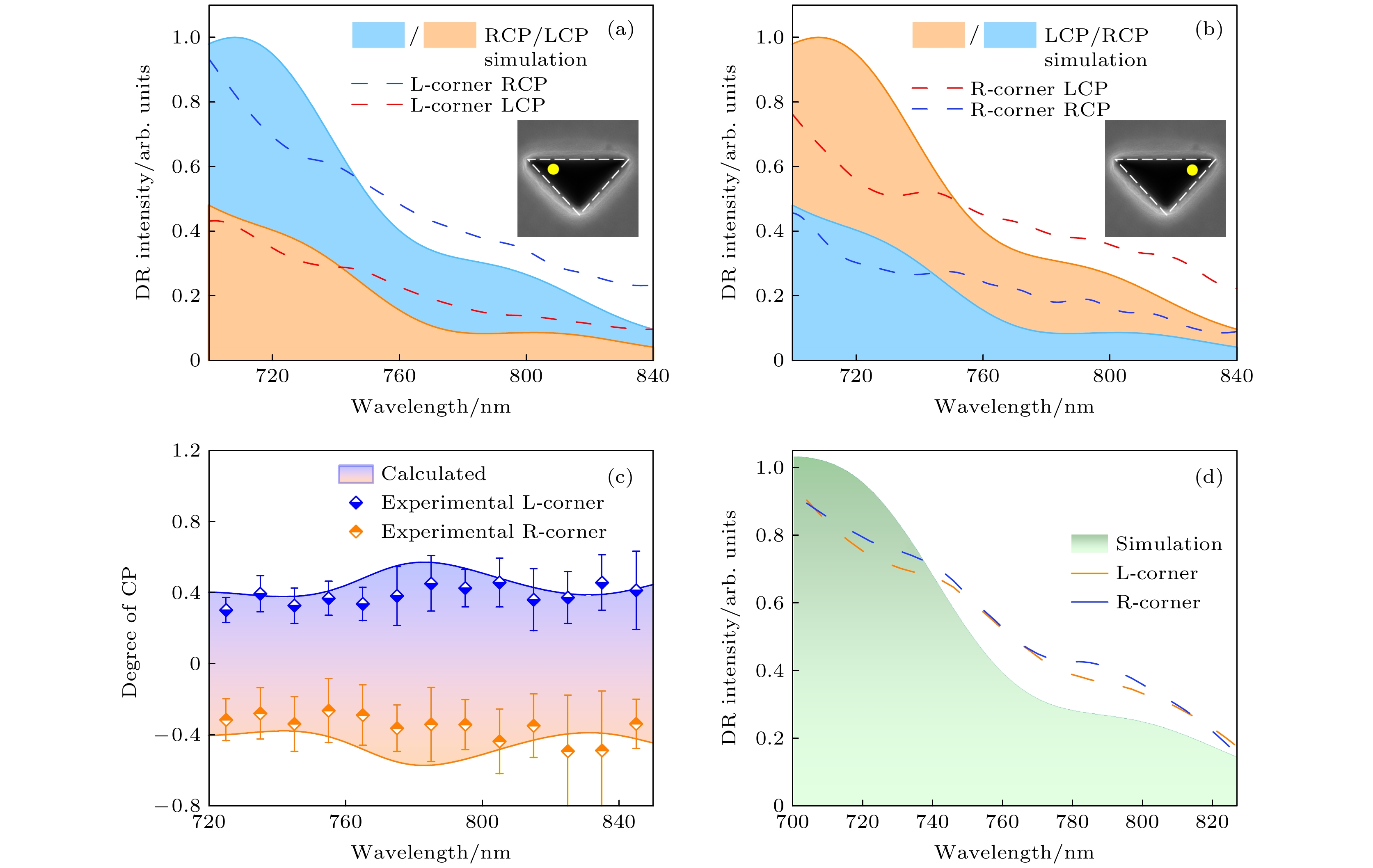
图 3 (a), (b) LCP和RCP分量的CL光谱, 波长为700—840 nm (插图黄点表示电子束的激发位置, 虚线和阴影分别对应实验结果和模拟结果); (c) 计算获得的手性度, 点和实线分别对应实验数据和模拟数据; (d) 两个激发位置下的衍射辐射光谱, 用全色模式测量, 并以短线表示 (阴影区域表示模拟结果)
Fig. 3. (a), (b) CL spectra of LCP and RCP components with the wavelength from 700–840 nm (The impinging point of the electron beam is shown in inset with yellow points. Dashed lines and shadows correspond to experimental and simulated results, respectively); (c) calculated chirality with dots and solid lines corresponding to experimental and simulated data, respectively; (d) total diffraction spectra of these two injection points, measured by panchromatic mode and illustrated as dots (The simulated spectrum is presented by shadows).
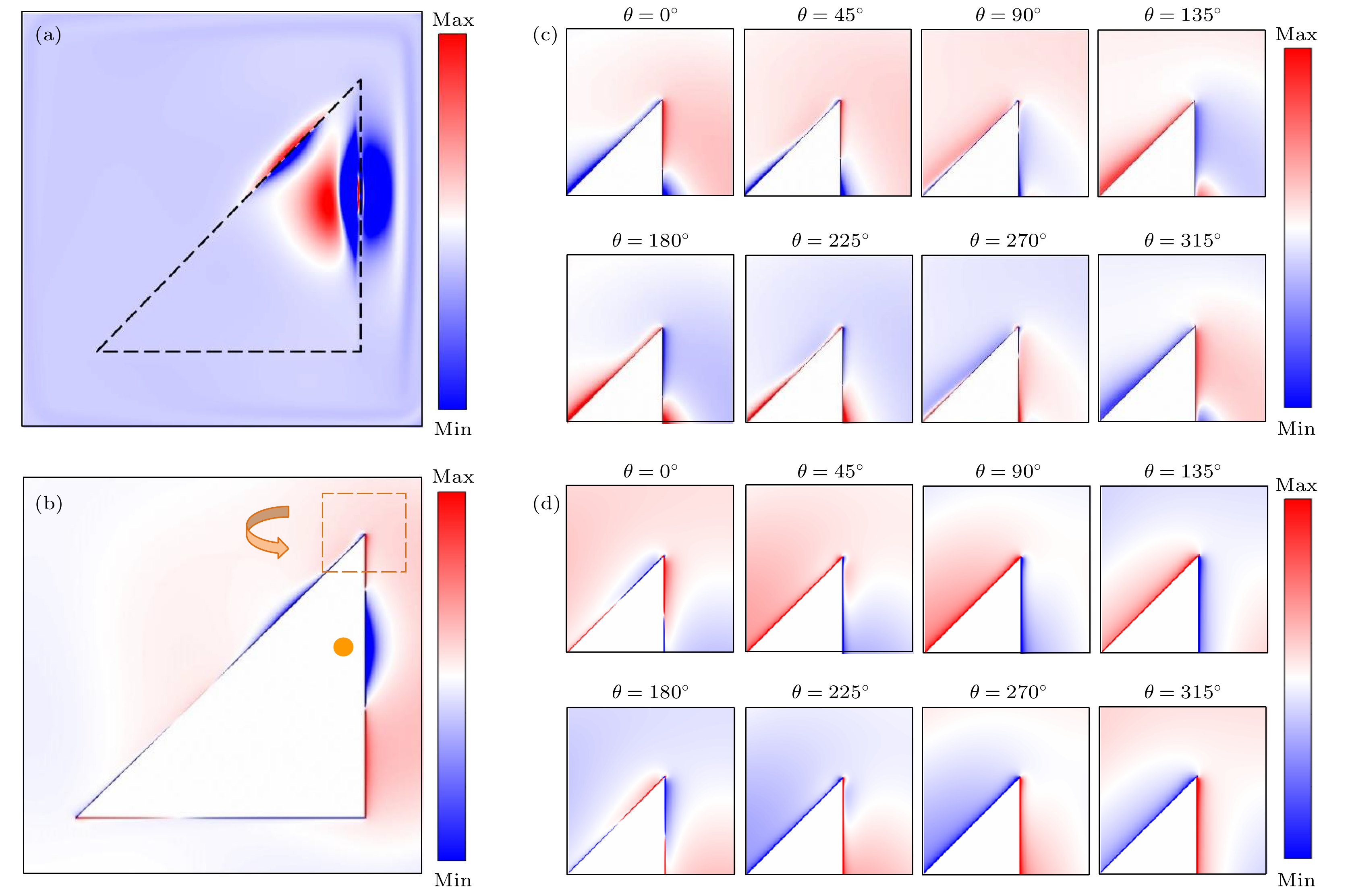
图 4 电子束在三角形光阱右上端入射点激发下电场和电荷分布模拟结果 (a) 780 nm探测波长下光阱表面的电场强度分布; (b)光阱结构最上层Au层的表面电荷分布, θ为相位角; (c) 780 nm探测波长下光阱结构右上角局部电荷分布随时间的变化, 在顶点处显示出了逆时针旋转的演变特征; (d) 780 nm波长的左旋圆偏振光激发下光阱结构最上层Au层的表面电荷随时间的动态演变
Fig. 4. Simulation of electric field and charge distribution of electron beam excited at right corner of light-well: (a) Electric field intensity distribution on the surface of the light-well at 780 nm detection wavelength; (b) surface charge distribution of the topside Au layer in the structure, and θ is the phase angle; (c) change of local charge distribution in the right corner of the light-well with time at the detection wavelength of 780 nm, and it shows a counterclockwise rotation at the vertex; (d) dynamic surface charge evolution of the topmost Au layer in light-well excited by 780 nm left-handed circularly polarized light.
-
[1] Zhang Y J, Oka T, Suzuki R, Ye J T, Iwasa Y 2014 Science 344 725
Google Scholar
[2] Takahashi S, Tajiri T, Ota Y, Tatebayashi J, Iwamoto S, Arakawa Y 2014 Appl. Phys. Lett. 105 051107
Google Scholar
[3] Kim J W, Carbone M, Dil J H, Tallarida M, Flammini R, Casaletto M P, Horn K, Piancastelli M N 2005 Phys. Rev. Lett. 95 107601
Google Scholar
[4] Mannini M, Pineider F, Sainctavit P, Joly L, Fraile-Rodríguez A, Arrio M A, Cartier dit Moulin C, Wernsdorfer W, Cornia A, Gatteschi D, Sessoli R 2009 Adv. Mater. 21 167
Google Scholar
[5] Lodahl P, Mahmoodian S, Stobbe S, Rauschenbeutel A, Schneeweiss P, Volz J, Pichler H, Zoller P 2017 Nature 541 473
Google Scholar
[6] Neufeld O, Cohen O 2018 Phys. Rev. Lett. 120 133206
Google Scholar
[7] Han T Y, Zu S, Li Z W, Jiang M L, Zhu X, Fang Z Y 2018 Nano Lett. 18 567
Google Scholar
[8] Zu S, Han T Y, Jiang M L, Lin F, Zhu X, Fang Z Y 2018 ACS Nano 12 3908
Google Scholar
[9] Potylitsyn A P 1998 Nucl. Instrum. Meth. B 145 169
[10] De Abajo F J G 2010 Rev. Mod. Phys. 82 209
Google Scholar
[11] Helliwell J R 1998 Nat. Struct. Biol. 5 614
Google Scholar
[12] Drummond W E, Rosenbluth M N 1960 Phys. Fluids 3 45
Google Scholar
[13] Ginis V, Danckaert J, Veretennicoff I, Tassin P 2014 Phys. Rev. Lett. 113 167402
Google Scholar
[14] Lin X, Easo S, Shen Y C, Chen H S, Zhang B L, Joannopoulos J D, Soljačić M, Kaminer I 2018 Nat. Phys. 14 816
Google Scholar
[15] Čerenkov P A 1937 Phys. Rev. 52 378
Google Scholar
[16] Urata J, Goldstein M, Kimmitt M F, Naumov A, Platt C, Walsh J E 1998 Phys. Rev. Lett. 80 516
Google Scholar
[17] Gardelle J, Modin P, Donohue J T 2010 Phys. Rev. Lett. 105 224801
Google Scholar
[18] So J K, García de Abajo F J, MacDonald K F, Zheludev N I 2015 ACS Photonics 2 1236
Google Scholar
[19] Kaminer I, Kooi S E, Shiloh R, Zhen B, Shen Y, López J J, Remez R, Skirlo S A, Yang Y, Joannopoulos J D, Arie A, Soljačić M 2017 Phys. Rev. X 7 011003
[20] Wang Z J, Yao K, Chen M, Chen H S, Liu Y M 2016 Phys. Rev. Lett. 117 157401
Google Scholar
[21] Remez R, Shapira N, Roques-Carmes C, Tirole R, Yang Y, Lereah Y, Soljačić M, Kaminer I, Arie A 2017 Phys. Rev. A 96 061801
Google Scholar
[22] Fang Y R, Sun M T 2015 Light Sci. Appl. 4 e294
Google Scholar
[23] Shen Y C, Harris N C, Skirlo S, et al. 2017 Nat. Photonics 11 441
Google Scholar
[24] Adamo G, MacDonald K F, Fu Y H, Wang C M, Tsai D P, De Abajo F J G, Zheludev N I 2009 Phys. Rev. Lett. 103 113901
Google Scholar
[25] Konishi K, Nomura M, Kumagai N, Iwamoto S, Arakawa Y, Kuwata-Gonokami M 2011 Phys. Rev. Lett. 106 057402
Google Scholar

 下载:
下载:
计量
- 文章访问数: 6177
- PDF下载量: 78
- 被引次数: 0
