


-
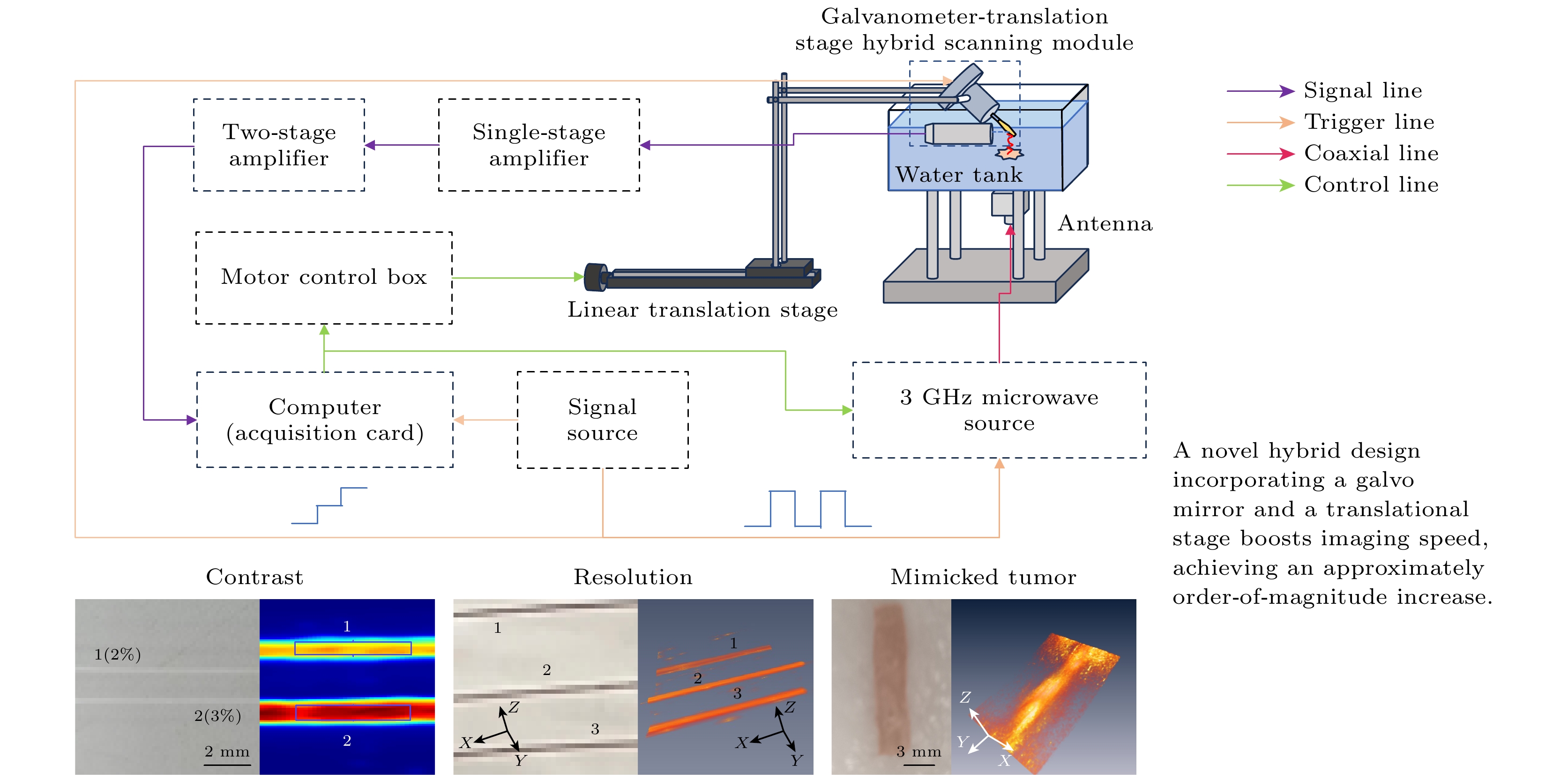
微波热声成像技术作为一种新兴生物医学成像方法, 融合了微波成像高对比度与超声成像高分辨率的优点. 微波热声显微成像作为其重要分支, 在继承其优点的基础上, 提供了观察更加精细组织特征的能力, 但传统栅型扫描机制因机械运动干扰微波场分布, 需要多次信号平均以保证信噪比, 且电机空程移动导致单次成像耗时较长, 制约了其实际应用. 本文提出并构建了一种基于一维振镜扫描的快速成像系统, 采用振镜-平移台混合架构, 通过优化扫描方式, 降低了微波场干扰, 同时减少了信号平均次数并缩减了空程耗时, 从而提升了成像速度. 设计的时序控制算法实现了微波激励、振镜运动与超声检测的精确同步, 适配扫描方式优化的图像重建算法有效校正了扫描过程中产生的畸变. 分辨率与对比度测试仿体实验, 与早期肿瘤模拟离体实验表明, 系统在速度提升超过10倍的同时, 保持了成像质量. 该方案有效提升了微波热声显微成像效率和稳定性, 为其实验室研究向临床应用转化奠定重要基础.Microwave-induced thermoacoustic imaging, as an emerging biomedical imaging technique, combines the high contrast of microwave imaging with the high spatial resolution of ultrasound imaging. Microwave-induced thermoacoustic microscopy, as an important branch of this technology, retains these advantages while possessing the ability to visualize finer tissue characteristics. However, traditional raster scanning mechanisms introduce interference into microwave field distribution due to mechanical motion, thus necessitating multiple signal average to maintain signal-to-noise ratio. Additionally, the idle time during motor movement results in extended single-scan durations, limiting its practical applications. To address these limitations, this work proposes a rapid imaging system based on one-dimensional galvanometer scanning. The system employs a hybrid galvanometer-translation stage architecture and an optimized scanning strategy to minimize microwave field interference, reduce the number of signal averages and shortens the idle time, ultimately achieving more than a tenfold improvement in imaging speed. A specially designed timing control algorithm ensures the precise synchronization of microwave excitation, galvanometer motion, and ultrasound detection, while the reconstruction algorithm suitable for the optimizing scanning method effectively corrects distortions generated in the scanning process. The system performance is assessed through phantom and ex vivo tissue experiments. Resolution tests show hundred-micrometer resolution along all three axes (332 μm × 324 μm × 79 μm), while contrast and depth imaging experiments confirm its ability to clearly distinguish targets with different conductivities, achieving an effective detection depth of at least 10 mm in tissue. Early tumor mimicking experiments further demonstrate the ability of the system to identify lesion boundaries, preliminarily revealing its potential for rapid tumor margin assessment. This approach maintains the imaging quality of microwave-induced thermoacoustic microscopy while enhancing imaging efficiency and system stability, thereby laying a crucial foundation for advancing the technology from laboratory research to clinical applications.
-
Keywords:
- microwave-induced thermoacoustic imaging /
- microwave-induced thermoacoustic microscopy /
- galvanometer scanning /
- imaging speed
-
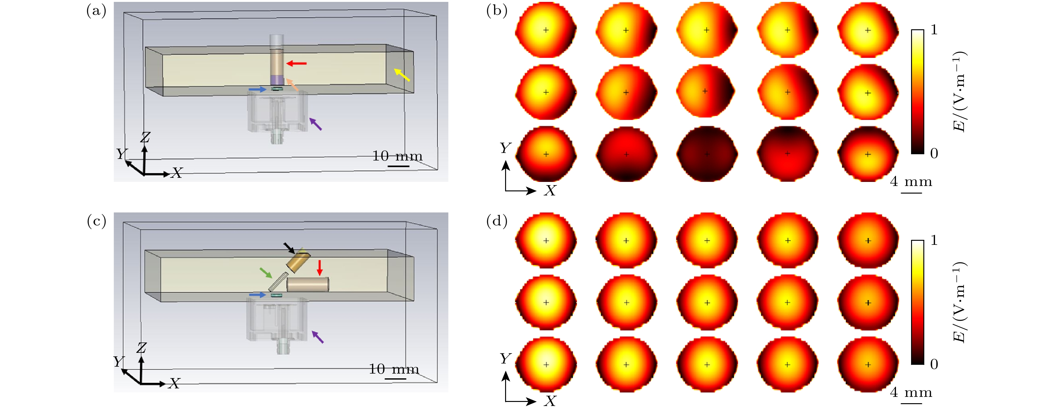
图 1 CST仿真模型及电场分布结果 (a)传统栅型扫描机制下的仿真模型; (b)对应(a) 的组织内电场分布; (c)振镜-平移台混合扫描机制下的仿真模型; (d)对应(c)的组织内电场分布
Fig. 1. CST simulation models and electric field (E-field) distribution results: (a) Simulation model under the conventional raster-scanning mechanism; (b) E-field distribution within the tissue corresponding to (a); (c) simulation model under the galvanometer-translational stage hybrid scanning mechanism; (d) E-field distribution within the tissue corresponding to (c).
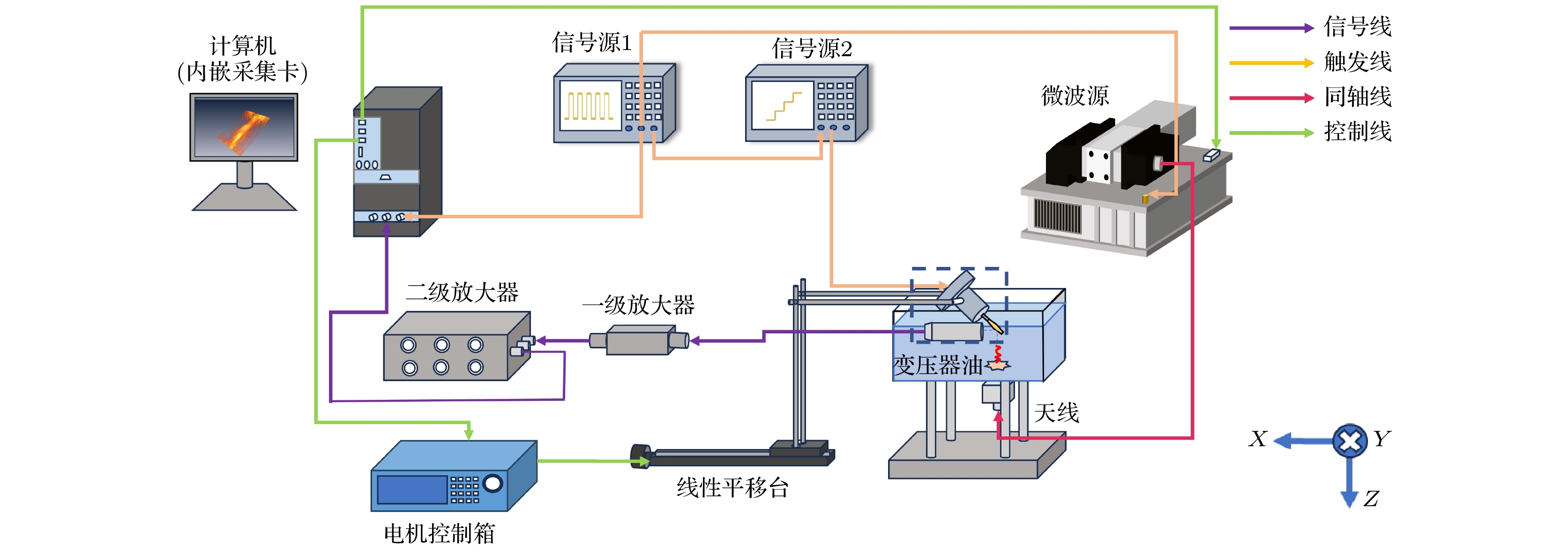
图 2 快速微波热声显微成像系统示意图
Fig. 2. Schematic of fast microwave-induced thermoacoustic microscopy imaging system.
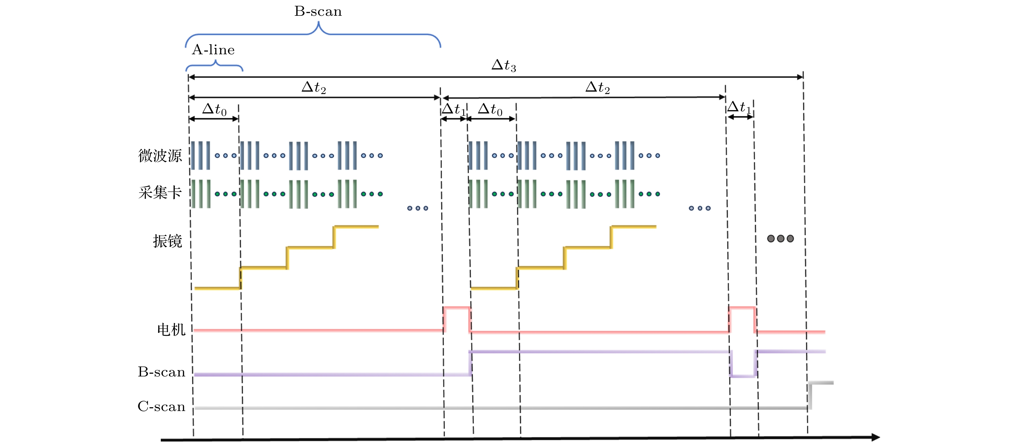
图 3 快速微波热声显微成像系统时序图
Fig. 3. Timing diagram for fast microwave-induced thermoacoustic microscopy system.
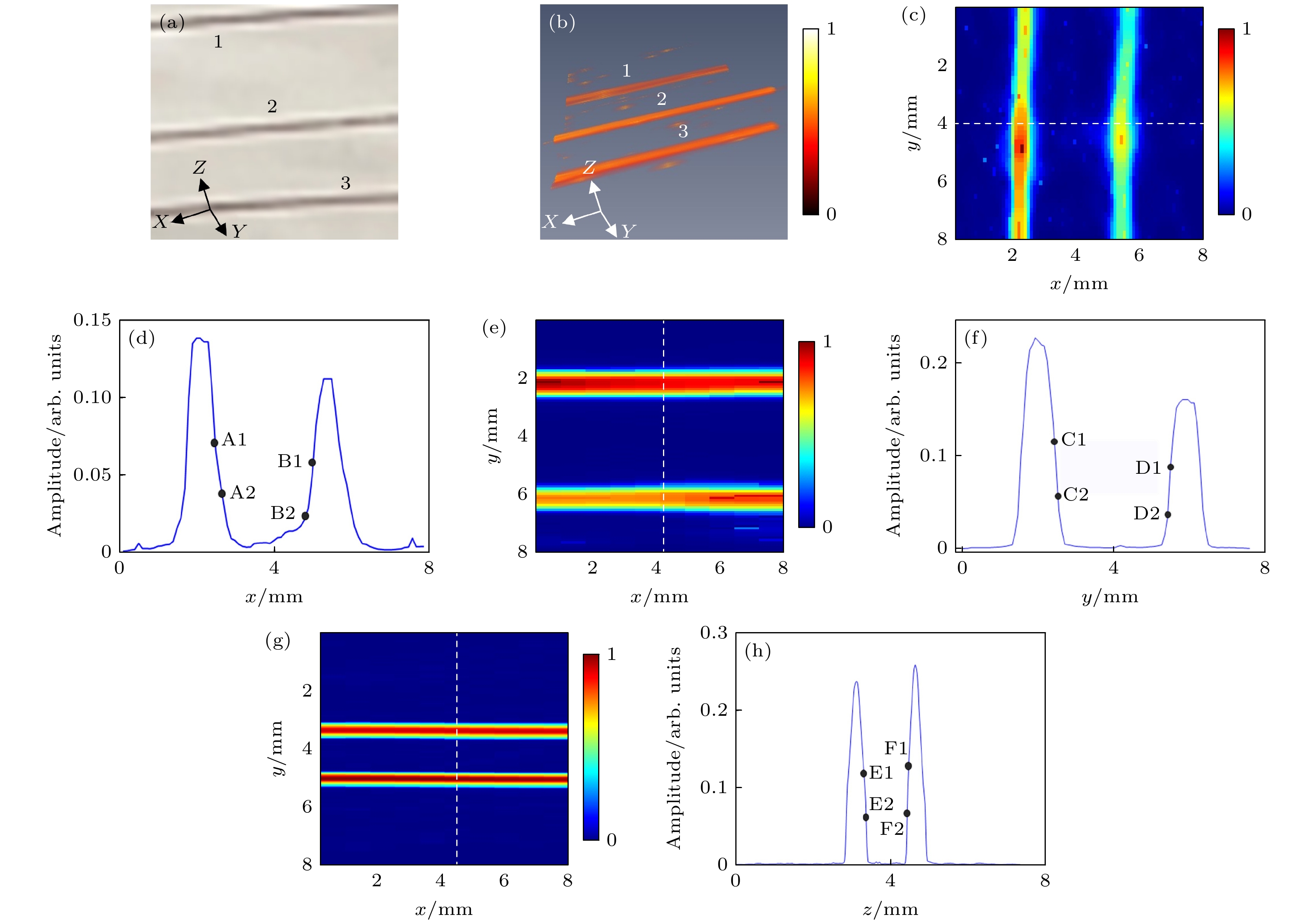
图 4 铜丝分辨率测试实验 (a)直径为90 μm的铜丝实物图; (b)三维热声图像; (c) X轴分辨率测试时沿Z轴方向投影的1号和2号铜丝的二维热声显微图像; (d) (c)图中沿白色虚线的热声信剖面图; (e) Y轴分辨率测试时沿Z轴方向投影的1号和2号铜丝的二维热声显微图像; (f) (e)图中沿白色虚线的热声信剖面图; (g) Z轴分辨率测试时2号与3号铜丝沿Y轴方向投影的二维热声显微图像; (h) (g)图中沿白色虚线的热声信剖面图
Fig. 4. Copper wire experiment: (a) Photograph of 90 μm-diameter copper wires; (b) 3D FTAM image; (c) 2D FTAM images of copper wires 1 and 2 along the z-axis for X-axis resolution testing; (d) thermoacoustic signal profile along the white dashed line in (c); (e) 2D FTAM images of copper wires 1 and 2 along the Z-axis for Y-axis resolution testing; (f) thermoacoustic signal profile along the white dashed line in (e); (g) 2D FTAM images of copper wires 2 and 3 along the Y-axis for Z-axis resolution testing; (h) thermoacoustic signal profile along the white dashed line in (g).
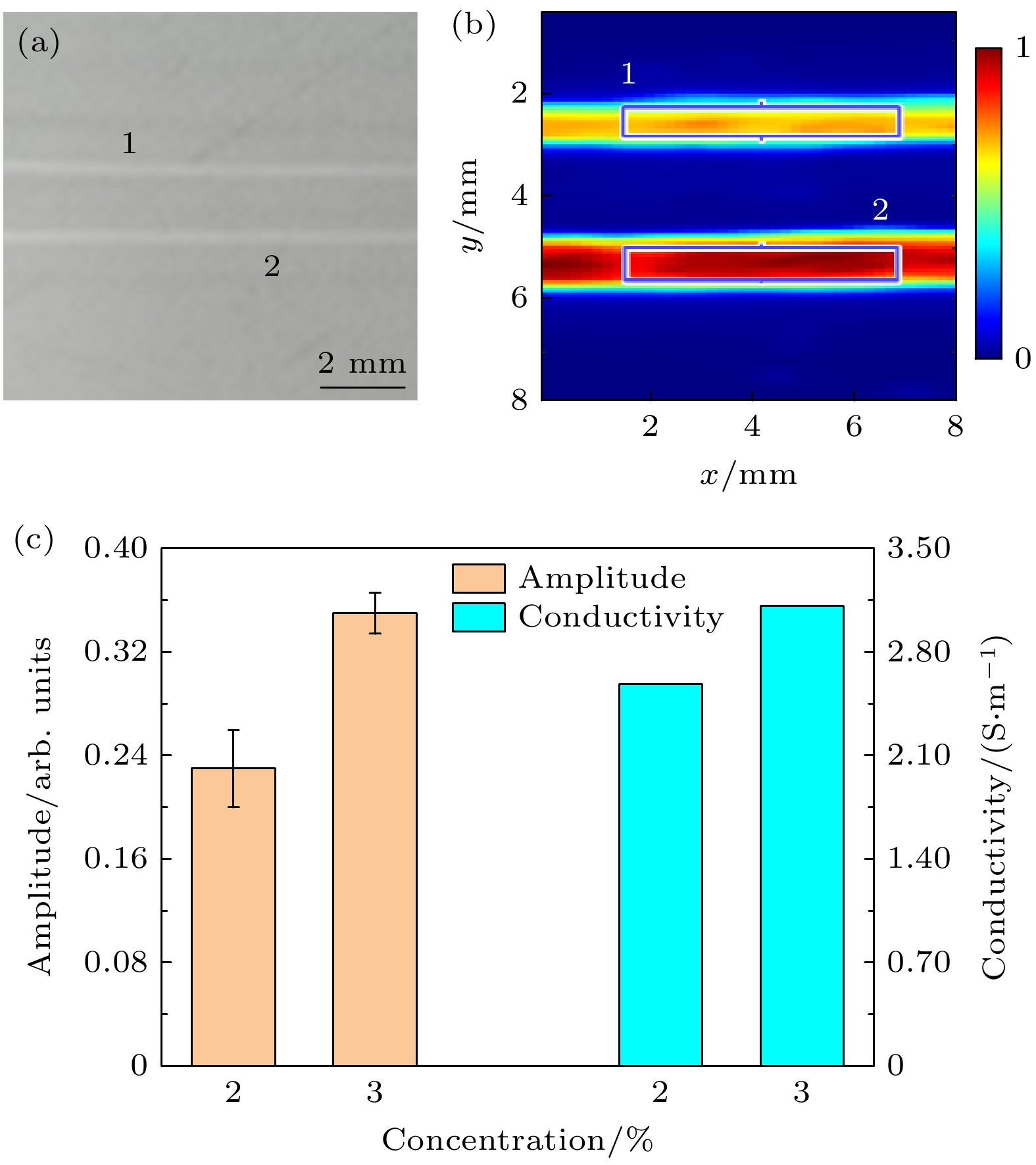
图 5 盐水管对比度测试实验 (a)内径0.2 mm的盐水管实物图; (b)二维热声显微图像; (c) (b)图中选中区域像素值的统计结果以及与2%, 3%浓度盐水管的理论电导率对比图
Fig. 5. Saline tube experiment: (a) Photograph of saline tubes with 0.2 mm inner diameter; (b) 2D FTAM image; (c) statistical results of pixel values in the selected region from (b), with theoretical electrical conductivity values for 2% and 3% saline concentration tubes.
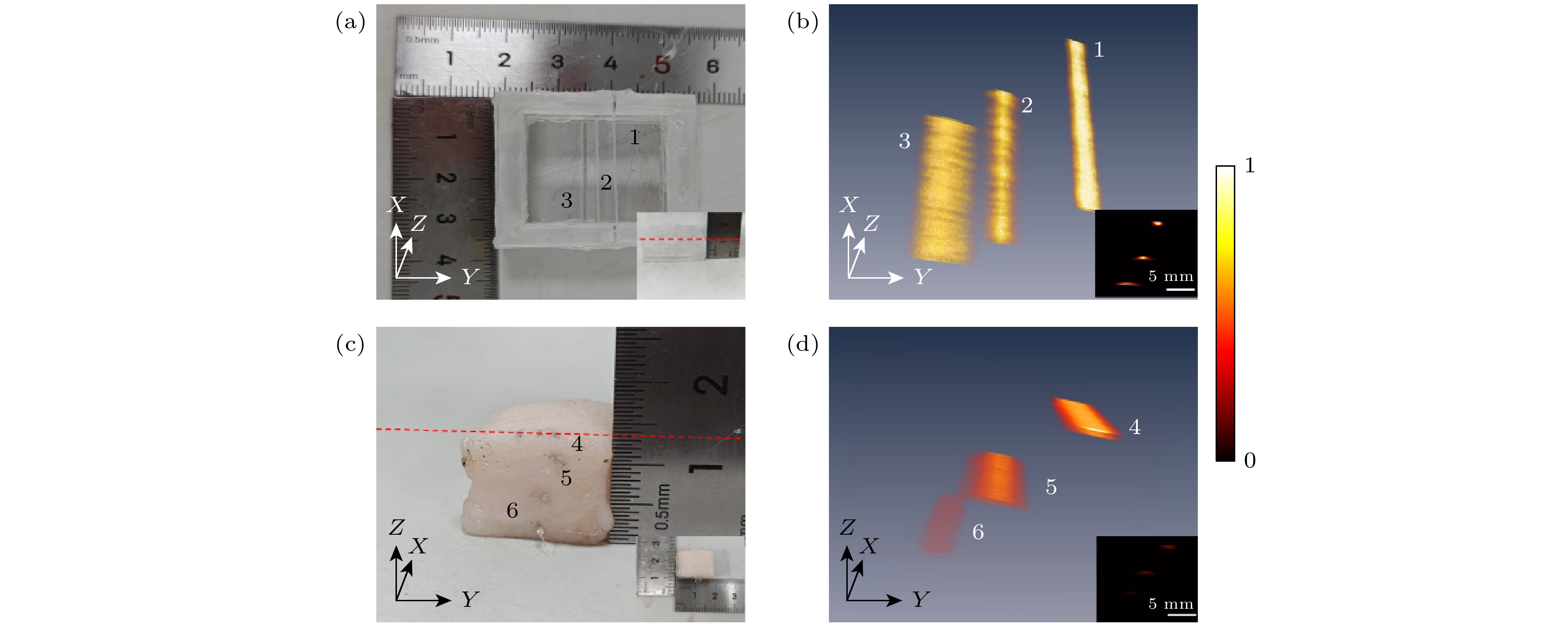
图 6 深度成像 (a)置于空气中的样品实物图; (b) (a)图中样品对应的三维及二维微波热声图像; (c)嵌入脂肪中的样品实物图; (d) (c)图中样品对应的三维及二维微波热声图像
Fig. 6. Depth imaging: (a) Photograph of the sample placed in air; (b) corresponding 3D and 2D microwave-induced thermoacoustic images of the sample in (a); (c) photograph of the sample embedded in fat; (d) corresponding 3D and 2D microwave-induced thermoacoustic images of the sample in (c).
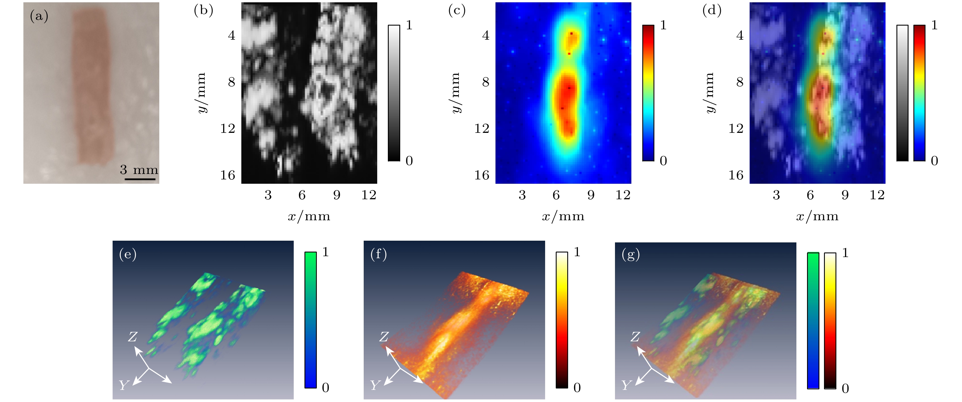
图 7 早期肿瘤模拟成像 (a)实物图; (b)二维超声显微图像; (c)二维热声显微图像; (d)二维热声/超声显微融合图像; (e)三维超声显微图像; (f)三维热声显微图像; (g)三维热声/超声显微融合图像
Fig. 7. Early-stage tumor simulation imaging: (a) Photograph of the phantom; (b) 2D ultrasound image; (c) 2D TAM image; (d) 2D thermoacoustic-ultrasound image; (e) 3D ultrasound image; (f) 3D TAM image; (g) 3D thermoacoustic-ultrasound image.
-
[1] Bell A G 1880 Am. J. Sci. s3-20 305
[2] Olsen R G, Lin J C 1983 Bioelectromagnetics 4 397
 Google Scholar
[3] Kruger R A, Kopecky K K, Aisen A M, Reinecke D R, Kruger G A, Kiser W L 1999 Radiology 211 275
Google Scholar
[4] Ku G, Wang L V 2000 Med. Phys. 27 1195
Google Scholar
[5] Kruger R A, Miller K D, Reynolds H E, Kiser W L, Reinecke D R, Kruger G A 2000 Radiology 216 279
Google Scholar
[6] Singhvi A, Boyle K C, Fallahpour M, Khuri-Yakub B T, Arbabian A 2019 IEEE Trans. Ultrason. Ferroelectr. Freq. Control 66 1587
Google Scholar
[7] Ren M Y, Cheng Z W, Wu L H, Zhang H M, Zhang S X, Chen X Y 2022 IEEE Trans. Biomed. Eng. 70 175
[8] Wu L H, Cheng Z W, Ma Y Z, Li Y J, Ren M Y, Xing D, Qin H 2021 IEEE Trans. Med. Imaging 41 1080
[9] Zhao S X, Wang H H, Li Y J, Nie L M, Zhang S X, Xing D, Qin H 2021 IEEE Trans. Biomed. Eng. 69 725
[10] Liang X, Guo H, Liu Q, Wu C F, Gong Y B, Xi L 2020 Appl. Phys. Lett. 116 013701
Google Scholar
[11] Chen Y, Chi Z H, Du S, Fang Q C, Jiang H B 2024 Appl. Phys. Lett. 124 163702
[12] Xu M H, Xu Y, Wang L H V 2003 IEEE Trans. Biomed. Eng. 50 1086
Google Scholar
[13] Wan P C, Liu S L, Tian R P, Shang X, Peng W T 2023 J. Appl. Phys. 133 103101
Google Scholar
[14] Liu S L, Zheng Z, Sun X X, Zhao Z Q, Zheng Y J, Jiang H B 2019 IEEE Trans. Biomed. Eng. 67 2206
[15] Luo Z X, Li C Z, Liu D T, Wang B S, Zhang L J, Ma Y X 2023 IEEE Trans. Microw. Theory Tech. 71 2652
Google Scholar
[16] Evans A L, Ma C, Hagness S C 2022 Biomed. Phys. Eng. Express 8 035020
Google Scholar
[17] Mast T D, Johnstone D A, Dumoulin C L, Lamba M A, Patch S K 2023 Phys. Med. Biol. 68 025003
Google Scholar
[18] Kruger R A, Kiser W L, Reinecke D R, Kruger G A, Miller K D 2003 Mol. Imaging 2 113
Google Scholar
[19] Chi Z H, Huang L, Wu D, Long X J, Xu X L, Jiang H B 2022 Med. Phys. 49 84
Google Scholar
[20] Huang L, Zheng Z, Chi Z H, Jiang H B 2021 Med. Phys. 48 4242
Google Scholar
[21] Xiang H J, Zheng Z, Huang L, Qiu T T, Luo Y, Jiang H B 2021 Med. Phys. 48 1608
Google Scholar
[22] Kellnberger S, Hajiaboli A, Razansky D, Ntziachristos V 2011 Phys. Med. Biol. 56 3433
Google Scholar
[23] Tamimi E A, Xin H, Witte R S 2020 Appl. Opt. 59 G255
Google Scholar
[24] Liu S L, Shang X, Lu Y X, Huang L 2022 Appl. Phys. Lett. 121 243701
Google Scholar
[25] Xi Z J, Wang X Y, Ye K, Wang X 2023 IEEE J. Electromagn. RF Microw. Med. Biol. 7 383
Google Scholar
[26] Wang X Y, Xi Z J, Ye K, Gong Z, Chen Y F, Wang X 2024 Sensors 24 2682
[27] Lou C G, Yang S H, Ji Z, Chen Q, Xing D 2012 Phys. Rev. Lett. 109 218101
Google Scholar
[28] Fang Q C, Chi Z H, Liu Y, Wang Y, Du S, Wu D, Jiang H B 2023 Med. Phys. 50 6036
Google Scholar
[29] Chen S L, Xie Z X, Ling T, Guo L J, Wei X B, Wang X D 2012 Opt. Lett. 37 4263
Google Scholar
[30] Yao J J, Huang C H, Wang L D, Yang J M, Gao L, Maslov K I, Zou J, Wang L H V 2012 J. Biomed. Opt. 17 080505.
[31] 2022 物理学报 71 054301
Xu S Z, Xie S M, Wu D, Chi Z H, Huang L 2022 Acta Phys. Sin. 71 054301
[32] Zhu K G, Popovic M 2009 IEEE Antennas Wirel. Propag. Lett. 8 1259
Google Scholar
[33] 王雨, 张慧敏, 覃欢 2023 物理学报 72 204301
Google Scholar
Wang Y, Zhang H M, Qin H 2023 Acta Phys. Sin. 72 204301
Google Scholar
[34] Wang L H V, Zhao X M, Sun H T, Ku G 1999 Rev. Sci. Instrum. 70 3744
Google Scholar
[35] Zhu G K, Popovic M 2011 Prog. Electromagn. Res. B 35 1
Google Scholar
[36] Calasso I G, Craig W, Diebold G J 2001 Phys. Rev. Lett. 86 3550
Google Scholar
[37] Gusev V E, Karabutov A A 1993 Laser Optoacoustics (New York: AIP Press) pp1-12
[38] Imasonic https://www.imasonic-ndt.com/online-design/ [2025-07-20].
[39] Sonic Concepts https://www.sonicconcepts.com/transducer-selection-guide-single-element/ [2025-07-25].
[40] Sun J, Wang W L, Yue Q Y 2016 Materials 9 231
Google Scholar
[41] Benny R, Anjit T A, Mythili P 2020 Prog. Electromagn. Res. B 87 61
Google Scholar
[42] Fishman E K, Ney D R, Heath D G, Corl F M, Horton K M, Johnson P T 2006 Radiographics 26 905
Google Scholar
[43] Liang Z, Wang W P, Qiao S Q, Huang L 2022 J. Innov. Opt. Health Sci. 15 2250015
Google Scholar
[44] Niskanen A O, Hassel J, Tikander M, Maijala P, Grönberg L, Helistö P 2009 Appl. Phys. Lett. 95 163701
Google Scholar
[45] 杨晓庆, 黄卡玛 2006 电子学报 34 356
Yang X Q, Huang K M 2006 Acta Electron. Sin. 34 356
[46] Zhang W T, Chen X, Wang Y, Wu L Y, Hu Y D 2010 Res. Explor. Lab. 29 159
[47] Du S, Qiang T, Chi Z H, Jiang H B 2024 J. Innov. Opt. Health Sci. 17 2450014
Google Scholar
[48] 杜劲松, 高扬, 毕欣, 齐伟智, 黄林, 荣健 2015 物理学报 64 034301
Google Scholar
Du J S, Gao Y, Bi X, Qi W Z, Huang L, Rong J 2015 Acta Phys. Sin. 64 034301
Google Scholar
[49] Xie S M, Huang L, Wang X, Chi Z H, Tang Y H, Zheng Z, Jiang H B 2021 J. Mech. Eng. 70 100701
[50] Cheng Z W, Wu L H, Qiu T S, Duan Y H, Qin H, Hu J 2021 IEEE Trans. Med. Imaging 40 3498
Google Scholar
[51] 张玉敏, 王富, 林俐, 叶坚 2024 分析测试学报 43 19
Zhang Y M, Wang F, Lin L, Ye J 2024 J. Instrum. Anal. 43 19
[52] Tang X Y, Fu J, Qin H 2023 J. Innov. Opt. Health Sci. 16 2230014
Google Scholar
[53] Sun M L, Li C Y, Chen R M, Shi J H 2024 Laser Optoelectron. Prog. 61 0618017
Google Scholar
[54] Jeon S, Kim J, Lee D, Baik J W, Kim C 2019 Photoacoustics 15 100141
Google Scholar
[55] Chen Z J, Yang S H, Xing D 2018 Chin. J. Lasers 45 0307008
Google Scholar
[56] Kim J Y, Lee C, Park K, Lim G, Kim C 2015 Sci. Rep. 5 7932
Google Scholar
[57] Qi W Z, Jin T, Rong J, Jiang H B, Xi L 2017 J. Biophotonics 10 1580
Google Scholar

 下载:
下载:
计量
- 文章访问数: 284
- PDF下载量: 11
- 被引次数: 0
