


-
The in-situ dynamic observation of ion migration and redox reactions during electrochemical reactions is critical for the understanding of the charging and discharging performance, ion migration characteristics, causes and preventives of defects in cells and electrolytic cells. For the convenience of parameter tuning, an electrolytic cell is adopted to investigate the electrochemical reaction. The processes of ion migration and redox reaction are investigated based on move contrast X-ray imaging. The experimental results demonstrate that the contrast-to-noise ratio of move contrast X-ray imaging is one order higher than that of the conventional temporal subtraction imaging. The initial status of the electrochemical reaction is successfully revealed by move contrast X-ray imaging. The images show that at the very beginning of the reaction, the signals of move contrast distribute almost evenly in the electrolytic cell, which implicates that the ion migration is initiated as soon as the cell is switched on and redox reaction occurs simultaneously all over the cell, other than the fact that ions are driven by electric field, approach to the cathode and then are reduced through electron gain. The signals of move contrast imaging are obviously stronger at positions inside the shadow of the electrodes than elsewhere. This means that the redox processes react densely at the electrodes. When the electrical voltage is adjusted to a critical value and the conventional methods are hard to observe ion migration or atom accumulation, the move contrast X-ray imaging can still disclose evidently the trace of ion migration or movement of atom clusters. Therefore, the move contrast X-ray imaging can improve significantly the sensitivity of observation to the trace of ions or atoms in the electrolyte and has great potentials in in-situ investigating the characteristics of electrochemical reactions.
-
Keywords:
- electrochemical reactions /
- move contrast /
- X-ray imaging /
- ion migration
[1] Kang B, Ceder G 2009 Nature 458 190
 Google Scholar
[2] Okubo M, Mizuno Y, Yamada H, Kim J, Hosono E, Zhou H S, Kudo T, Honma I 2010 ACS Nano 4 741
Google Scholar
[3] Ellis B, Perry L K, Ryan D H, Nazar L F 2006 J. Am. Chem. Soc. 128 11416
Google Scholar
[4] Zhao W Y, Sakurai K 2019 J. Synchrotron. Radiat. 26 230
Google Scholar
[5] Chen S L, Zhang Y, Zhao J J, Mi Z, Zhang J M, Cao J, Feng J C, Zhang G L, Qi J L, Li J Y, Gao P 2020 Sci. Bull. 65 1643
Google Scholar
[6] 陈树林, 高鹏 2019 物理 48 168
Google Scholar
Chen S L, Gao P 2019 Physics 48 168
Google Scholar
[7] 刘玄玄, 国洪轩, 徐涛, 尹奎波, 孙立涛 2021 物理学报 70 086701
Google Scholar
Liu X X, Guo H X, Xu T, Yin K B, Sun L T 2021 Acta Phys. Sin. 70 086701
Google Scholar
[8] Yang Y C, Gao P, Gaba S, Chang T, Pan X, Lu W 2012 Nat. Commun. 3 732
Google Scholar
[9] 陆敬予, 柯承志, 龚正良, 李德平, 慈立杰, 张力, 张桥保 2021 物理学报 70 198102
Google Scholar
Lu J Y, Ke C Z, Gong Z L, Li D P, Ci L J, Zhang L, Zhang Q B 2021 Acta Phys. Sin. 70 198102
Google Scholar
[10] 郭祝崑, 李香庭 1983 物理学报 32 406
Google Scholar
Guo Z K, Li X T 1983 Acta Phys. Sin. 32 406
Google Scholar
[11] 杨同华, 包宗渝 1984 物理学报 33 1149
Google Scholar
Yang T H, Bao Z Y 1984 Acta Phys. Sin. 33 1149
Google Scholar
[12] Warren J M, Bilheux H Z, Kang M, Voisin S 2013 Plant Soil 366 683
Google Scholar
[13] Ilott A J, Trease N M, Grey C P, Jerschow A 2014 Nat. Commun. 5 4536
Google Scholar
[14] Zhou L, Leskes M, Liu T, Grey C P 2015 Angew. Chem. Int. Edit. 54 14782
Google Scholar
[15] Zheng J, Tang M X, Hu Y-Y 2016 Angew Chem. Int. Edit. 55 12538
Google Scholar
[16] Takanashi T, Kawamura H 2019 World Congress on Medical Physics and Biomedical Engineering 2018 Prague, Czech Republic, June 3–8, 2018 p35
[17] 安汉文, 莫生凯, 李梦璐, 王家钧 2022 储能科学与技术 11 834
An H W, Mo S K, Li M L, Wang J J 2022 Energy Storage Science and Technology 11 834
[18] 周逸凡, 杨慕紫, 佘峰权, 龚力, 张晓琪, 陈建, 宋树芹, 谢方艳 2021 物理学报 70 178801
Google Scholar
Zhou Y F, Yang M Z, She F Q, Gong L, Zhang X Q, Chen J, Song S Q, Xie F Y 2021 Acta Phys. Sin. 70 178801
Google Scholar
[19] Cheng L, Tscheuschner S, Paulus F, Hopkinson P E, Kieling J, Khler A, Vaynzof Y, Huettner S 2016 Adv. Mater. 28 2446
Google Scholar
[20] 王继飞, 林东旭, 袁永波 2019 物理学报 68 158801
Google Scholar
Wang J F, Lin D X, Yuan Y B 2019 Acta Phys. Sin. 68 158801
Google Scholar
[21] 果辰, 蔡欣炜, 罗文浩, 黄子耕, 冯庆荣, 甘子钊 2021 物理学报 70 197401
Google Scholar
Guo C, Cai X W, Luo W H, Huang Z G, Feng Q R, Gan Z Z 2021 Acta Phys. Sin. 70 197401
Google Scholar
[22] 王丽, 王海波, 王涛, 李发伸 2006 物理学报 55 6515
Google Scholar
Wang L, Wang H B, Wang T, Li F S 2006 Acta Phys. Sin. 55 6515
Google Scholar
[23] Zhao W Y, Sakurai K 2017 ACS Omega 2 4363
Google Scholar
[24] 王飞翔 2019 博士学位论文 (北京: 中国科学院大学 (中国科学院上海应用物理研究所))
Wang F X 2019 Ph. D. Dissertation (Beijing: Shanghai Institute of Applied Physics, University of Chinese Academy of Sciences) (in Chinese)
[25] Wang F X, Zhou P T, Li K, Mamtilahun M, Tang Y H, Du G H, Deng B, Xie H L, Yang G Y, Xiao T Q 2020 IUCrJ 7 1
Google Scholar
[26] 李可 2021 博士学位论文 (北京: 中国科学院大学 (中国科学院上海应用物理研究所))
Li K 2021 Ph. D. Dissertation (Beijing: Shanghai Institute of Applied Physics, University of Chinese Academy of Sciences) (in Chinese)
[27] Song X M, Pogue B W, Jiang S D, Doyley M M, Dehghani H, Tosteson T D, Paulsen K D 2004 Appl. Optics 43 1053
Google Scholar
[28] Xie H L, Deng B, Du G H, Fu Y N, Guo H, Xue Y L, Peng G Y, Tao F, Zhang L, Xiao T Q 2020 Nucl. Sci. Tech. 31 102
Google Scholar
[29] 郭荣怡, 马红娟, 薛艳玲, 谢红兰, 邓彪, 杜国浩, 王敏, 肖体乔 2010 光学学报 30 2898
Google Scholar
Guo R Y, Ma H J, Xue Y L, Xie H L, Deng B, Du G H, Wang M, Xiao T Q 2010 Acta Optica Sin. 30 2898
Google Scholar
[30] Ju X L, Deng B, Li K, Yu F C, Zhang H P, Xu M W, Du G H, Xie H L, Li B, Xiao T Q 2022 Nucl. Sci. Tech. 33 1
Google Scholar
-
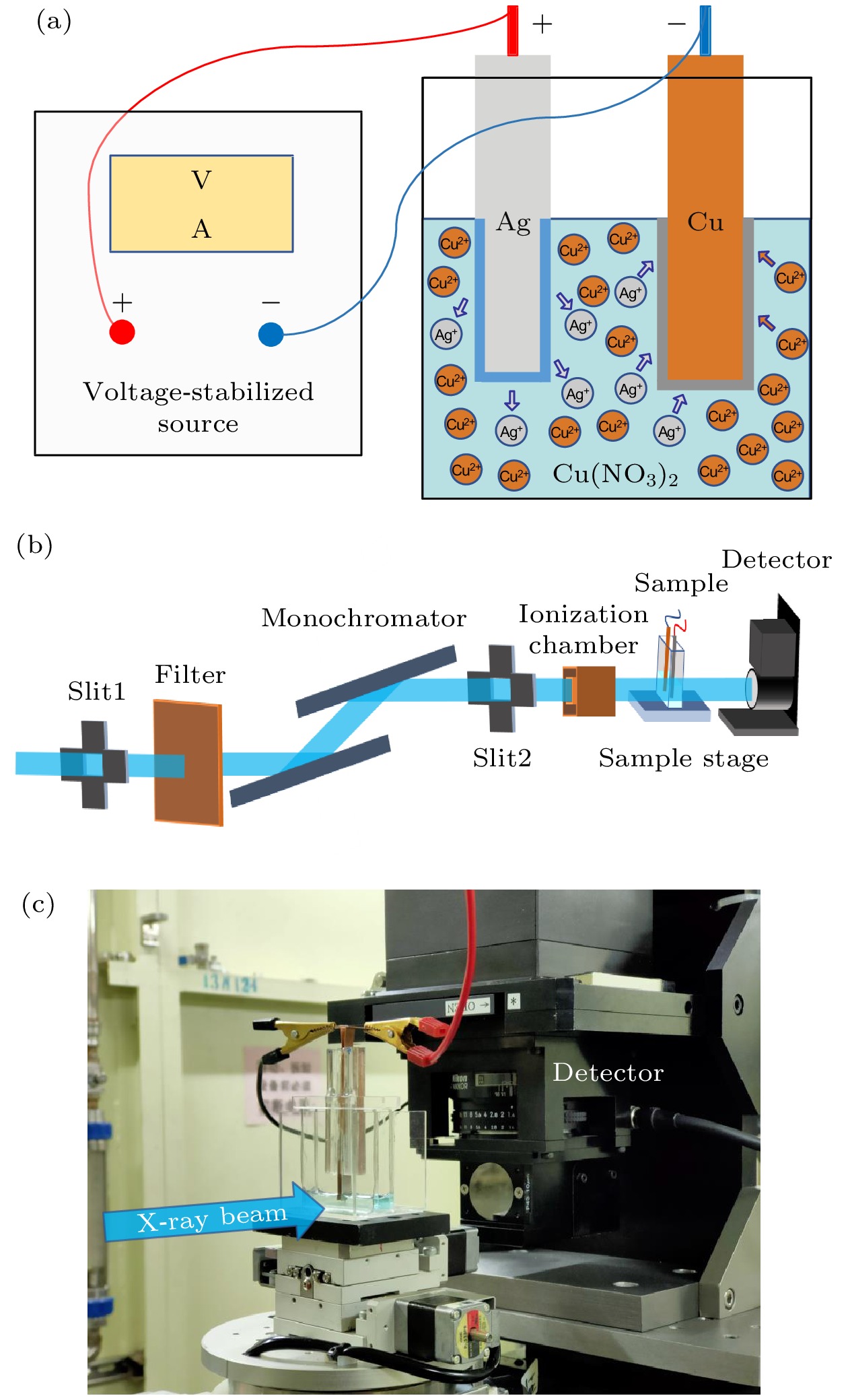
图 1 实验原理和装置 (a)电解池内离子迁移示意图; (b) 成像光路示意图; (c)包含电解池和探测器的实验装置照片
Figure 1. Experimental setup for the electrochemical reaction: (a) Schematic diagram of ion migration; (b) schematic diagram of optical path for X-ray imaging; (c) photo for the experimental equipment including electrolytic cell and X-ray detector.
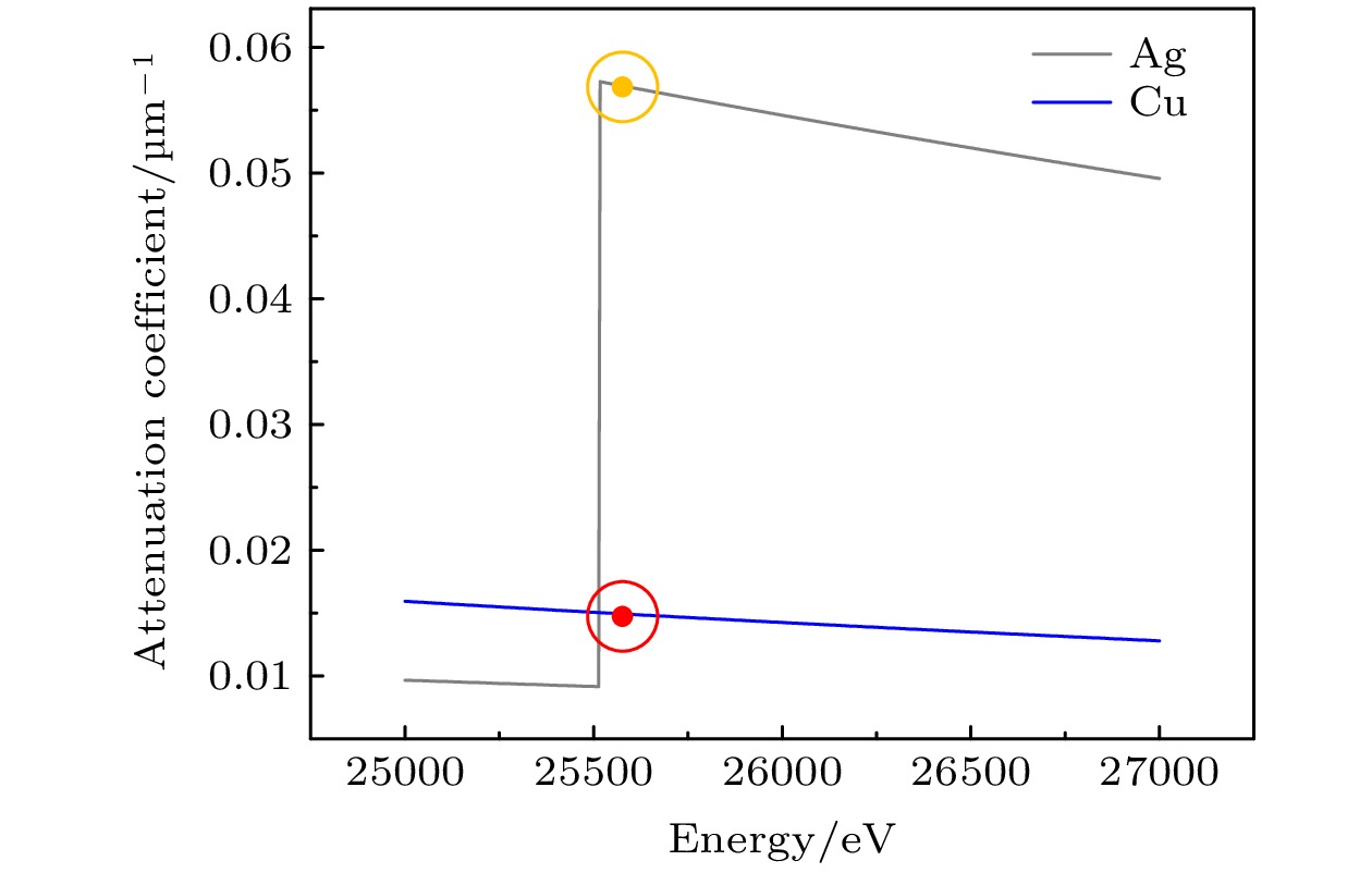
图 2 Ag元素和Cu元素在25—27 keV能量下的线性吸收系数
Figure 2. Linear absorption coefficients of Ag and Cu elements at the energy range of 25–27 keV.

图 3 电解池0.7 V电压通电后化学反应过程动态成像 (a) 传统时间减影成像1—12 s 关键帧; (b)对应的运动衬度成像关键帧
Figure 3. Dynamic X-ray imaging of electrochemical reaction after electrolytic cell is powered on at a voltage of 0.7 V: (a) Keyframes of traditional temporal subtraction imaging at the time period of 1–12 s; (b) the corresponding keyframes of move contrast imaging.

图 4 电解池0.7 V电压通电初期800 ms内的电化学反应 (a) 时间减影成像关键帧; (b)运动衬度成像关键帧
Figure 4. The initial stage of electrochemical reaction in the electrolytic cell with the voltage of power supply set to 0.7 V: (a) Keyframes of 300, 400, 500, 600, 700, 800 ms respectively obtained with temporal subtraction X-ray imaging; (b) the correspondent keyframes of move contrast X-ray imaging.
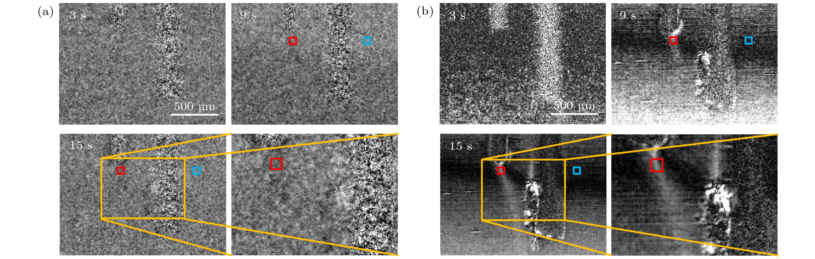
图 5 电解池0.5 V电压通电后的电化学反应过程成像 (a)传统时间减影成像3, 9, 15 s关键帧及在15 s时的局部区域的放大图; (b)运动衬度成像关键帧
Figure 5. X-ray imaging of electrochemical reaction after electrolytic cell is switched on at a voltage of 0.5 V: (a) Keyframes of 3, 9, 15 s respectively obtained with traditional temporal subtraction imaging supplied with a magnified view of the selected area at 15 s; (b) the corresponding keyframes of move contrast imaging.
-
[1] Kang B, Ceder G 2009 Nature 458 190
Google Scholar
[2] Okubo M, Mizuno Y, Yamada H, Kim J, Hosono E, Zhou H S, Kudo T, Honma I 2010 ACS Nano 4 741
Google Scholar
[3] Ellis B, Perry L K, Ryan D H, Nazar L F 2006 J. Am. Chem. Soc. 128 11416
Google Scholar
[4] Zhao W Y, Sakurai K 2019 J. Synchrotron. Radiat. 26 230
Google Scholar
[5] Chen S L, Zhang Y, Zhao J J, Mi Z, Zhang J M, Cao J, Feng J C, Zhang G L, Qi J L, Li J Y, Gao P 2020 Sci. Bull. 65 1643
Google Scholar
[6] 陈树林, 高鹏 2019 物理 48 168
Google Scholar
Chen S L, Gao P 2019 Physics 48 168
Google Scholar
[7] 刘玄玄, 国洪轩, 徐涛, 尹奎波, 孙立涛 2021 物理学报 70 086701
Google Scholar
Liu X X, Guo H X, Xu T, Yin K B, Sun L T 2021 Acta Phys. Sin. 70 086701
Google Scholar
[8] Yang Y C, Gao P, Gaba S, Chang T, Pan X, Lu W 2012 Nat. Commun. 3 732
Google Scholar
[9] 陆敬予, 柯承志, 龚正良, 李德平, 慈立杰, 张力, 张桥保 2021 物理学报 70 198102
Google Scholar
Lu J Y, Ke C Z, Gong Z L, Li D P, Ci L J, Zhang L, Zhang Q B 2021 Acta Phys. Sin. 70 198102
Google Scholar
[10] 郭祝崑, 李香庭 1983 物理学报 32 406
Google Scholar
Guo Z K, Li X T 1983 Acta Phys. Sin. 32 406
Google Scholar
[11] 杨同华, 包宗渝 1984 物理学报 33 1149
Google Scholar
Yang T H, Bao Z Y 1984 Acta Phys. Sin. 33 1149
Google Scholar
[12] Warren J M, Bilheux H Z, Kang M, Voisin S 2013 Plant Soil 366 683
Google Scholar
[13] Ilott A J, Trease N M, Grey C P, Jerschow A 2014 Nat. Commun. 5 4536
Google Scholar
[14] Zhou L, Leskes M, Liu T, Grey C P 2015 Angew. Chem. Int. Edit. 54 14782
Google Scholar
[15] Zheng J, Tang M X, Hu Y-Y 2016 Angew Chem. Int. Edit. 55 12538
Google Scholar
[16] Takanashi T, Kawamura H 2019 World Congress on Medical Physics and Biomedical Engineering 2018 Prague, Czech Republic, June 3–8, 2018 p35
[17] 安汉文, 莫生凯, 李梦璐, 王家钧 2022 储能科学与技术 11 834
An H W, Mo S K, Li M L, Wang J J 2022 Energy Storage Science and Technology 11 834
[18] 周逸凡, 杨慕紫, 佘峰权, 龚力, 张晓琪, 陈建, 宋树芹, 谢方艳 2021 物理学报 70 178801
Google Scholar
Zhou Y F, Yang M Z, She F Q, Gong L, Zhang X Q, Chen J, Song S Q, Xie F Y 2021 Acta Phys. Sin. 70 178801
Google Scholar
[19] Cheng L, Tscheuschner S, Paulus F, Hopkinson P E, Kieling J, Khler A, Vaynzof Y, Huettner S 2016 Adv. Mater. 28 2446
Google Scholar
[20] 王继飞, 林东旭, 袁永波 2019 物理学报 68 158801
Google Scholar
Wang J F, Lin D X, Yuan Y B 2019 Acta Phys. Sin. 68 158801
Google Scholar
[21] 果辰, 蔡欣炜, 罗文浩, 黄子耕, 冯庆荣, 甘子钊 2021 物理学报 70 197401
Google Scholar
Guo C, Cai X W, Luo W H, Huang Z G, Feng Q R, Gan Z Z 2021 Acta Phys. Sin. 70 197401
Google Scholar
[22] 王丽, 王海波, 王涛, 李发伸 2006 物理学报 55 6515
Google Scholar
Wang L, Wang H B, Wang T, Li F S 2006 Acta Phys. Sin. 55 6515
Google Scholar
[23] Zhao W Y, Sakurai K 2017 ACS Omega 2 4363
Google Scholar
[24] 王飞翔 2019 博士学位论文 (北京: 中国科学院大学 (中国科学院上海应用物理研究所))
Wang F X 2019 Ph. D. Dissertation (Beijing: Shanghai Institute of Applied Physics, University of Chinese Academy of Sciences) (in Chinese)
[25] Wang F X, Zhou P T, Li K, Mamtilahun M, Tang Y H, Du G H, Deng B, Xie H L, Yang G Y, Xiao T Q 2020 IUCrJ 7 1
Google Scholar
[26] 李可 2021 博士学位论文 (北京: 中国科学院大学 (中国科学院上海应用物理研究所))
Li K 2021 Ph. D. Dissertation (Beijing: Shanghai Institute of Applied Physics, University of Chinese Academy of Sciences) (in Chinese)
[27] Song X M, Pogue B W, Jiang S D, Doyley M M, Dehghani H, Tosteson T D, Paulsen K D 2004 Appl. Optics 43 1053
Google Scholar
[28] Xie H L, Deng B, Du G H, Fu Y N, Guo H, Xue Y L, Peng G Y, Tao F, Zhang L, Xiao T Q 2020 Nucl. Sci. Tech. 31 102
Google Scholar
[29] 郭荣怡, 马红娟, 薛艳玲, 谢红兰, 邓彪, 杜国浩, 王敏, 肖体乔 2010 光学学报 30 2898
Google Scholar
Guo R Y, Ma H J, Xue Y L, Xie H L, Deng B, Du G H, Wang M, Xiao T Q 2010 Acta Optica Sin. 30 2898
Google Scholar
[30] Ju X L, Deng B, Li K, Yu F C, Zhang H P, Xu M W, Du G H, Xie H L, Li B, Xiao T Q 2022 Nucl. Sci. Tech. 33 1
Google Scholar
-
 144101-20220339补充材料.pdf


 DownLoad:
Catalog
Metrics
- Abstract views: 9294
- PDF Downloads: 134
- Cited By: 0
