


-
In 2005, the FLASH soft X-ray free-electron laser (FEL) in Hamburg, Germany, achieved its first lasing, which began an intensive phase of global FEL construction. Subsequently, the United States, Japan, South Korea, China, Italy, and Switzerland all began building such photon facilities. Recently, the new generation of FEL has started to utilize superconducting acceleration technology to achieve high-repetition-rate pulse output, thereby improving experimental efficiency. Currently completed facility is the European XFEL, ongoing constructions are the LCLS-II in the United States and the SHINE facility in Shanghai, and the facility in preparation is the Shenzhen superconducting soft X-ray free-electron laser (S3FEL). These FEL facilities generate coherent and tunable ultrashort pulses ranging from the extreme ultraviolet to hard X-ray spectrum, which advances the FEL-based scattering techniques such as ultrafast X-ray scattering, spectroscopy, and X-ray nonlinear optics, thereby transforming the way we study correlated quantum materials on an ultrafast timescale. The self-amplified spontaneous emission (SASE) process in FEL leads to timing jitter between FEL pulses and the synchronized pump laser, influencing the accuracy of ultrafast time-resolved measurements. To address this issue, timing tools have been developed to measure these jitters and reindexed each pump-probe signal after measurement. This success enables ultrafast X-ray diffraction (UXRD) to be first realized, and a systematic study of Peierls distorted materials is demonstrated. In addition, the high flux of FEL pulses enables Fourier transform inelastic X-ray scattering (FT-IXS) method, which can extract the phonon dispersion curve of the entire Brillouin zone by performing the Fourier transform on the measured momentum dependent coherent phonon scattering signals, even when the system is in a non-equilibrium state. The UXRD is typically used to study ultrafast lattice dynamics, which requires hard X-ray wavelengths. In contrast, time resolved resonant elastic X-ray scattering (tr-REXS) in the soft X-ray regime has become a standard method of investigating nano-sized charge and spin orders in correlated quantum materials on an ultrafast time scale. In correlated quantum materials, the interplay between electron dynamics and lattice dynamics represents another important research direction. In addition to Zhi-Xun Shen's successful demonstration of the combined tr-ARPES and UXRD method at SLAC, this paper also reports the attempts to integrate UXRD with resonant X-ray emission spectroscopy (RXES) for the simultaneous measurement of electronic and lattice dynamics. Resonant inelastic X-ray scattering (RIXS) is a powerful tool for studying elementary and collective excitations in correlated quantum materials. However, in FEL-based soft X-ray spectroscopy, the wavefront tilt introduced by the widely used grating monochromators inevitably stretches the FEL pulses, which degrades the time resolution. Therefore, the new design at FEL beamlines adopts low line density gratings with long exit arms to reduce pulse stretch and achieve relatively high energy resolution. For example, the Heisenberg-RIXS instrument at the European XFEL achieves an energy resolution of 92 meV at the Cu L3 edge and approximately 150 fs time resolution. In recent years, scientists at SwissFEL’s Furka station have drawn inspiration from femtosecond optical covariance spectroscopy to propose a new method of generating two-dimensional time-resolved resonant inelastic X-ray scattering (2D tr-RIXS) spectra. This method involves real-time detection of single-shot FEL incident and scattered spectra, followed by deconvolution calculation to avoid photon waste and wavefront tilt caused by monochromator slits. The SQS experimental station at European XFEL, built in 2023, features a 1D-XUV spectrometer that utilizes subtle variations in photon energy absorption across the sample to induce spatial energy dispersion. Using Wolter mirrors, it directly images spatially resolved fluorescence emission from the sample onto the detector to generate 2D tr-RIXS spectra without the need for deconvolution. However, this design is limited to specific samples. Currently, the S3FEL under designing has a novel 2D tr-RIXS instrument that uses an upstream low line density grating monochromator to generate spatial dispersion of the beam spot, allowing the full bandwidth of SASE to project spatially dispersed photon energy onto the sample. Subsequently, an optical design similar to the 1D-XUV spectrometer will be employed to achieve 2D tr-RIXS spectra, thereby expanding the applicability beyond specific liquid samples. These new instruments are designed to minimize pulse elongation by fully utilizing SASE’s full bandwidth, approaching Fourier-transform-limited RIXS spectra in both time and energy resolution. Nonlinear X-ray optical techniques, such as sum-frequency generation (SFG) and second-harmonic generation, are adapting to X-ray wavelengths and opening up new avenues for detecting elementary excitations. The X-ray transient grating spectroscopy extends its capabilities to studying charge transport and spin dynamics on an ultrafast timescale. The future development of these scattering methods provides unique opportunities for detecting dynamical events in various systems, including surface and interface processes, chirality, nanoscale transport, and so-called multidimensional core-level spectroscopy. -
Keywords:
- free electron laser /
- ultrafast X-ray scattering /
- ultrafast X-ray spectroscopy /
- X-ray nonlinear optics
[1] Rossbach J, Schneider J R, Wurth W 2019 Phys. Rep. 808 1
 Google Scholar
[2] Emma P, Akre R, Arthur J, et al. 2010 Nat. Photonics 4 641
Google Scholar
[3] Ishikawa T, Aoyagi H, Asaka T, et al. 2012 Nat. Photonics 6 540
Google Scholar
[4] Allaria E, Castronovo D, Cinquegrana P, et al. 2013 Nat. Photonics 7 913
Google Scholar
[5] Wang H L, Yu Y, Chang Y, et al. 2018 J. Chem. Phys. 148 124301
Google Scholar
[6] Kang H S, Min C K, Heo H, et al. 2017 Nat. Photonics 11 708
Google Scholar
[7] Milne C J, Schietinger T, Aiba M, et al. 2017 Appl. Sci. 7 720
Google Scholar
[8] Zhao Z T, Wang D, Gu Q, Yin L X, Fang G P, Gu M, Leng Y B, Zhou Q G, Liu B, Tang C X, Huang W H, Liu Z, Jiang H D 2017 Synchrotron Radiat. News 30 29
Google Scholar
[9] Ball P 2017 Nature 548 7669
Google Scholar
[10] Halavanau A, Decker F J, Emma C, Sheppard J, Pellegrini C 2019 J. Synchrotron Radiat. 26 635
Google Scholar
[11] Zhu Z Y, Zhao Z T, Wang D, Liu Z, Li R X, Yin L X, Yang Z H 2017 Proceedings of the 38th International Free-Electron Laser Conference Santa Fe, NeW Mexico, August 20–25, MOP055
[12] Simmermacher M, Moreno Carrascosa A, E Henriksen N, B Møller K, Kirrander A 2019 J. Chem. Phys. 151 174302
Google Scholar
[13] Reich C, Gibbon P, Uschmann I, Förster E 2000 Phys. Rev. Lett. 84 4846
Google Scholar
[14] Corde S, Ta Phuoc K, Lambert G, Fitour R, Malka V, Rousse A, Beck A, Lefebvre E 2013 Rev. Mod. Phys. 85 1
Google Scholar
[15] Zamponi F, Ansari Z, V. Korff Schmising C, Rothhardt P, Zhavoronkov N, Woerner M, Elsaesser T, Bargheer M, Trobitzsch-Ryll T, Haschke M 2009 Appl. Phys. A 96 51
Google Scholar
[16] Rose-Petruck C, Jimenez R, Guo T, Cavalleri A, Siders C W, Rksi F, Squier J A, Walker B C, Wilson K R, Barty C P J 1999 Nature 398 310
Google Scholar
[17] Cavalleri A, Tóth C, Siders C W, Squier J A, Ráksi F, Forget P, Kieffer J C 2001 Phys. Rev. Lett. 87 237401
Google Scholar
[18] Sokolowski-Tinten K, Blome C, Blums J, Cavalleri A, Dietrich C, Tarasevitch A, Uschmann I, Förster E, Kammler M, Horn-von-Hoegen M, Von der Linde D 2003 Nature 422 287
Google Scholar
[19] Fabricius N, Hermes P, Von der Linde D, Pospieszczyk A, Stritzker B 1986 Solid State Commun. 58 239
Google Scholar
[20] Rethfeld B, Sokolowski-Tinten K, Von der Linde D, Anisimov S I 2002 Phys. Rev. B 65 092103
Google Scholar
[21] Huang N S, Deng H X, Liu B, Wang D, Zhao Z T 2021 The Innovation 2 100097
Google Scholar
[22] Zhao Z T, Wang D, Chen J H, et al. 2012 Nat. Photonics 6 360
Google Scholar
[23] Kondratenko A M, Saldin E L 1980 Part. Accel. 10 207
[24] Fritz D M, Reis D A, Adams B, et al. 2007 Science 315 633
Google Scholar
[25] Harmand, M, Coffee R, Bionta M R, et al. 2013 Nat. Photonics 7 215
Google Scholar
[26] Epp S W, Hada M, Zhong Y, et al. 2017 Struct. Dyn. 4 054308
Google Scholar
[27] Krasniqi F S, Zhong Y, Epp S W, Foucar L, Trigo M, Chen J, Reis D A, Wang H L, Zhao J H, Lemke H T, Zhu D, Chollet M, Fritz D M, Hartmann R, Englert L, Strüder L, Schlichting I, Ullrich J 2018 Phys. Rev. Lett. 120 105501
Google Scholar
[28] Chuang Y D, Lee W S, Kung Y F, et al. 2013 Phys. Rev. Lett. 110 127404
Google Scholar
[29] Först M, Beyerlein K R, Mankowsky R, Hu W, Mattoni G, Catalano S, Gibert M, Yefanov O, Clark J N, Frano A, Glownia J M, Chollet M, Lemke H, Moser B, Collins S P, Dhesi S S, Caviglia A D, Triscone J M, Cavalleri A 2017 Phys. Rev. Lett. 118 027401
Google Scholar
[30] Doering D, Chuang Y D, Andresen N, Chow K, Contarato D, Cummings C, Domning E, Joseph J, Pepper J S, Smith B, Zizka G, Ford C, Lee W S, Weaver M, Patthey L, Weizeorick J, Hussain Z, Denes P 2011 Rev. Sci. Instrum. 82 073303
Google Scholar
[31] Jang H Y, Kim H D, Kim M, et al. 2020 Rev. Sci. Instrum. 91 083904
Google Scholar
[32] Trigo M, Fuchs M, Chen J, et al. 2013 Nat. Phys. 9 790
Google Scholar
[33] Zhu D L, Robert A, Henighan T, T. Lemke H, Chollet M, Glownia J M, A. Reis D, Trigo M 2015 Phys. Rev. B 92 054303
Google Scholar
[34] Engel R Y, Miedema P S, Turenne D, Vaskivskyi I, Brenner G, Dziarzhytski S, Kuhlmann M, Schunck J O, Döring F, Styervoyedov A, Parkin S S P, David C, Schüßler-Langeheine C, Dürr H A, Beye M 2020 Appl. Sci. 10 6947
Google Scholar
[35] Gerber S, Yang S L, Zhu D, et al. 2017 Science 357 71
Google Scholar
[36] Krasniqi F S, Zhong Y P, Reis D A, et al. 2012 Research in Optical Sciences, IW1D. 2
[37] Zastrau U, Appel K, Baehtz C, et al. 2021 J. Synchrotron Radiat. 28 1393
Google Scholar
[38] Wollenweber L, Preston T R, Descamps A, et al. 2021 Rev. Sci. Instrum. 92 013101
Google Scholar
[39] Dean M P M, Cao Y, Liu X, et al. 2016 Nat. Mater. 15 601
Google Scholar
[40] Gerasimova N, La Civita D, Samoylova L, et al. 2022 J. Synchrotron Radiat. 29 1299
Google Scholar
[41] Tollerud J O, Sparapassi G, Montanaro A, Asban S, Glerean F, Giusti F, Marciniak A, Kourousias G, Billè F, Cilento F, Mukamel S, Fausti D 2019 Proc. Natl. Acad. Sci. U. S. A 116 5383
Google Scholar
[42] Chergui M, Beye M, Mukamel S, Svetina C, Masciovecchio C 2023 Nat. Rev. Phys. 5 578
Google Scholar
[43] Harter J W, Zhao Z Y, Yan J Q, Mandrus D G, Hsieh D 2017 Science 356 295
Google Scholar
[44] Lam R K, Raj S L, Pascal T A, et al. 2018 Phys. Rev. Lett. 120 23901
Google Scholar
[45] Zhao L, Belvin C A, Liang R, Bonn D A, Hardy W N, Armitage N P, Hsieh D 2017 Nat. Phys. 13 250
Google Scholar
[46] Uzundal C B, Jamnuch S, Berger E, et al. 2021 Phys. Rev. Lett. 127 237402
Google Scholar
[47] Patterson B D 2010 SLAC Technical Note SLAC-TN-10-026
[48] Glover T E, Fritz D M, Cammarata M, et al. 2012 Nature 488 603
Google Scholar
[49] Rottke H, Engel R Y, Schick D, Schunck J O, Miedema P S, Borchert M C, Kuhlmann M, Ekanayake N, Dziarzhytski S, Brenner G, Eichmann U, von Korff Schmising C, Beye M, Eisebitt S 2022 Sci. Adv. 8 5127
Google Scholar
[50] Beye M 2021 Nat. Photonics 15 490
Google Scholar
[51] Bencivenga F, Mincigrucci R, Capotondi F, et al. 2019 Sci. Adv. 5 5805
Google Scholar
[52] Mincigrucci R, Foglia L, Naumenko D, et al. 2018 Nucl. Instrum. Meth. A 907 132
Google Scholar
[53] Rouxel J R, Fainozzi D, Mankowsky R, et al. 2021 Nat. Photonics 15 499
Google Scholar
[54] Jonnard P, André J M, Le Guen K, Le, Wu M Y, Principi E, Simoncig A, Gessini A, Mincigrucci R, Masciovecchio C, Peyrusse O 2017 Struct. Dyn. 4 054306
Google Scholar
[55] Foglia L, Mincigrucci R, Maznev A A, et al. 2023 Photoacoustics 29 100453
Google Scholar
[56] Sumi T, Horio M, Senoo T, et al. 2022 E-J. Surf. Sci. Nanotechnol. 20 31
Google Scholar
[57] Robin Y E, Oliver A, Kaan A, et al. 2023 Struct. Dyn. 10 054501
Google Scholar
-

图 1 基于FEL的UXRD实验布局, 铍透镜组被用来实现FEL聚焦
Figure 1. Schematic layout of UXRD set up based on FEL, a group of Be lens is used for focusing the FEL.
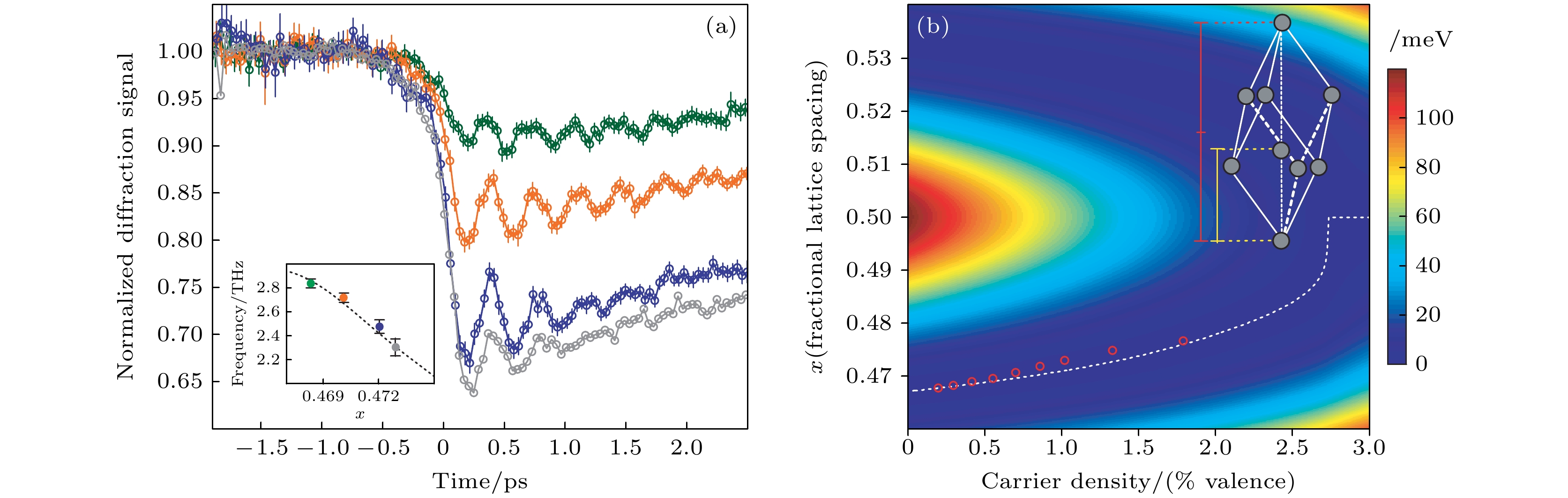
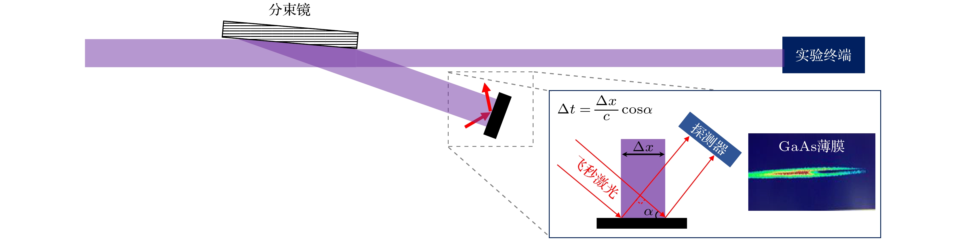
图 4 FEL与飞秒激光到达时间的差表现为飞秒激光经GaAs反射后的光斑在空间上被调制. 小插图为本文在SACLA装置上的单脉冲实测数据, 明亮区别即为FEL激发产生的飞秒激光反射斑的空间调制
Figure 4. Time difference between the FEL and the femtosecond laser is manifested as a spatial modulation of the femtosecond laser spot reflected by GaAs. The inset shows single-shot experimental data from the SACLA facility, where the bright contrast represents the spatial modulation of the femtosecond laser reflection spot induced by FEL excitation.
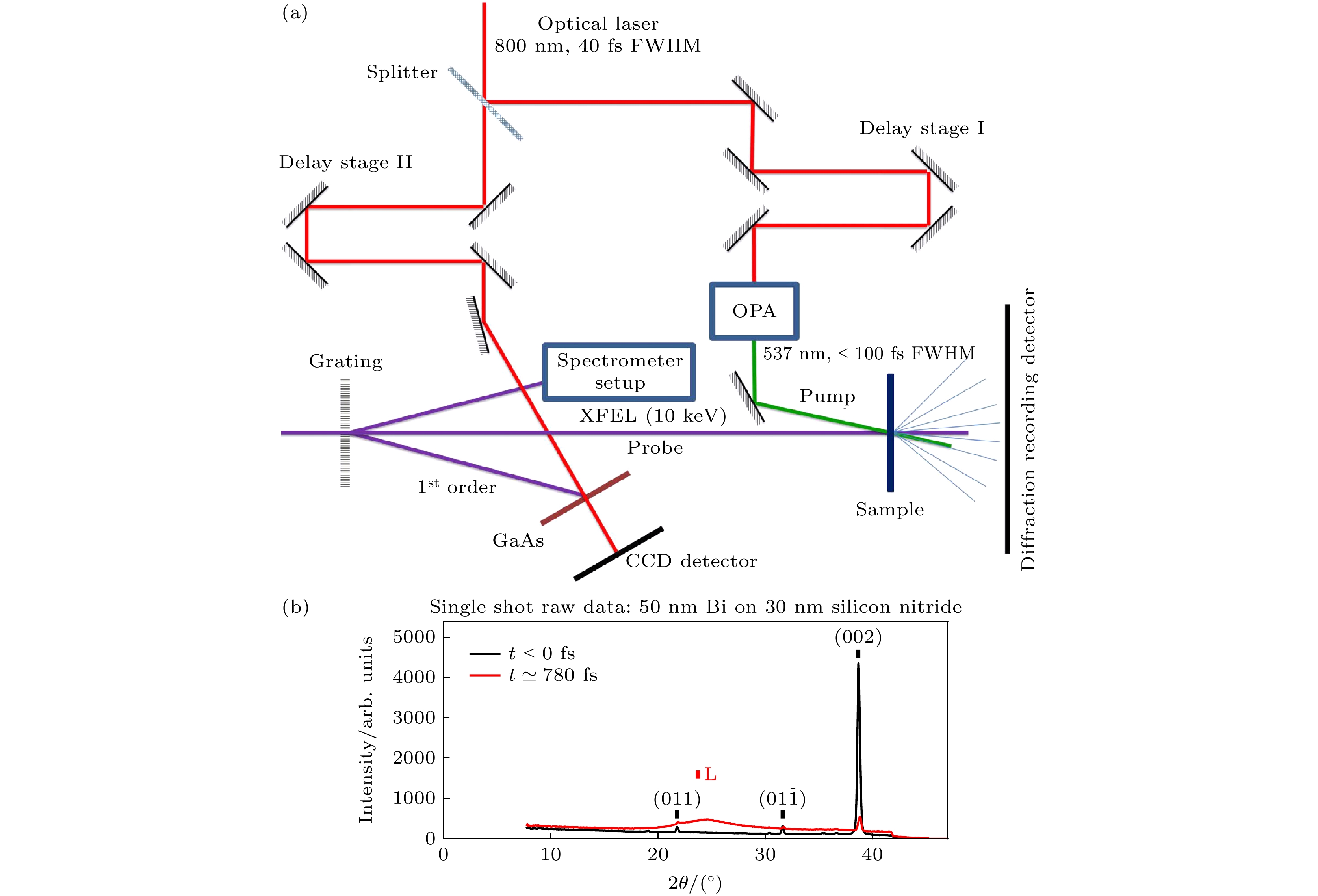
图 5 (a) 劳厄情形下时间零点的确定. FEL经过透射光栅产生的1级衍射被用来做时间工具, 和用于实验的0级光自然同步[26]. (b) 泵浦激光超快熔化30 nm铋薄膜, 引起固态铋(002)X射线衍射峰强度下降和产生新的液态铋衍射环L[26]
Figure 5. (a) Determination of the time zero in the Laue geometry. The first-order diffraction of FEL light through a transmission grating is used as a timing tool, synchronized naturally with the zero-order light used for the experiment[26]. (b) Pump laser induces ultrafast melting of a 30 nm Bi film, causing a decrease in the intensity of the solid Bi (002) X-ray diffraction peak and the formation of a new liquid Bi diffraction ring L[26].
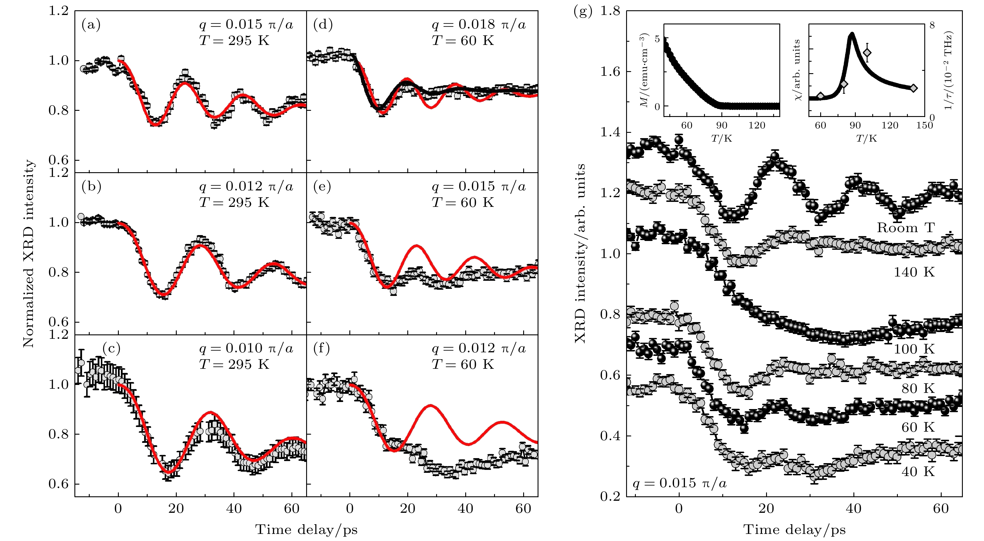
图 6 Ga0.91Mn0.09As样品在不同温度不同声子波长下测量(实心圆)和模拟(实线)XRD随时间变化[27] (a)—(c)室温; (d)—(f) 60 K; (g) 居里温度前后测量得到的UXRD信号, 小插图是自旋关联函数
Figure 6. Ga0.91Mn0.09As was measured (solid circles) and simulated (solid lines) using XRD over time at different temperatures and phonon wavelengths[27]: (a)–(c) At room temperature; (d)–(f) at 60 K; (g) UXRD signals measured below and above the Curie temperature, and the inset shows the spin correlation function.
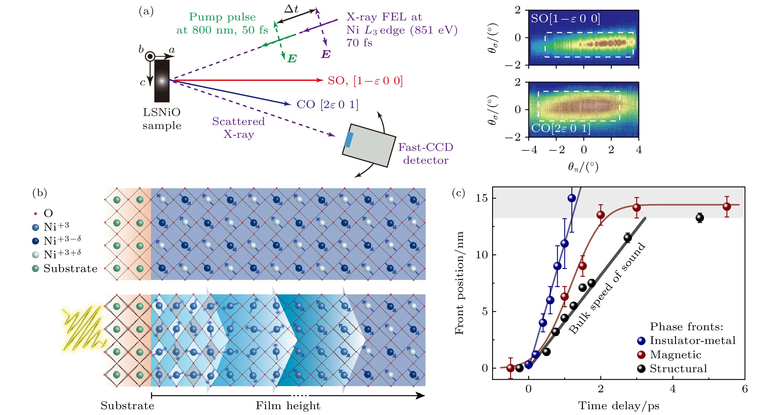
图 7 (a) 典型的时间分辨共振弹性X射线散射(time resolved resonant elastic X-ray scattering, tr-REXS)测量设置[28]; (b) 在LaAlO3/NdNiO3异质结构中飞秒中红外激发诱导的各相前沿传播的示意图[29]; (c) 电荷激发(绝缘体-金属相变)、磁有序的熔化(tr-REXS测量)以及晶格变化(UXRD)在图(b)中薄膜的差异化传播速度[29]
Figure 7. (a) Typical tr-REXS measurement setup[28]; (b) schematic of the propagation of various phase fronts induced by femtosecond mid-infrared excitation in the LaAlO3/NdNiO3 heterostructure[29]; (c) the different propagation speeds of electronic excitation (insulator to metal transition), magnetic order melting (tr-REXS measurement), and lattice changes (UXRD) in the film shown in panel (b) [29].
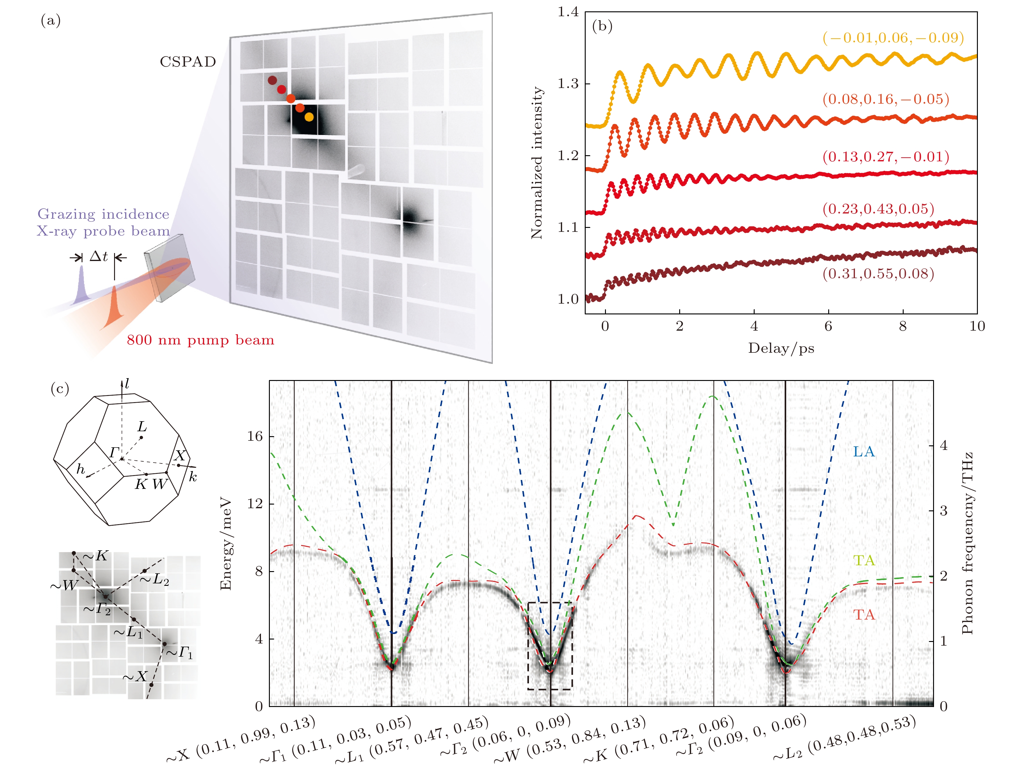
图 8 (a) 傅里叶变换X射线非弹性散射示意图. 灰度图像为多次平均的热漫散射图案[33]; (b) 不同动量空间位置处声子强度随泵浦-探测延迟时间的演变[33]; (c) 将图(b)测量曲线经傅里叶变换得到的声子色散关系[33]
Figure 8. (a) Schematic of FT-IXS. The grayscale image shows a multi-shot averaged thermal diffuse scattering pattern[33]; (b) evolution of phonon intensity as a function of pump-probe delay time at different momentum space positions[33]; (c) phonon dispersion relations obtained by performing a Fourier transform on the measurement curves from panel (b) [33].
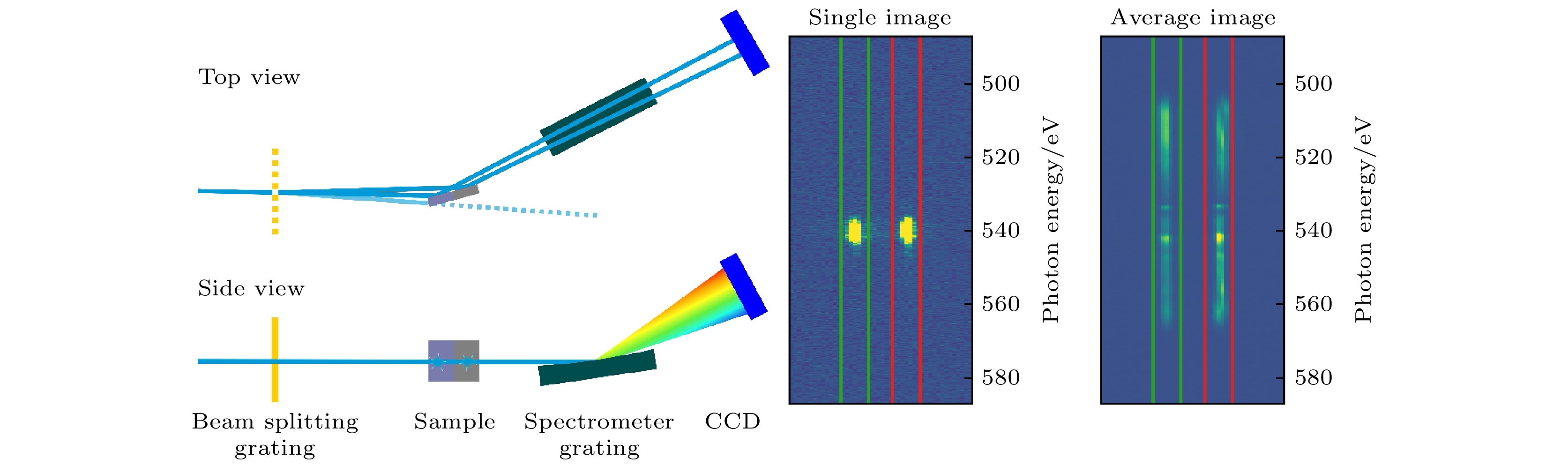
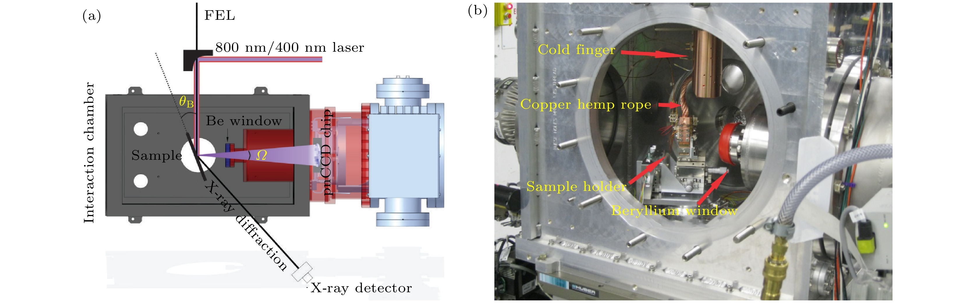
图 10 (a) UXRD和tr-RXES测量装置. pnCCD用来实现tr-RXES的采集, 铍窗用来隔开pnCCD与实验腔体的真空, 同时保持较高的X射线荧光透过率[36]. (b) 实验腔体内部环境, 闭循环氦冷指通过铜辫与样品架连接, 降温的同时隔振[36]
Figure 10. (a) The UXRD and tr-RXES measurement setup. The pnCCD is used for tr-RXES collection, and the Be window separates the pnCCD from the vacuum of the experimental chamber while maintaining a high X-ray fluorescence transmission rate[36]. (b) Internal environment of the experimental chamber: the closed-loop helium cooling system is connected to the sample holder via copper braids, providing cooling while also isolating vibrations[36].
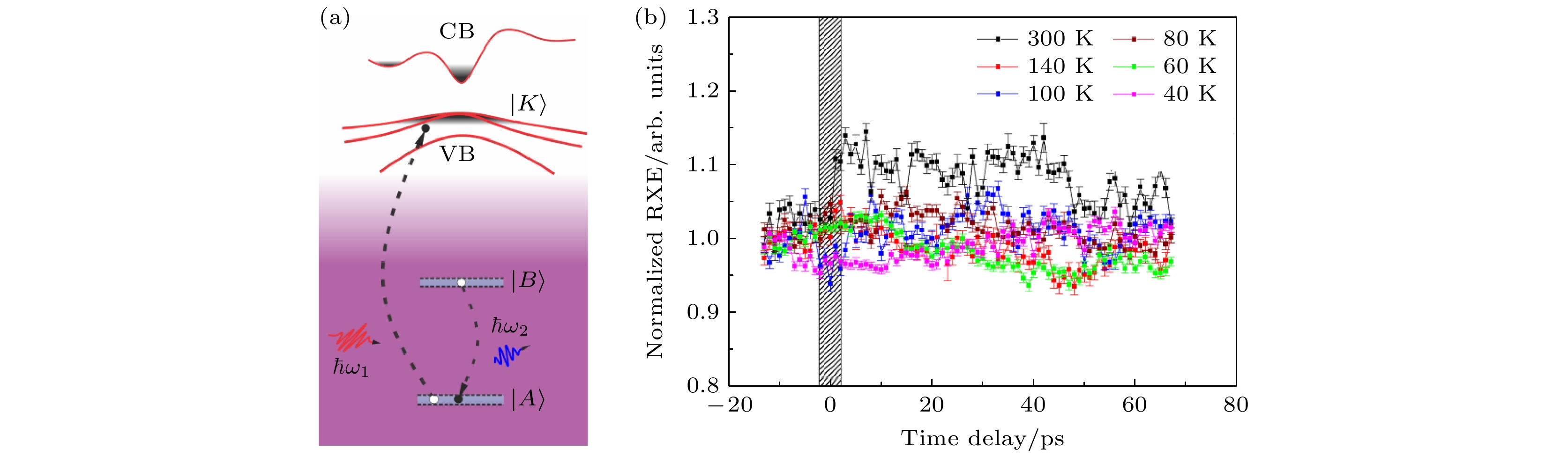
图 11 (a) tr-RXES测量原理, 当能带被占据时, 跃迁(红色脉冲)是禁止的. 而当泵浦激光激发产生带隙跃迁后产生空穴, 则引起吸收增强. 随后产生从B能级到A跃迁的发射谱, B能级的位置可以在内壳层, 也可以在带隙附近. (b) 实验观测的时间分辨RXE数据, 可见在时间零点之后的变化
Figure 11. (a) Principle of tr-RXES measurement: when the valence band is occupied, transitions (represented by the red pulse) are forbidden. However, when pump laser excitation induces a bandgap transition and creates holes, it leads to enhanced absorption. This is followed by an emission spectrum resulting from the transition from energy level B to A. The B energy level can be in the inner shell or near the bandgap. (b) Experimental observation of RXE data showing changes after time zero.
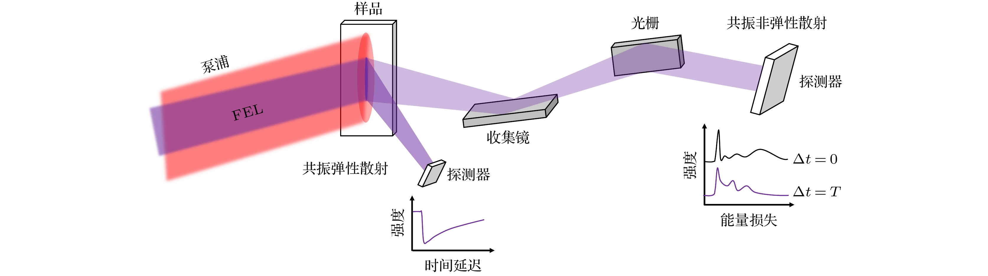
图 12 共振弹性散射和非弹性散射的同时测量设想
Figure 12. Simultaneous measurement concept of resonant elastic scattering and inelastic scattering.
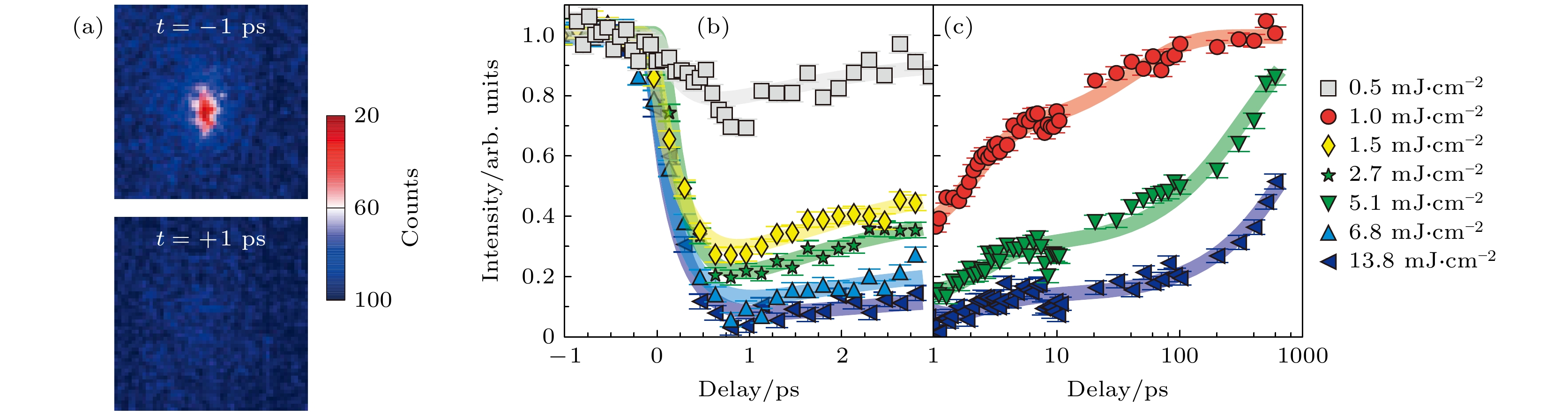
图 13 在激光泵浦作用下, Sr2IrO4中三维磁有序的动态变化过程[39] (a) 在激光泵浦前后1 ps时间内, (–3, –2, 28)磁有序布拉格峰的强度变化; (b), (c) 在不同强度的激光泵浦后, 磁有序布拉格峰强度随时间演化的过程
Figure 13. Dynamic changes of three-dimensional magnetic ordering in Sr2IrO4 under laser pumping[39]: (a) Intensity variation of the (–3, –2, 28) magnetically ordered Bragg peak within 1 ps before and after laser pumping; (b), (c) the evolution of magnetic ordered Bragg peak intensity over time after laser pumping at different intensities.
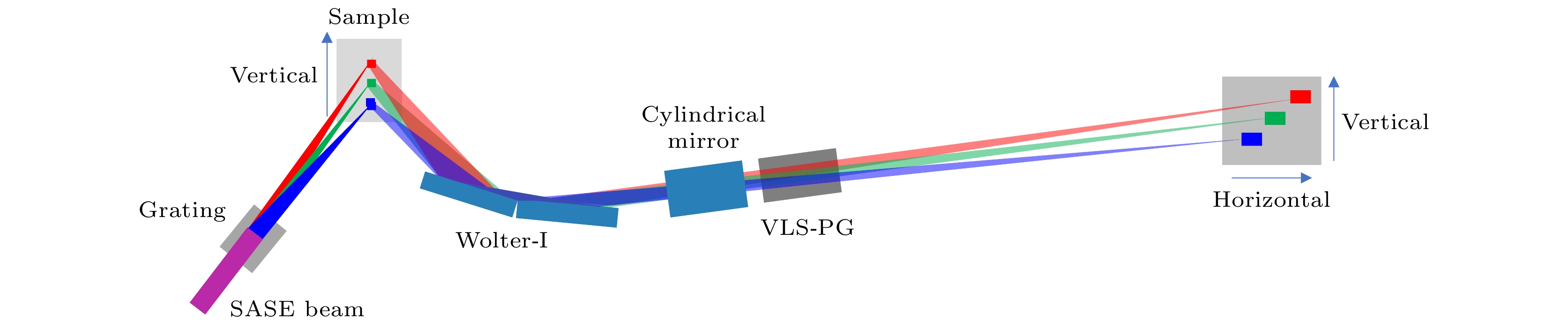
图 14 软X射线2D-RIXS装置光路示意图
Figure 14. Schematic of the optical layout for a soft X-ray 2D-RIXS spectrometer.
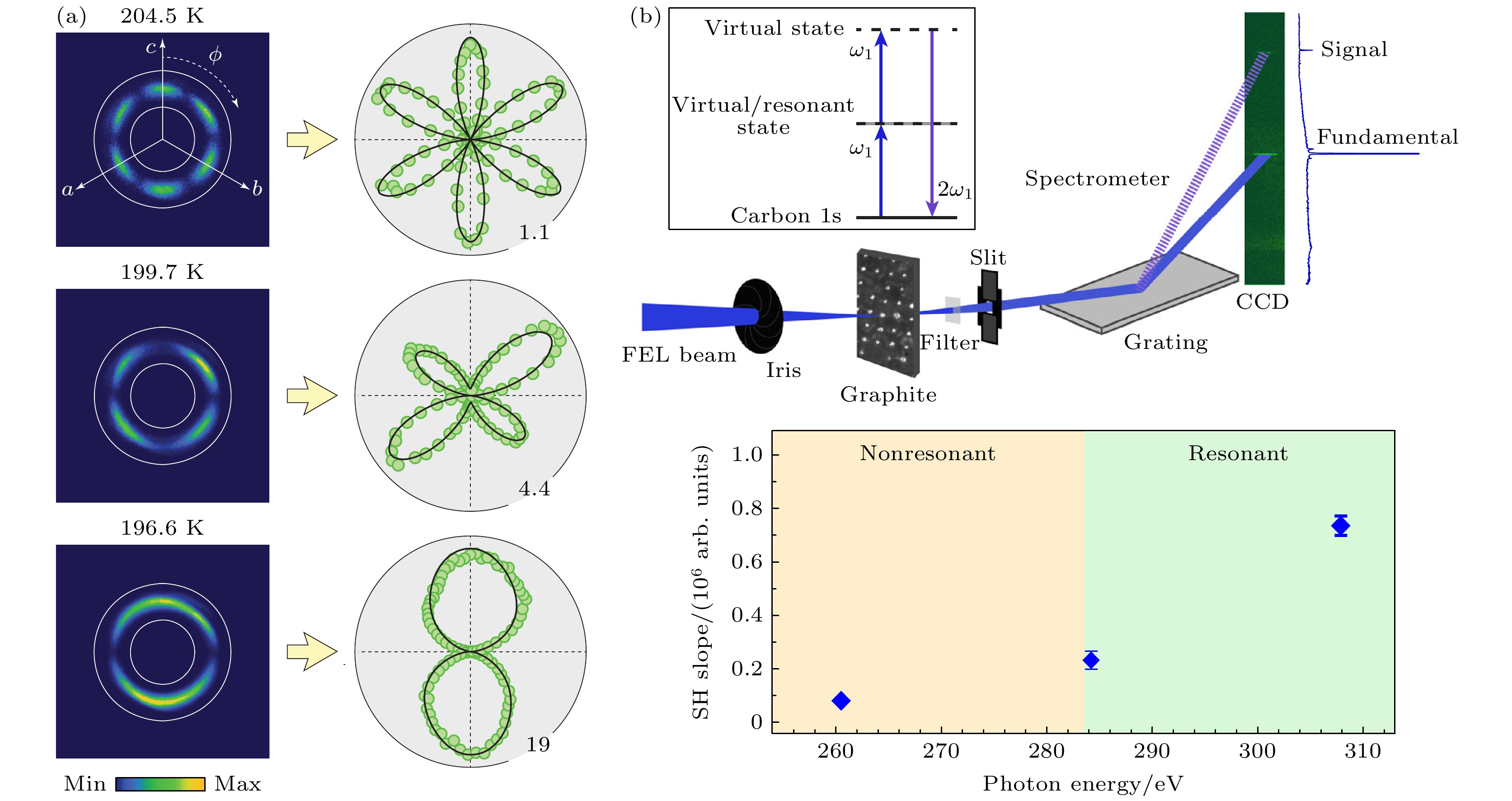
图 15 (a) 由800 nm激光在自旋轨道耦合金属Cd2Re2O7中产生的SHG信号, SHG信号的角分布代表了材料中的极化方向[43]; (b) 软X射线FEL作用于石墨产生SHG(上图), 波长调至共振边使得SHG信号增强[44]
Figure 15. (a) SHG signal generated by the 800 nm laser in the spin-orbit coupled metal Cd2Re2O7, and the angular distribution of SHG represents the polarization direction in the material[43]; (b) soft X-ray FEL is applied to the graphite generating SHG (above), and the resonance enhances the generated SHG signal[44].
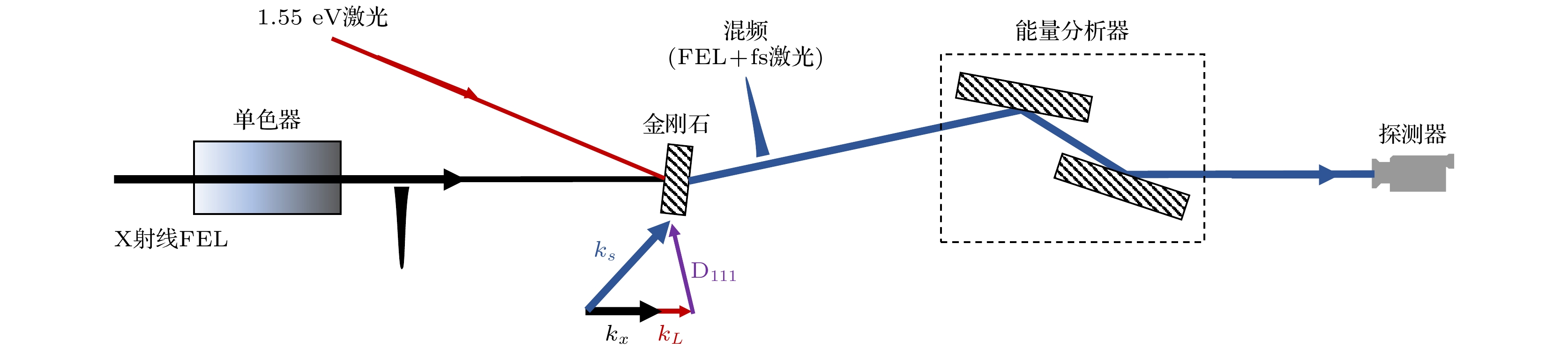
图 16 LCLS实现硬X射线与光学波段混频的装置
Figure 16. LCLS setup for hard X-ray and optical wave mixing.
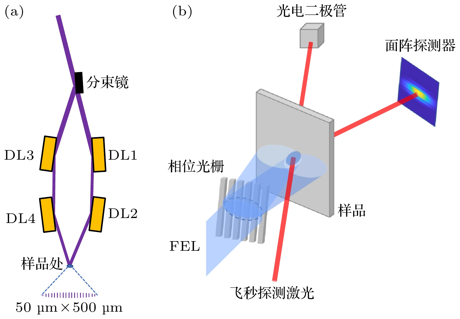
图 18 TG装置原理 (a) 入射的FEL光经分束镜后由DL1—4镜组形成两束时间延迟可调的束线, 于样品表面重合形成TG[52]; (b) 入射的FEL脉冲(蓝色部分)被一个传输相位光栅衍射. 衍射阶之间的干涉产生了周期结构. 探测光脉冲(红色)被瞬态光栅衍射到面阵探测器上, 通过调节探测光的时间延迟来测量TG激发后样品上的动力学[53]
Figure 18. Schematic of the X-ray TG set-up: (a) The incident FEL light is split by a splitting mirror and then reflected by the DL1—4 mirror group to form two beams with adjustable time delays, which overlap on the sample surface to form the TG[52]; (b) the incident FEL pulse (blue) is diffracted by a transmission phase grating. Interference between the diffraction orders creates a periodic structure. The probe pulse (red) is diffracted by the transient grating onto the area detector. By adjusting the time delay of the probe pulse, the dynamics of the sample following TG excitation can be measured[53].
-
[1] Rossbach J, Schneider J R, Wurth W 2019 Phys. Rep. 808 1
Google Scholar
[2] Emma P, Akre R, Arthur J, et al. 2010 Nat. Photonics 4 641
Google Scholar
[3] Ishikawa T, Aoyagi H, Asaka T, et al. 2012 Nat. Photonics 6 540
Google Scholar
[4] Allaria E, Castronovo D, Cinquegrana P, et al. 2013 Nat. Photonics 7 913
Google Scholar
[5] Wang H L, Yu Y, Chang Y, et al. 2018 J. Chem. Phys. 148 124301
Google Scholar
[6] Kang H S, Min C K, Heo H, et al. 2017 Nat. Photonics 11 708
Google Scholar
[7] Milne C J, Schietinger T, Aiba M, et al. 2017 Appl. Sci. 7 720
Google Scholar
[8] Zhao Z T, Wang D, Gu Q, Yin L X, Fang G P, Gu M, Leng Y B, Zhou Q G, Liu B, Tang C X, Huang W H, Liu Z, Jiang H D 2017 Synchrotron Radiat. News 30 29
Google Scholar
[9] Ball P 2017 Nature 548 7669
Google Scholar
[10] Halavanau A, Decker F J, Emma C, Sheppard J, Pellegrini C 2019 J. Synchrotron Radiat. 26 635
Google Scholar
[11] Zhu Z Y, Zhao Z T, Wang D, Liu Z, Li R X, Yin L X, Yang Z H 2017 Proceedings of the 38th International Free-Electron Laser Conference Santa Fe, NeW Mexico, August 20–25, MOP055
[12] Simmermacher M, Moreno Carrascosa A, E Henriksen N, B Møller K, Kirrander A 2019 J. Chem. Phys. 151 174302
Google Scholar
[13] Reich C, Gibbon P, Uschmann I, Förster E 2000 Phys. Rev. Lett. 84 4846
Google Scholar
[14] Corde S, Ta Phuoc K, Lambert G, Fitour R, Malka V, Rousse A, Beck A, Lefebvre E 2013 Rev. Mod. Phys. 85 1
Google Scholar
[15] Zamponi F, Ansari Z, V. Korff Schmising C, Rothhardt P, Zhavoronkov N, Woerner M, Elsaesser T, Bargheer M, Trobitzsch-Ryll T, Haschke M 2009 Appl. Phys. A 96 51
Google Scholar
[16] Rose-Petruck C, Jimenez R, Guo T, Cavalleri A, Siders C W, Rksi F, Squier J A, Walker B C, Wilson K R, Barty C P J 1999 Nature 398 310
Google Scholar
[17] Cavalleri A, Tóth C, Siders C W, Squier J A, Ráksi F, Forget P, Kieffer J C 2001 Phys. Rev. Lett. 87 237401
Google Scholar
[18] Sokolowski-Tinten K, Blome C, Blums J, Cavalleri A, Dietrich C, Tarasevitch A, Uschmann I, Förster E, Kammler M, Horn-von-Hoegen M, Von der Linde D 2003 Nature 422 287
Google Scholar
[19] Fabricius N, Hermes P, Von der Linde D, Pospieszczyk A, Stritzker B 1986 Solid State Commun. 58 239
Google Scholar
[20] Rethfeld B, Sokolowski-Tinten K, Von der Linde D, Anisimov S I 2002 Phys. Rev. B 65 092103
Google Scholar
[21] Huang N S, Deng H X, Liu B, Wang D, Zhao Z T 2021 The Innovation 2 100097
Google Scholar
[22] Zhao Z T, Wang D, Chen J H, et al. 2012 Nat. Photonics 6 360
Google Scholar
[23] Kondratenko A M, Saldin E L 1980 Part. Accel. 10 207
[24] Fritz D M, Reis D A, Adams B, et al. 2007 Science 315 633
Google Scholar
[25] Harmand, M, Coffee R, Bionta M R, et al. 2013 Nat. Photonics 7 215
Google Scholar
[26] Epp S W, Hada M, Zhong Y, et al. 2017 Struct. Dyn. 4 054308
Google Scholar
[27] Krasniqi F S, Zhong Y, Epp S W, Foucar L, Trigo M, Chen J, Reis D A, Wang H L, Zhao J H, Lemke H T, Zhu D, Chollet M, Fritz D M, Hartmann R, Englert L, Strüder L, Schlichting I, Ullrich J 2018 Phys. Rev. Lett. 120 105501
Google Scholar
[28] Chuang Y D, Lee W S, Kung Y F, et al. 2013 Phys. Rev. Lett. 110 127404
Google Scholar
[29] Först M, Beyerlein K R, Mankowsky R, Hu W, Mattoni G, Catalano S, Gibert M, Yefanov O, Clark J N, Frano A, Glownia J M, Chollet M, Lemke H, Moser B, Collins S P, Dhesi S S, Caviglia A D, Triscone J M, Cavalleri A 2017 Phys. Rev. Lett. 118 027401
Google Scholar
[30] Doering D, Chuang Y D, Andresen N, Chow K, Contarato D, Cummings C, Domning E, Joseph J, Pepper J S, Smith B, Zizka G, Ford C, Lee W S, Weaver M, Patthey L, Weizeorick J, Hussain Z, Denes P 2011 Rev. Sci. Instrum. 82 073303
Google Scholar
[31] Jang H Y, Kim H D, Kim M, et al. 2020 Rev. Sci. Instrum. 91 083904
Google Scholar
[32] Trigo M, Fuchs M, Chen J, et al. 2013 Nat. Phys. 9 790
Google Scholar
[33] Zhu D L, Robert A, Henighan T, T. Lemke H, Chollet M, Glownia J M, A. Reis D, Trigo M 2015 Phys. Rev. B 92 054303
Google Scholar
[34] Engel R Y, Miedema P S, Turenne D, Vaskivskyi I, Brenner G, Dziarzhytski S, Kuhlmann M, Schunck J O, Döring F, Styervoyedov A, Parkin S S P, David C, Schüßler-Langeheine C, Dürr H A, Beye M 2020 Appl. Sci. 10 6947
Google Scholar
[35] Gerber S, Yang S L, Zhu D, et al. 2017 Science 357 71
Google Scholar
[36] Krasniqi F S, Zhong Y P, Reis D A, et al. 2012 Research in Optical Sciences, IW1D. 2
[37] Zastrau U, Appel K, Baehtz C, et al. 2021 J. Synchrotron Radiat. 28 1393
Google Scholar
[38] Wollenweber L, Preston T R, Descamps A, et al. 2021 Rev. Sci. Instrum. 92 013101
Google Scholar
[39] Dean M P M, Cao Y, Liu X, et al. 2016 Nat. Mater. 15 601
Google Scholar
[40] Gerasimova N, La Civita D, Samoylova L, et al. 2022 J. Synchrotron Radiat. 29 1299
Google Scholar
[41] Tollerud J O, Sparapassi G, Montanaro A, Asban S, Glerean F, Giusti F, Marciniak A, Kourousias G, Billè F, Cilento F, Mukamel S, Fausti D 2019 Proc. Natl. Acad. Sci. U. S. A 116 5383
Google Scholar
[42] Chergui M, Beye M, Mukamel S, Svetina C, Masciovecchio C 2023 Nat. Rev. Phys. 5 578
Google Scholar
[43] Harter J W, Zhao Z Y, Yan J Q, Mandrus D G, Hsieh D 2017 Science 356 295
Google Scholar
[44] Lam R K, Raj S L, Pascal T A, et al. 2018 Phys. Rev. Lett. 120 23901
Google Scholar
[45] Zhao L, Belvin C A, Liang R, Bonn D A, Hardy W N, Armitage N P, Hsieh D 2017 Nat. Phys. 13 250
Google Scholar
[46] Uzundal C B, Jamnuch S, Berger E, et al. 2021 Phys. Rev. Lett. 127 237402
Google Scholar
[47] Patterson B D 2010 SLAC Technical Note SLAC-TN-10-026
[48] Glover T E, Fritz D M, Cammarata M, et al. 2012 Nature 488 603
Google Scholar
[49] Rottke H, Engel R Y, Schick D, Schunck J O, Miedema P S, Borchert M C, Kuhlmann M, Ekanayake N, Dziarzhytski S, Brenner G, Eichmann U, von Korff Schmising C, Beye M, Eisebitt S 2022 Sci. Adv. 8 5127
Google Scholar
[50] Beye M 2021 Nat. Photonics 15 490
Google Scholar
[51] Bencivenga F, Mincigrucci R, Capotondi F, et al. 2019 Sci. Adv. 5 5805
Google Scholar
[52] Mincigrucci R, Foglia L, Naumenko D, et al. 2018 Nucl. Instrum. Meth. A 907 132
Google Scholar
[53] Rouxel J R, Fainozzi D, Mankowsky R, et al. 2021 Nat. Photonics 15 499
Google Scholar
[54] Jonnard P, André J M, Le Guen K, Le, Wu M Y, Principi E, Simoncig A, Gessini A, Mincigrucci R, Masciovecchio C, Peyrusse O 2017 Struct. Dyn. 4 054306
Google Scholar
[55] Foglia L, Mincigrucci R, Maznev A A, et al. 2023 Photoacoustics 29 100453
Google Scholar
[56] Sumi T, Horio M, Senoo T, et al. 2022 E-J. Surf. Sci. Nanotechnol. 20 31
Google Scholar
[57] Robin Y E, Oliver A, Kaan A, et al. 2023 Struct. Dyn. 10 054501
Google Scholar

 DownLoad:
Catalog
Metrics
- Abstract views: 6585
- PDF Downloads: 242
- Cited By: 0
