


-
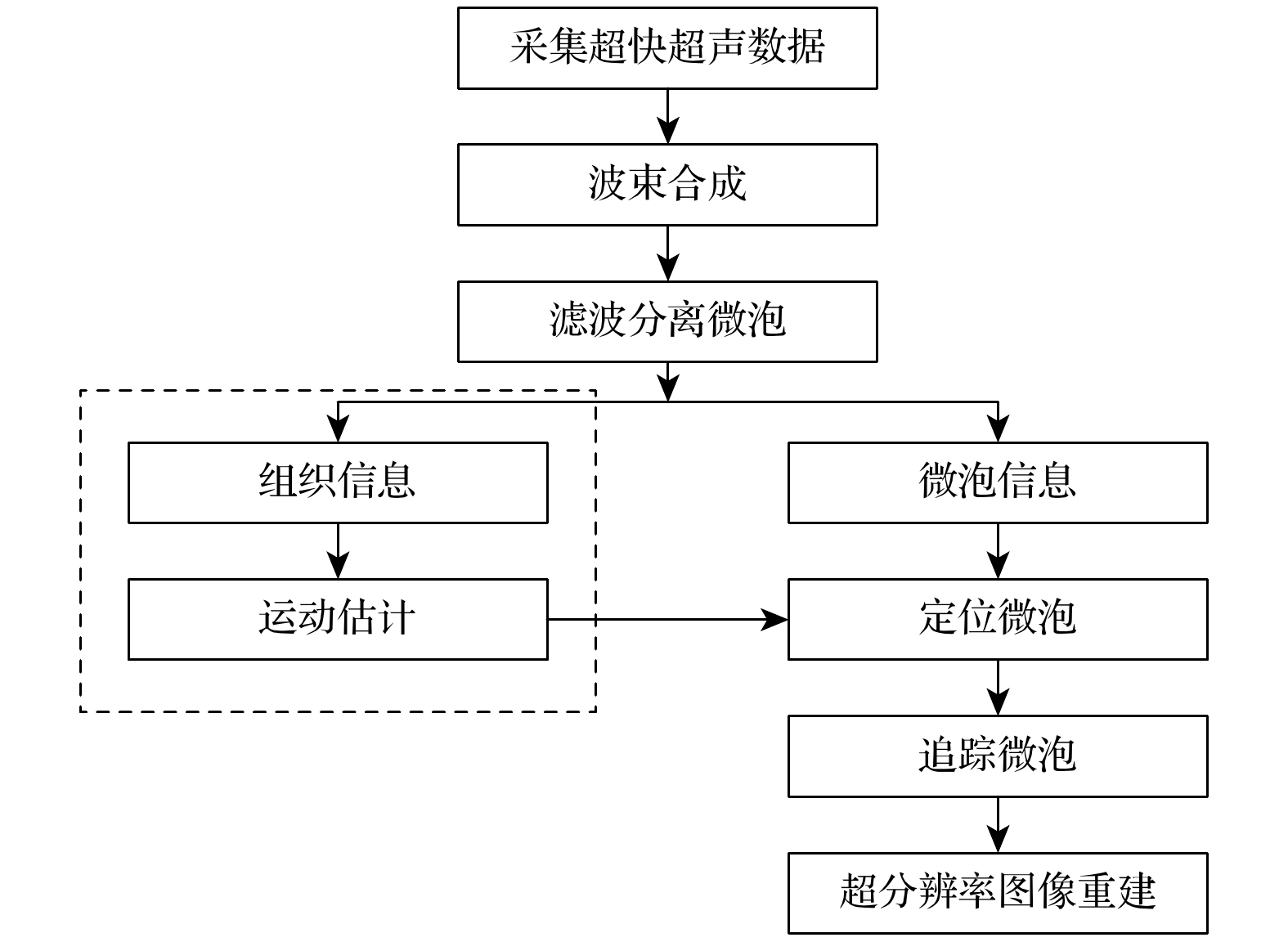
脊髓功能对神经传导通路至关重要, 脊髓血管受损及伴随的继发性损伤与脊髓功能状态密切相关. 因而, 脊髓内微血管网络结构和血流状态在脊髓功能在体、精准与实时评价中具有重要前景. 临床常用的血管造影手段存在分辨率低、放射性、设备笨重和使用不便等问题, 无法全面满足脊髓血流术中检查与预后跟踪的需求. 本文以基于多角度复合平面波的超快超声技术为基础, 应用超分辨率定位显微技术(ULM), 实现了大鼠脊髓内微血管成像. 基本原理为应用基于鲁棒主成分分析(RPCA)的滤波方法, 分离脊髓组织信号和运动的造影微泡信号, 通过微泡定位、轨迹追踪, 实现亚波长分辨率的超分辨率超声成像. 随后, 引入基于傅里叶环相关系数方法, 对成像分辨率进行量化分析; 进而对微泡数量、有效轨迹、血管饱和度、血流速度和半高全宽范围等进行了定量评价. 在体成像实验结果表明, ULM可获得清晰的大鼠脊髓内微血流图像. 定量分析表明, 发射频率为15.625 MHz的超声探头可实现13—16 μm范围的分辨率, 远小于100 μm成像波长. 综上, ULM可被应用于脊髓内微血管精准成像, 相关结果可为超分辨率脊髓功能监测与动态评价的进一步研究提供借鉴, 对于脊髓损伤诊断、应急治疗与预后恢复等临床研究亦有一定的借鉴意义.
-
关键词:
- 超声定位显微(ULM) /
- 超分辨率 /
- 超快超声 /
- 脊髓微血管
Function of spinal cord is crucial to nerve conduction pathway. Traumatic spinal cord injury often results in a vasculature disruption after primary insult and further leads to abnormal responses of the intact vessels in neighboring tissue during secondary injury. Therefore, the vasculature and blood supply play significant roles in evaluating the spinal cord function . Ultrasound localization microscopy (ULM) overcomes the shortcomings of extensively used angiography, such as computed tomography angiography (CTA) and magnetic resonance angiography (MRA), in terms of limited resolution, radiation and poor-portability, which meets the needs of comprehensive intraoperative examination and prognosis tracking. In this study, an L22-14vX probe with a transmission frequency of 15.625 MHz is utilized, yielding an imaging wavelength of 100 μm. The ULM is conducted based on ultrafast ultrasound technology with multiple tilted plane-wave illuminations. Robust principal component analysis (RPCA) based spatial-temporal clutter filtering method is used for separating the microbubble signals from tissue signals and high frequency noise. Through microbubble localization, trajectory tracking and mapping, subwavelength super-resolution ultrasound imaging is finally achieved. The whole process of microbubble localization and vessel reconstruction are monitored through measuring the time dependent microbubble detections and saturation. Saturation curve corresponds to the time dependent total area covered by microbubble detections on the image. Quantification analysis is carried out for evaluating the imaging results including resolution measurements based on the Fourier ring correlation (FRC) and full-width at half-maximum (FWHM). The in-vivo experimental results show that ULM can be used to obtain super-resolution vasculature imaging in rat spinal cord. The velocity distributed from 1 mm/s to 50 mm/s can be detected. Within the same vessel, the velocity of a point is inversely correlated with the distance from the point to the center of the vessel. The velocity in the center of the vessel is larger than that at the wall of the vessel. The larger vessels support higher flow in the center of the vessel. The FWHM results indicate that ultrafast Doppler displays vessels in diameters between 135 μm and 270 μm while ULM displays them in diameters between 28 μm and 35 μm. The FRC-based resolution evaluation shows that the ULM achieves a super resolution of 16 μm, much less than the imaging wavelength of 100 μm. Yet, long acquisition time is required to detect microbubbles in the smallest vessels, leading to long reconstruction of the microvasculature, which is still a problem worth studying . Compromise between saturation and acquisition time needs considering. Generally speaking, microbubbles are more likely to flow in large vessels, leading to relatively short reconstruction time of large vessels. When saturation curve almost converges, the imaging improvement with new vessels is not so significant that the detail sacrifice of some small microvessels can reduce acquisition time (i.e. most of microvasculature can still be gained when the saturation curve does not converge). Besides, the increase of microbubble concentration and advanced track identification and extraction may also accelerate the saturation rate of convergence with acquisition time decreasing. In conclusion, ULM can be used to obtain a super-resolution imaging of spinal cord microvasculature, giving a 10-fold improvement in resolution in comparison with ultrafast Doppler imaging. Relevant results can facilitate the super-resolution ULM imaging of spinal cord which may promote the function diagnosis, treatment intervention, disability prevention, and prognosis recovery of spinal cord injury.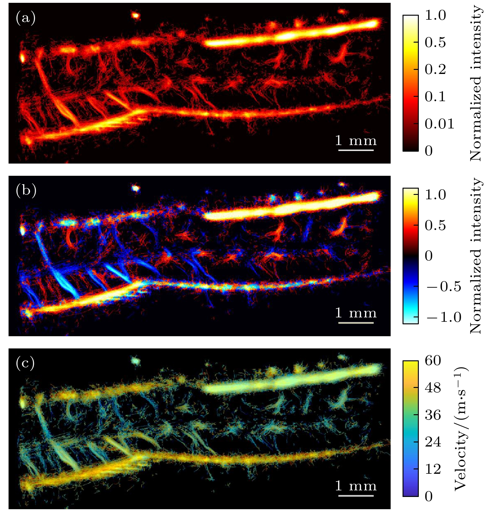
-
Keywords:
- ultrasound localization microscopy (ULM) /
- ultrafast ultrasound /
- super-resolution /
- spinal cord microvasculature
[1] Kwon B K, Tetzlaff W, Grauer J N, Beiner J, Vaccaro A R 2004 Spine J. 4 451
 Google Scholar
[2] Ahuja C S, Wilson J R, Nori S, Kotter M R N, Druschel C, Curt A, Fehlings M G 2017 Nat. Rev. Dis. Primers 3 17018
Google Scholar
[3] Fawcett J W, Schwab M E, Montani L, Brazda N, Muller H W 2012 Handb. Clin. Neurol. 109 503
[4] Ruedinger K L, Schafer S, Speidel M A, Strother C M 2021 AJNR Am. J. Neuroradiol. 42 214
Google Scholar
[5] Vargas M I, Bing F, Gariani J, Dietemann J L 2016 Neurovascular Imaging (New York: Springer) pp. 1063-1093
[6] Tanter M, Fink M 2014 IEEE Trans. Ultrason. Ferroelectr. Freq. Control 61 102
Google Scholar
[7] Betzig E, Patterson G H, Sougrat R, Lindwasser O W, Olenych S, Bonifacino J S, Davidson M W, Lippincott-Schwartz J H, Hess H F 2006 Science 313 1642
Google Scholar
[8] Couture O, Besson B, Montaldo G, Fink M, Tanter M 2011 IEEE International Ultrasonics Symposium (IUS) Caribe Royale, Orlando, Florida, USA, October 18–21, 2011, p1285
[9] 钟传钰, 郑元义 2021 中国医学影像技术 37 1799
Google Scholar
Zhong C, Zheng Y 2021 Chin. J. Med. Imaging Technol. 37 1799
Google Scholar
[10] Couture O, Hingot V, Heiles B, Muleki-Seya P, Tanter M 2018 IEEE Trans. Ultrason. Ferroelectr. Freq. Control 65 1304
Google Scholar
[11] Errico C, Pierre J, Pezet S, Desailly Y, Lenkei Z, Couture O, Tanter M 2015 Nature 527 499
Google Scholar
[12] Christensen-Jeffries K, Browning R J, Tang M X, Dunsby C, Eckersley R J 2015 IEEE Trans. Med. Imaging 34 433
Google Scholar
[13] Opacic T, Dencks S, Theek B, Piepenbrock M, Ackermann D, Rix A, Lammers T, Stickeler E, Delorme S, Schmitz G, Kiessling F 2018 Nat. Commun. 9 1527
Google Scholar
[14] Andersen S B, Taghavi I, Hoyos C A V, Sogaard S B, Gran F, Lonn L, Hansen K L, Jensen J A, Nielsen M B, Sorensen C M 2020 Diagnostics 10 862
[15] Ghosh D, Peng J, Brown K, Sirsi S, Mineo C, Shaul P W, Hoyt K 2019 J. Ultrasound Med. 38 2589
Google Scholar
[16] Zhu J, Rowland E M, Harput S, Riemer K, Leow C H, Clark B, Cox K, Lim A, Christensen-Jeffries K, Zhang G, Brown J, Dunsby C, Eckersley R J, Weinberg P D, Tang M X 2019 Radiology 291 642
Google Scholar
[17] Qian X, Huang C, Li R, Song B, Tchelepi H, Shung K K, Chen S, Humayun M, Zhou Q 2021 IEEE Trans. Biomed. Eng. 69 1585
[18] Song P, Manduca A, Trzasko J D, Daigle R E, Chen S 2018 IEEE Trans. Ultrason. Ferroelectr. Freq. Control 65 2264
Google Scholar
[19] Hingot V, Errico C, Heiles B, Rahal L, Tanter M, Couture O 2019 Sci. Rep. 9 2456
Google Scholar
[20] Hingot V, Chavignon A, Heiles B, Couture O 2021 IEEE Trans. Med. Imaging 40 3812
Google Scholar
[21] Liu X, Zhou T, Lu M, Yang Y, He Q, Luo J 2020 IEEE Trans. Med. Imaging 39 3064
Google Scholar
[22] Xu K, Guo X, Sui Y, Hingot V, Couture O, Ta D, Wang W 2021 IEEE International Ultrasonics Symposium (IUS) Xi’an, China, September 11–16, 2021 p1
[23] Soloukey S, Vincent A, Satoer D D, Mastik F, Smits M, Dirven C M F, Strydis C, Bosch J G, van der Steen A F W, De Zeeuw C I, Koekkoek S K E, Kruizinga P 2019 Front. Neurosci. 13 1384
Google Scholar
[24] Khaing Z Z, Cates L N, DeWees D M, Hannah A, Mourad P, Bruce M, Hofstetter C P 2018 J. Neurosurg. Spine 29 306
Google Scholar
[25] 臧佳琦,许凯亮,韩清见,陆起涌,梅永丰,他得安 2021 物理学报 70 114304
Google Scholar
Zang J Q, Xu K L, Han Q J, Lu Q Y, Mei Y F, Ta D A 2021 Acta Phys. Sin. 70 114304
Google Scholar
[26] Sui Y, Yan S, Zang J, Liu X, Ta D, Wang W, Xu K 2021 IEEE International Ultrasonics Symposium (IUS) Xi’an, China, September 11–16, 2021 p1
[27] Pezet S, Beliard B, Ahmanna C, Tiran E, Kanté K, Deffieux T, Tanter M, Nothias F, Soares S 2022 Sci. Rep. 12 6574
[28] Desailly Y, Tissier A M, Correas J M, Wintzenrieth F, Tanter M, Couture O 2017 Phys. Med. Biol. 62 31
Google Scholar
[29] Hingot V, Errico C, Tanter M, Couture O 2017 Ultrasonics 77 17
Google Scholar
[30] Candès E J, Li X, Ma Y, Wright J 2011 J. ACM 58 1
[31] Bayat M, Fatemi M 2018 IEEE International Conference on Acoustics, Speech and Signal Processing (ICASSP) Calgary, AB, Canada, April 15–20, 2018 p1080
[32] Boyd S 2010 Foundations and Trends® in Machine Learning 3 1
Google Scholar
[33] Christensen-Jeffries K, Couture O, Dayton P A, Eldar Y C, Hynynen K, Kiessling F, O'Reilly M, Pinton G F, Schmitz G, Tang M X, Tanter M, van Sloun R J G 2020 Ultrasound Med. Biol. 46 865
Google Scholar
[34] Heiles B, Correia M, Hingot V, Pernot M, Provost J, Tanter M, Couture O 2019 IEEE Trans. Med. Imaging 38 2005
Google Scholar
[35] Nieuwenhuizen R P, Lidke K A, Bates M, Puig D L, Grunwald D, Stallinga S, Rieger B 2013 Nat. Methods 10 557
Google Scholar
[36] Banterle N, Bui K H, Lemke E A, Beck M 2013 J. Struct. Biol. 183 363
Google Scholar
[37] Viessmann O M, Eckersley R J, Christensen-Jeffries K, Tang M X, Dunsby C 2013 Phys. Med. Biol. 58 6447
Google Scholar
[38] Tang J, Kilic K, Szabo T L, Boas D A 2021 IEEE Trans. Med. Imaging 40 758
Google Scholar
[39] Bar-Zion A, Solomon O, Tremblay-Darveau C, Adam D, Eldar Y C 2018 IEEE Trans. Ultrason. Ferroelectr. Freq. Control 65 2365
Google Scholar
[40] Milecki L, Poree J, Belgharbi H, Bourquin C, Damseh R, Delafontaine-Martel P, Lesage F, Gasse M, Provost J 2021 IEEE Trans. Med. Imaging 40 1428
Google Scholar
[41] van Sloun R J G, Solomon O, Bruce M, Khaing Z Z, Wijkstra H, Eldar Y C, Mischi M 2021 IEEE Trans. Med. Imaging 40 829
Google Scholar
[42] Guasch L, Calderon Agudo O, Tang M X, Nachev P, Warner M 2020 NPJ Digit. Med. 3 28
Google Scholar
[43] 李云清, 江晨, 李颖, 徐峰, 许凯亮, 他得安, 黎仲勋 2019 物理学报 68 184302
Google Scholar
Li Y Q, Jiang C, Li Y, Xu F, Xu K L, Ta D A, Le L H 2019 Acta Phys. Sin. 68 184302
Google Scholar
[44] Jiang C, Li Y, Xu K, Ta D 2021 IEEE Trans. Ultrason. Ferroelectr. Freq. Control 68 72
Google Scholar
-

图 3 ULM处理过程结果 (a) 第150个数据块中第200帧回波信号的B超图像; (b) 第150个数据块中第200帧分离出的微泡回波信号; (c) 第150个数据块中第300帧微泡定位结果; (d) 第150个数据块中第301帧微泡定位结果
Fig. 3. Results during ULM processing: (a) B-mode image of the 200th frame of block 150; (b) isolated signal of microbubbles after filtering from the 200th frame of block 150; (c) localization of microbubble centers in the 300th frame of block 150; (d) localization of microbubble centers in the 301th frame of block 150.

图 4 超快超分辨率超声成像结果 (a) 脊髓血流密度图; (b) 脊髓血流方向图; (c) 脊髓血流速度图
Fig. 4. ULM Results: (a) Intensity map of spinal cord; (b) direction map of spinal cord; (c) velocity map of spinal cord.
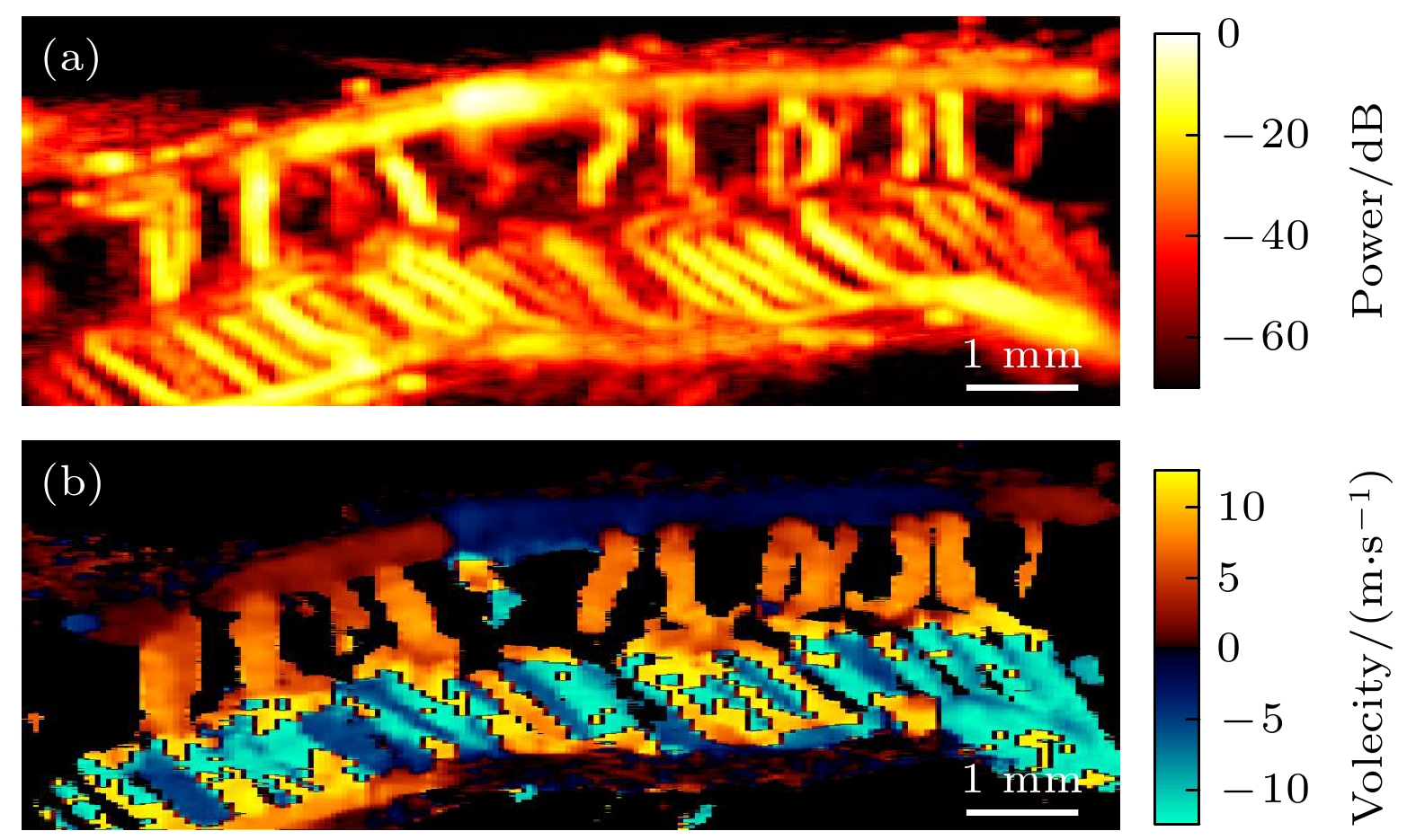
图 5 超快多普勒超声成像结果 (a) 功率多普勒血流图; (b) 彩色多普勒血流图
Fig. 5. Results of ultrafast Doppler imaging: (a) Power Doppler; (b) color Doppler.
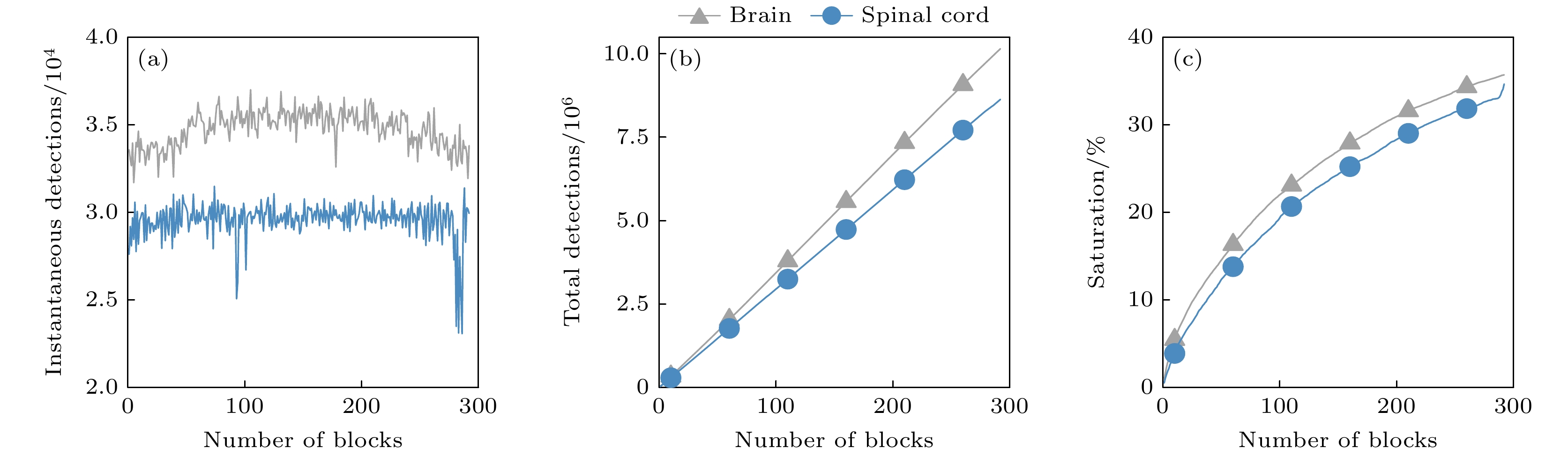
图 6 微泡定位统计 (a) 瞬时微泡数量; (b) 累计微泡数量; (c) 饱和度随时间变化图曲线
Fig. 6. Quantification of microbubble localization: (a) Instantaneous detections; (b) accumulated detections; (c) saturation curve along time.

图 7 超快多普勒与超快超分辨率超声成像结果分辨率测算 (a) 脊髓超快功率多普勒血流局部放大图; (b) 图(a)中部分血管剖面FWHM结果; (c) 脊髓超分辨率血流密度局部放大图; (d) 图(c)中部分血管剖面FWHM结果; (e) 脊髓超分辨率血流方向局部图; (f) 超分辨率血流密度图基于FRC的分辨率结果
Fig. 7. Resolution measurements of ultrafast Doppler imaging and ULM: (a) Zoom in of power Doppler; (b) FWHM of vessels from panel (a); (c) zoom in of ULM intensity map; (d) FWHM of vessels from panel (c); (e) zoom in of ULM direction map; (f) resolution of ULM intensity map based on FRC curve.
表 1 ULM参数统计结果
Table 1. Results of ULM parameter measurement.
参数 值 微泡保留比例/% 14.7 半高全宽/μm 28—50 轨迹保留比例/% 1.7 传统定义分辨率/μm 28 饱和度/% 32 FRC分辨率 –2$ \sigma $/μm 13 血流速度/(mm·s–1) 1—50 FRC分辨率 –1/2 bit/μm 16  下载: 导出CSV
-
[1] Kwon B K, Tetzlaff W, Grauer J N, Beiner J, Vaccaro A R 2004 Spine J. 4 451
Google Scholar
[2] Ahuja C S, Wilson J R, Nori S, Kotter M R N, Druschel C, Curt A, Fehlings M G 2017 Nat. Rev. Dis. Primers 3 17018
Google Scholar
[3] Fawcett J W, Schwab M E, Montani L, Brazda N, Muller H W 2012 Handb. Clin. Neurol. 109 503
[4] Ruedinger K L, Schafer S, Speidel M A, Strother C M 2021 AJNR Am. J. Neuroradiol. 42 214
Google Scholar
[5] Vargas M I, Bing F, Gariani J, Dietemann J L 2016 Neurovascular Imaging (New York: Springer) pp. 1063-1093
[6] Tanter M, Fink M 2014 IEEE Trans. Ultrason. Ferroelectr. Freq. Control 61 102
Google Scholar
[7] Betzig E, Patterson G H, Sougrat R, Lindwasser O W, Olenych S, Bonifacino J S, Davidson M W, Lippincott-Schwartz J H, Hess H F 2006 Science 313 1642
Google Scholar
[8] Couture O, Besson B, Montaldo G, Fink M, Tanter M 2011 IEEE International Ultrasonics Symposium (IUS) Caribe Royale, Orlando, Florida, USA, October 18–21, 2011, p1285
[9] 钟传钰, 郑元义 2021 中国医学影像技术 37 1799
Google Scholar
Zhong C, Zheng Y 2021 Chin. J. Med. Imaging Technol. 37 1799
Google Scholar
[10] Couture O, Hingot V, Heiles B, Muleki-Seya P, Tanter M 2018 IEEE Trans. Ultrason. Ferroelectr. Freq. Control 65 1304
Google Scholar
[11] Errico C, Pierre J, Pezet S, Desailly Y, Lenkei Z, Couture O, Tanter M 2015 Nature 527 499
Google Scholar
[12] Christensen-Jeffries K, Browning R J, Tang M X, Dunsby C, Eckersley R J 2015 IEEE Trans. Med. Imaging 34 433
Google Scholar
[13] Opacic T, Dencks S, Theek B, Piepenbrock M, Ackermann D, Rix A, Lammers T, Stickeler E, Delorme S, Schmitz G, Kiessling F 2018 Nat. Commun. 9 1527
Google Scholar
[14] Andersen S B, Taghavi I, Hoyos C A V, Sogaard S B, Gran F, Lonn L, Hansen K L, Jensen J A, Nielsen M B, Sorensen C M 2020 Diagnostics 10 862
[15] Ghosh D, Peng J, Brown K, Sirsi S, Mineo C, Shaul P W, Hoyt K 2019 J. Ultrasound Med. 38 2589
Google Scholar
[16] Zhu J, Rowland E M, Harput S, Riemer K, Leow C H, Clark B, Cox K, Lim A, Christensen-Jeffries K, Zhang G, Brown J, Dunsby C, Eckersley R J, Weinberg P D, Tang M X 2019 Radiology 291 642
Google Scholar
[17] Qian X, Huang C, Li R, Song B, Tchelepi H, Shung K K, Chen S, Humayun M, Zhou Q 2021 IEEE Trans. Biomed. Eng. 69 1585
[18] Song P, Manduca A, Trzasko J D, Daigle R E, Chen S 2018 IEEE Trans. Ultrason. Ferroelectr. Freq. Control 65 2264
Google Scholar
[19] Hingot V, Errico C, Heiles B, Rahal L, Tanter M, Couture O 2019 Sci. Rep. 9 2456
Google Scholar
[20] Hingot V, Chavignon A, Heiles B, Couture O 2021 IEEE Trans. Med. Imaging 40 3812
Google Scholar
[21] Liu X, Zhou T, Lu M, Yang Y, He Q, Luo J 2020 IEEE Trans. Med. Imaging 39 3064
Google Scholar
[22] Xu K, Guo X, Sui Y, Hingot V, Couture O, Ta D, Wang W 2021 IEEE International Ultrasonics Symposium (IUS) Xi’an, China, September 11–16, 2021 p1
[23] Soloukey S, Vincent A, Satoer D D, Mastik F, Smits M, Dirven C M F, Strydis C, Bosch J G, van der Steen A F W, De Zeeuw C I, Koekkoek S K E, Kruizinga P 2019 Front. Neurosci. 13 1384
Google Scholar
[24] Khaing Z Z, Cates L N, DeWees D M, Hannah A, Mourad P, Bruce M, Hofstetter C P 2018 J. Neurosurg. Spine 29 306
Google Scholar
[25] 臧佳琦,许凯亮,韩清见,陆起涌,梅永丰,他得安 2021 物理学报 70 114304
Google Scholar
Zang J Q, Xu K L, Han Q J, Lu Q Y, Mei Y F, Ta D A 2021 Acta Phys. Sin. 70 114304
Google Scholar
[26] Sui Y, Yan S, Zang J, Liu X, Ta D, Wang W, Xu K 2021 IEEE International Ultrasonics Symposium (IUS) Xi’an, China, September 11–16, 2021 p1
[27] Pezet S, Beliard B, Ahmanna C, Tiran E, Kanté K, Deffieux T, Tanter M, Nothias F, Soares S 2022 Sci. Rep. 12 6574
[28] Desailly Y, Tissier A M, Correas J M, Wintzenrieth F, Tanter M, Couture O 2017 Phys. Med. Biol. 62 31
Google Scholar
[29] Hingot V, Errico C, Tanter M, Couture O 2017 Ultrasonics 77 17
Google Scholar
[30] Candès E J, Li X, Ma Y, Wright J 2011 J. ACM 58 1
[31] Bayat M, Fatemi M 2018 IEEE International Conference on Acoustics, Speech and Signal Processing (ICASSP) Calgary, AB, Canada, April 15–20, 2018 p1080
[32] Boyd S 2010 Foundations and Trends® in Machine Learning 3 1
Google Scholar
[33] Christensen-Jeffries K, Couture O, Dayton P A, Eldar Y C, Hynynen K, Kiessling F, O'Reilly M, Pinton G F, Schmitz G, Tang M X, Tanter M, van Sloun R J G 2020 Ultrasound Med. Biol. 46 865
Google Scholar
[34] Heiles B, Correia M, Hingot V, Pernot M, Provost J, Tanter M, Couture O 2019 IEEE Trans. Med. Imaging 38 2005
Google Scholar
[35] Nieuwenhuizen R P, Lidke K A, Bates M, Puig D L, Grunwald D, Stallinga S, Rieger B 2013 Nat. Methods 10 557
Google Scholar
[36] Banterle N, Bui K H, Lemke E A, Beck M 2013 J. Struct. Biol. 183 363
Google Scholar
[37] Viessmann O M, Eckersley R J, Christensen-Jeffries K, Tang M X, Dunsby C 2013 Phys. Med. Biol. 58 6447
Google Scholar
[38] Tang J, Kilic K, Szabo T L, Boas D A 2021 IEEE Trans. Med. Imaging 40 758
Google Scholar
[39] Bar-Zion A, Solomon O, Tremblay-Darveau C, Adam D, Eldar Y C 2018 IEEE Trans. Ultrason. Ferroelectr. Freq. Control 65 2365
Google Scholar
[40] Milecki L, Poree J, Belgharbi H, Bourquin C, Damseh R, Delafontaine-Martel P, Lesage F, Gasse M, Provost J 2021 IEEE Trans. Med. Imaging 40 1428
Google Scholar
[41] van Sloun R J G, Solomon O, Bruce M, Khaing Z Z, Wijkstra H, Eldar Y C, Mischi M 2021 IEEE Trans. Med. Imaging 40 829
Google Scholar
[42] Guasch L, Calderon Agudo O, Tang M X, Nachev P, Warner M 2020 NPJ Digit. Med. 3 28
Google Scholar
[43] 李云清, 江晨, 李颖, 徐峰, 许凯亮, 他得安, 黎仲勋 2019 物理学报 68 184302
Google Scholar
Li Y Q, Jiang C, Li Y, Xu F, Xu K L, Ta D A, Le L H 2019 Acta Phys. Sin. 68 184302
Google Scholar
[44] Jiang C, Li Y, Xu K, Ta D 2021 IEEE Trans. Ultrason. Ferroelectr. Freq. Control 68 72
Google Scholar

 下载:
下载:

计量
- 文章访问数: 10945
- PDF下载量: 246
- 被引次数: 0
