


-
太赫兹成像技术是医学成像的候选技术之一, 尤其是连续太赫兹反射和衰减全反射成像有望实现术中快速成像, 是目前研究的热点课题. 本文理论研究了成像角度对反射信号和衰减全反射穿透深度的影响, 获得了适用于反射和衰减全反射成像的太赫兹波成像角度. 在此基础上设计了全反射棱镜, 通过反射窗口和全反射棱镜快速切换的方式, 实现了共光路连续太赫兹反射和衰减全反射双模式成像. 以蒸馏水和猪肉为样品, 实验研究了连续太赫兹反射和衰减全反射成像. 结果表明, 与反射成像相比, 连续太赫兹衰减全反射成像具有成像分辨率高、图像对比度高和信号稳定性高的优点, 且可以准确获得样品反射率. 太赫兹衰减全反射成像技术更有助于实现样品的高灵敏度成像.Terahertz imaging technology is one of the candidate technologies for medical imaging. In particular, continuous terahertz reflection and attenuated total reflection imaging are expected to achieve rapid intraoperative imaging, which is hot research topic at present. In order to realize the rapid multi-dimensional and high-quality terahertz imaging detection of sample, it is necessary to study the common optical path continuous terahertz reflection/attenuated total reflection dual-mode imaging system based on point scanning. By using the Fresnel formula and the penetration depth formula of evanescent wave, the influence of imaging angle on the reflected signal and the penetration depth of attenuated total reflection are studied theoretically in this paper. The imaging angle of terahertz wave suitable for both reflection and attenuation total reflection imaging is obtained. Based on this, an isoscele total reflection prism with a base angle of 49° is designed. The dual-mode imaging of common optical path continuous terahertz reflection and attenuated total reflection is realized by quickly switching between reflection window and total reflection prism. The reflection and attenuation total reflection imaging modes have imaging resolutions of 400 μm and 500 μm, respectively. Continuous terahertz reflection and attenuated total reflection imaging are experimentally studied by using distilled water and pork as samples. The results show that the relative reflectance of the sample obtained in the attenuated total reflection imaging mode fluctuates within a range of 1%, and the image contrast is 9 times that of the reflection imaging mode. Moreover, attenuated total reflection imaging can effectively identify the sample with the length less than 1 mm. Thus, compared with reflection imaging, continuous terahertz attenuated total reflection imaging has the advantages of high image resolution, high image contrast and highr signal stability, and can accurately obtain the reflectivity of sample. The terahertz attenuated total reflection imaging technology is more helpful in achieving high sensitivity imaging of samples. By combining reflection and attenuated total reflection imaging modes, the advantages of different imaging modes can be compensated for and the performance of the imaging system can be further improved. This common path continuous terahertz reflection and attenuated total reflection dual mode imaging system is expected to achieve a high sensitivity detection of sample.
[1] Son J H 2009 J. Appl. Phys. 105 10
 Google Scholar
[2] Hu B B, Nuss M C 1995 Opt. Lett. 20 16
Google Scholar
[3] Bowman T, Shenawee M, Campbell L K 2016 Biomed. Opt. Express 7 9
Google Scholar
[4] Ji Y B, Park C H, Kim H, Kim S H, Lee G M, Noh S K, Jeon T I, Son J H, Huh Y M, Haam S, Oh S J, Lee S K, Suh J S 2015 Biomed. Opt. Express 6 4
Google Scholar
[5] Ishikawa Y, Minamide H, Ikari T, Miura Y, Ito H 2005 Proceedings of the International Quantum Electronics Conference San Jose, USA, July 11−11, 2005, p1236
[6] Nishizawa J, Sasaki T, Suto K, Yamada T, Tanabe T, Tanno T, Sawai T, Miura Y 2005 Opt. Commun. 244 1
Google Scholar
[7] 杨昆, 赵国忠, 梁承森, 武利忠 2009 中国激光 25 29
Google Scholar
Yang K, Zhao G, Liang C S, Wu L Z 2009 J. Lasers 25 29
Google Scholar
[8] Wahaia F, Kasalynas I, Venckevicius R, Seliuta D, Granja P L 2016 J. mol. Struct. 5 1107
Google Scholar
[9] Hartwick T S, Hodges D T, Barker D H, Foote F B 1976 Appl. Optics 15 8
Google Scholar
[10] Park J Y, Choi H J, Cho K S, Kim K R, Son J H 2011 J. Appl. Phys. 109 6
Google Scholar
[11] Liu H, Wang Y, Xu D, Wu L, Yan C, Yan D, Tang L, He Y, Feng H, Yao J 2017 J. Phys. D Appl. Phys. 50 37
Google Scholar
[12] Gerasimov V V, Knyazev B A and Cherkassky V S 2010 Opt. Spectrosc. 108 6
Google Scholar
[13] Bowman T, Walter A, EI-Shenawee M 2016 Proceedings Volume 9700, Design and Quality for Biomedical Technologies IX San Francisco, California, United States, February 13−14, 2016 p97000J-1–5
[14] Wallace V P, Fitzgerald A J, Shankar S, Flanagan N, Arnone D D 2015 Brit J. of Dermatol. 151 2
Google Scholar
[15] Sim Y C, Park J Y, Ahn K M, Park C, Son J H 2013 Biomed. Opt. Express 4 8
Google Scholar
[16] Wang Y, Chen L, Chen T, Jia S, Ren Y, Li C, Chao Z, Liu H, Wu L 2018 J. Phys. D Appl. Phys. 51 32
Google Scholar
[17] Chan K L A and Kazarian S G 2003 Appl. Spectrosc. 57 4
Google Scholar
[18] Wojdyla A, Gallot G 2013 Opt. Lett. 38 2
Google Scholar
[19] Catherine Z 2003 Nature 14 721
[20] Lee A W, Hu Q 2005 Opt. Lett. 30 19
Google Scholar
[21] Watts C M, Shrekenhamer D, Montoya J, Lipworth G, Hunt J, Sleasman T, Krishna S, Smith D R, Padilla W J 2014 Nat. Photonics 8 8
Google Scholar
[22] Doradla P, Alavi K, Joseph C S, Giles R 2013 J. Biomed. Opt. 18 9
Google Scholar
[23] Chernomyrdin N V, Kucheryavenko A S, Kolontaeva G S, G M Katyba, I N Dolganova, P A Karalkin, D S Ponomarev, V N Kurlov, I V Reshetov, Skorobogatiy M 2018 Appl. Phys. Lett. 113 11
Google Scholar
[24] Wu L, Xu D, Wang Y, Zhang Y, Wang H, Liao B, Gong S, Chen T, Wu N, Feng H, Yao J 2020 Neurophotonics 7 2
Google Scholar
[25] Johnk C T 1988 Engineering Electromagnetic Fields and Waves (2nd Ed.) (Hoboken, NJ, USA: Wiley) pp247−251
[26] Wang Y, Wang Y, Xu D, Wu L, Wang G, Jiang B, Yu T, Chang C, Chen T, Yao J 2020 Opt. Express 28 15
Google Scholar
[27] Shikata J, Handal H, Nawaharal A, Minamide H, Ito H 2007 Conference on Lasers and Electro Optics Pacific Rim, Seoul, South Korea, August 26–31, 2007 p1406
[28] Liu H, Wang Y, Xu D, Jiang Z, Wu L, Yan C, Tang L, He Y, Yan D, Ding X, Feng H, Yao J 2018 Opt. Express 26 16
Google Scholar
-

图 1 太赫兹波在成像窗口中传输示意图(此处以反射为例)
Fig. 1. Schematic diagram of terahertz wave propagation in the imaging window (take reflection as an example).
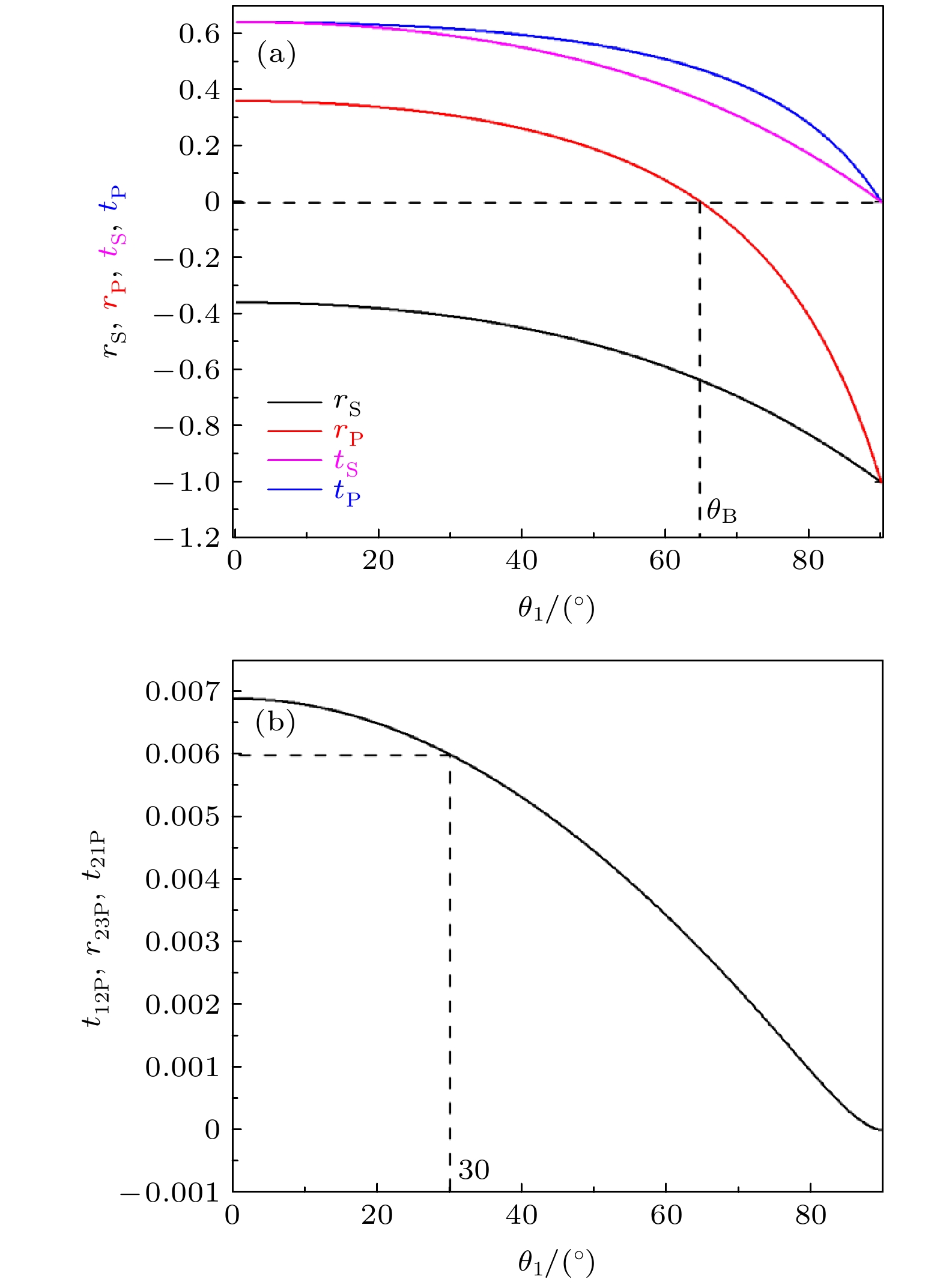
图 2 (a) S和P偏振波在界面处的反射系数和透射系数与入射角θ1的关系; (b)太赫兹波经反射窗口前表面透射、后表面反射和前表面透射后的反射系数r与入射角θ1的关系
Fig. 2. (a) The relation between the reflection coefficient and transmission coefficient of S and P polarized waves at the interface and the incident angle θ1; (b) the relation between the reflection coefficient r and incident angle θ1 of terahertz wave after the front surface transmission, back surface reflection and front surface transmission through reflection window.

图 3 采用高阻硅全反射棱镜时, 倏逝波在不同样品中的穿透深度与入射角的关系
Fig. 3. The relationship between the penetration depth and the incident angle of evanescent wave in different samples, when using the high resistance silicon total reflection prism.
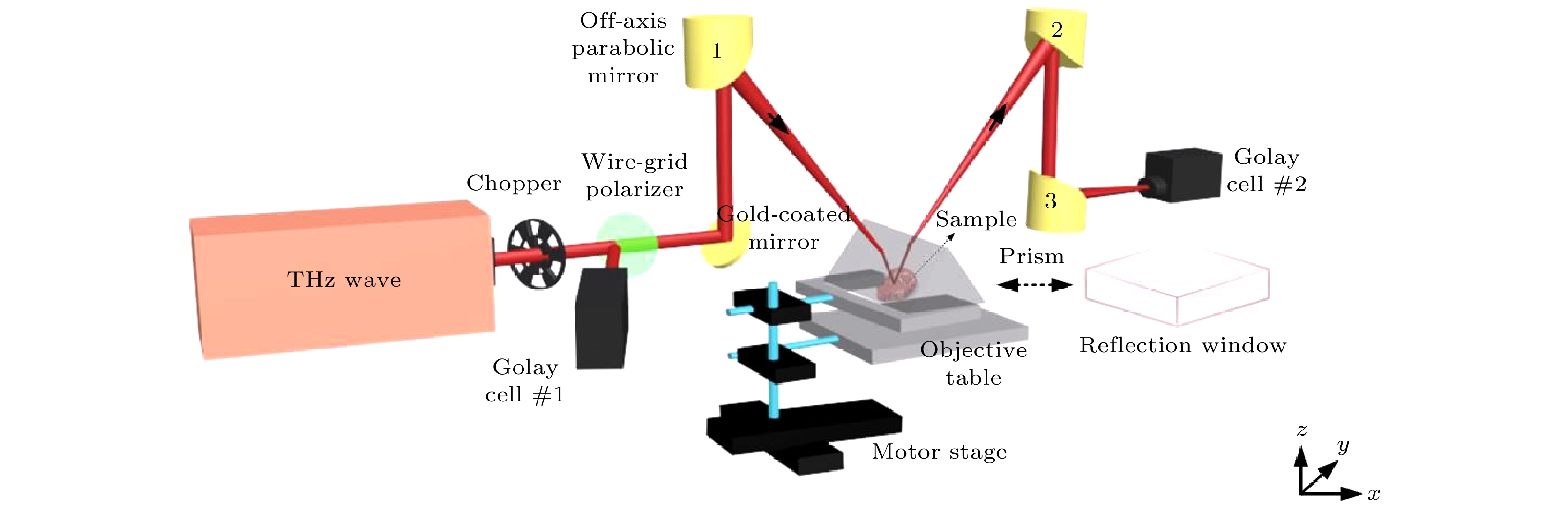
图 4 共光路连续太赫兹反射和衰减全反射成像系统示意图
Fig. 4. Schematic diagram of common path continuous terahertz reflection and attenuation total reflection imaging system.

图 5 采用石英和高阻硅材料为成像窗口时, 连续太赫兹反射和衰减全反射成像系统的分辨率
Fig. 5. The resolution of continuous terahertz reflection and attenuated total reflection imaging systems, when the quartz and the high resistance silicon materials are used as imaging windows.
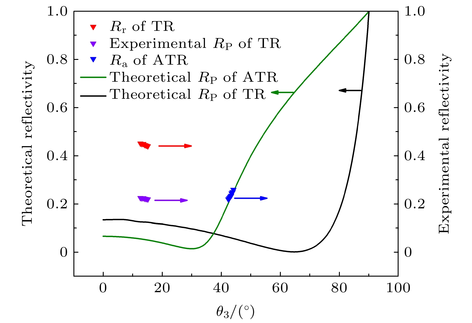
图 6 反射和衰减全反射模式下, 基于理论和实验获得蒸馏水的反射率和相对反射率
Fig. 6. The reflectivity and relative reflectivity of distilled water obtained theoretically and experimentally under the reflection and attenuation total reflection modes.
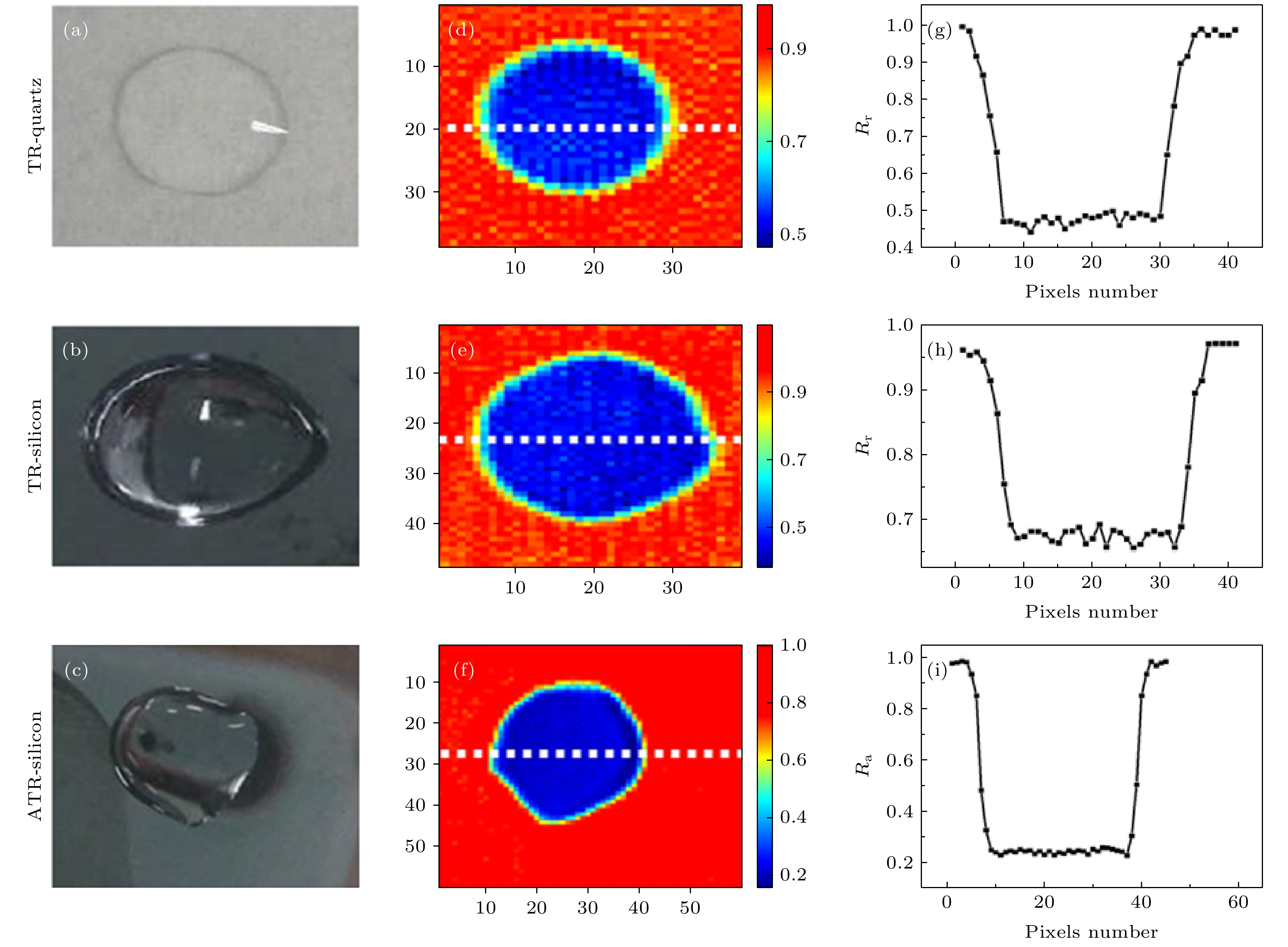
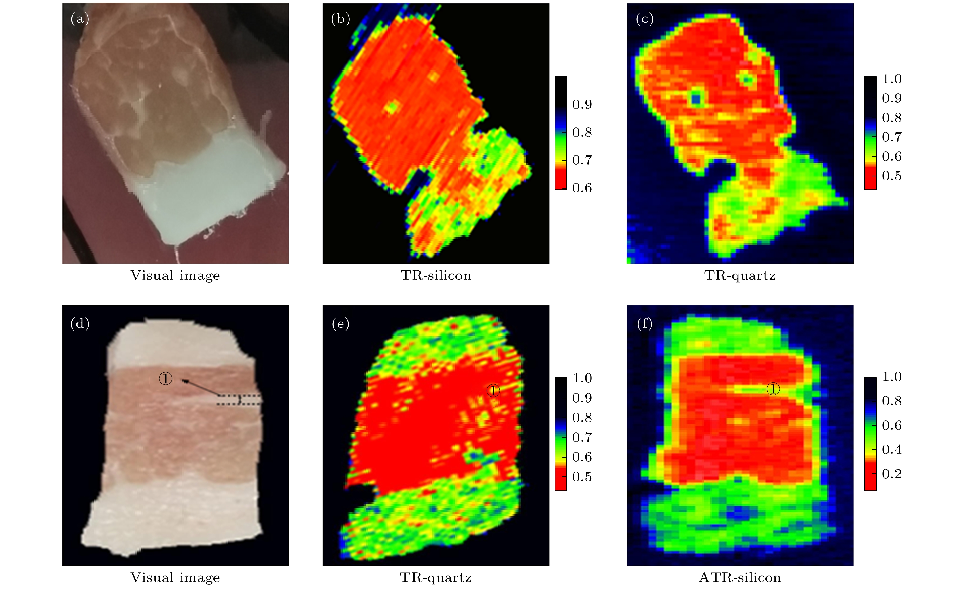
图 8 (a) 猪肉组织与反射窗口紧密接触可见光图; (b), (c) 采用高阻硅和石英材料为反射窗口时的太赫兹成像图; (d) 未覆盖成像窗口时猪肉组织可见光图; (e), (f) 反射和衰减全反射模式下猪肉组织的太赫兹成像图
Fig. 8. (a) Visible image of pork tissue in close contact with the reflection window; (b), (c) the terahertz images of pork tissue in reflective and attenuated total reflection modes using high resistance silicon and quartz materials as reflection windows, (d) visible image of pork tissue when the imaging window is not covered; (e), (f) the terahertz images of pork tissue in reflective and attenuated total reflection modes respectively.
-
[1] Son J H 2009 J. Appl. Phys. 105 10
Google Scholar
[2] Hu B B, Nuss M C 1995 Opt. Lett. 20 16
Google Scholar
[3] Bowman T, Shenawee M, Campbell L K 2016 Biomed. Opt. Express 7 9
Google Scholar
[4] Ji Y B, Park C H, Kim H, Kim S H, Lee G M, Noh S K, Jeon T I, Son J H, Huh Y M, Haam S, Oh S J, Lee S K, Suh J S 2015 Biomed. Opt. Express 6 4
Google Scholar
[5] Ishikawa Y, Minamide H, Ikari T, Miura Y, Ito H 2005 Proceedings of the International Quantum Electronics Conference San Jose, USA, July 11−11, 2005, p1236
[6] Nishizawa J, Sasaki T, Suto K, Yamada T, Tanabe T, Tanno T, Sawai T, Miura Y 2005 Opt. Commun. 244 1
Google Scholar
[7] 杨昆, 赵国忠, 梁承森, 武利忠 2009 中国激光 25 29
Google Scholar
Yang K, Zhao G, Liang C S, Wu L Z 2009 J. Lasers 25 29
Google Scholar
[8] Wahaia F, Kasalynas I, Venckevicius R, Seliuta D, Granja P L 2016 J. mol. Struct. 5 1107
Google Scholar
[9] Hartwick T S, Hodges D T, Barker D H, Foote F B 1976 Appl. Optics 15 8
Google Scholar
[10] Park J Y, Choi H J, Cho K S, Kim K R, Son J H 2011 J. Appl. Phys. 109 6
Google Scholar
[11] Liu H, Wang Y, Xu D, Wu L, Yan C, Yan D, Tang L, He Y, Feng H, Yao J 2017 J. Phys. D Appl. Phys. 50 37
Google Scholar
[12] Gerasimov V V, Knyazev B A and Cherkassky V S 2010 Opt. Spectrosc. 108 6
Google Scholar
[13] Bowman T, Walter A, EI-Shenawee M 2016 Proceedings Volume 9700, Design and Quality for Biomedical Technologies IX San Francisco, California, United States, February 13−14, 2016 p97000J-1–5
[14] Wallace V P, Fitzgerald A J, Shankar S, Flanagan N, Arnone D D 2015 Brit J. of Dermatol. 151 2
Google Scholar
[15] Sim Y C, Park J Y, Ahn K M, Park C, Son J H 2013 Biomed. Opt. Express 4 8
Google Scholar
[16] Wang Y, Chen L, Chen T, Jia S, Ren Y, Li C, Chao Z, Liu H, Wu L 2018 J. Phys. D Appl. Phys. 51 32
Google Scholar
[17] Chan K L A and Kazarian S G 2003 Appl. Spectrosc. 57 4
Google Scholar
[18] Wojdyla A, Gallot G 2013 Opt. Lett. 38 2
Google Scholar
[19] Catherine Z 2003 Nature 14 721
[20] Lee A W, Hu Q 2005 Opt. Lett. 30 19
Google Scholar
[21] Watts C M, Shrekenhamer D, Montoya J, Lipworth G, Hunt J, Sleasman T, Krishna S, Smith D R, Padilla W J 2014 Nat. Photonics 8 8
Google Scholar
[22] Doradla P, Alavi K, Joseph C S, Giles R 2013 J. Biomed. Opt. 18 9
Google Scholar
[23] Chernomyrdin N V, Kucheryavenko A S, Kolontaeva G S, G M Katyba, I N Dolganova, P A Karalkin, D S Ponomarev, V N Kurlov, I V Reshetov, Skorobogatiy M 2018 Appl. Phys. Lett. 113 11
Google Scholar
[24] Wu L, Xu D, Wang Y, Zhang Y, Wang H, Liao B, Gong S, Chen T, Wu N, Feng H, Yao J 2020 Neurophotonics 7 2
Google Scholar
[25] Johnk C T 1988 Engineering Electromagnetic Fields and Waves (2nd Ed.) (Hoboken, NJ, USA: Wiley) pp247−251
[26] Wang Y, Wang Y, Xu D, Wu L, Wang G, Jiang B, Yu T, Chang C, Chen T, Yao J 2020 Opt. Express 28 15
Google Scholar
[27] Shikata J, Handal H, Nawaharal A, Minamide H, Ito H 2007 Conference on Lasers and Electro Optics Pacific Rim, Seoul, South Korea, August 26–31, 2007 p1406
[28] Liu H, Wang Y, Xu D, Jiang Z, Wu L, Yan C, Tang L, He Y, Yan D, Ding X, Feng H, Yao J 2018 Opt. Express 26 16
Google Scholar

 下载:
下载:
计量
- 文章访问数: 6674
- PDF下载量: 121
- 被引次数: 0
