


-
X射线荧光CT(X-ray fluorescence computed tomography, XFCT)是一种使用X射线荧光(X-ray fluorescence, XRF)实现功能性成像的新技术, 在生物医学成像中表现出较大潜力. 但是, X射线穿过生物体的同时还会产生大量康普顿散射光子, 对XRF信号的采集形成很强的背景噪声; 因此, 如何有效消除康普顿散射噪声对于提高XFCT成像质量至关重要. 本文研究总结了XFCT成像过程中涉及的物理过程, 包括: 荧光的产额、退激发时间、荧光发射角分布、荧光偏振态、康普顿散射角分布与散射光偏振态, 并通过研究荧光与散射光物理性质的差异寻找去除康普顿散射噪声的方法. 经过物理过程推导和分析计算, 发现: 1) 高原子序数元素的K层荧光退激发时间极短, 在现有探测器的时间分辨率条件下, 无法分辨散射光与荧光; 2) K层发射荧光的角分布各向同性, 康普顿散射角分布在与入射光偏振方向附近取得最小值, 而且入射光线偏振度越高, 散射光的微分截面越小, 偏振光源将有利于减少康普顿散射噪声; 3) K层荧光线偏振度为零, 而康普顿散射光子在一些散射方向上具有一定线偏振度, 因此偏振态的差异可能用于降低康普顿散射噪声.X-ray fluorescence computed tomography (XFCT) is a molecular imaging technique with great potential applications in biomedical imaging, in which used is the primary X-ray to excite element probes with high atomic number inside samples or tissues for functional imaging. However, owing to the limitation of molecular sensitivity and spatial resolution, the XFCT has not been widely used in the molecular imaging. A large number of Compton scattering photons are produced as the broadband primary X-ray passes through the samples or tissues, forming a strong noise background in the collected XRF signal, which is a major cause of limited molecular sensitivity. Therefore, eliminating the Compton scattering noise is very important for improving molecular sensitivity. In this paper, we summarize the main physical processes involved in the imaging process of XFCT, including the angle distribution and polarization state of the fluorescence and Compton scattering photons, fluorescence yield and deexcitation time of K-shell vacancy. The above physical processes are the main limitations of the imaging quality of XFCT. Through the derivation and analysis of physical processes, we explore the possibility of using these physical effects to reduce the Compton scattering noise and draw some conclusions below. 1) The deexcitation time of K-shell vacancy of the element with high atomic number is very short, consequently the scattered light and fluorescence cannot be distinguished between each other under the time resolution condition of the existing detector. 2) The angular distribution of the K-shell fluorescence emission is isotropic, and the differential cross section of Compton scattering reaches a minimum value near the polarization direction of the incident light of which the minimum decreases as the linear polarization degree of the incident light increases. Therefore, the polarized light source is beneficial to reducing the Compton scattering noise. 3) The linear degree of polarization of K-shell fluorescence is zero, while the Compton scattering photons possess a certain linear degree of polarization in some scattering directions, so the difference between polarization states may be helpful in reducing the Compton scattering noise.
-
Keywords:
- X-ray fluorescence /
- computed tomography /
- polarized X-rays /
- Compton scattering /
- functional imaging
[1] Boisseau P, Grodzins L 1987 Hyperfine Interact. 33 283
 Google Scholar
[2] Cheong S K, Jones B L, Siddiqi A K, Liu F, Manohar N, Cho S H 2010 Phys. Med. Biol. 55 647
Google Scholar
[3] Bazalova M, Kuang Y, Pratx G, Xing L 2012 IEEE Trans. Med. Imaging 31 1620
Google Scholar
[4] Sjölin M, Danielsson M 2014 Phys. Med. Biol. 59 6507
Google Scholar
[5] Li L, Zhang S Y, Li R Z, Chen Z Q 2017 Opt. Eng. 56 043106
Google Scholar
[6] Zhang S Y, Li L, Chen Z Q 2019 IEEE Access 7 113589
Google Scholar
[7] Li L, Li R Z, Zhang S Y, Chen Z Q 2016 Proc. of SPIE 9967 99670F
Google Scholar
[8] Zhang S Y, Li L, Chen J Y, Chen Z Q, Zhang W L, Lu H B 2019 Int. J. Mol. Sci. 20 2315
Google Scholar
[9] Ahmad M, Bazalova M, Xiang L, Xing L 2014 IEEE Trans. Med. Imaging 33 1119
Google Scholar
[10] Sasaya T, Sunaguchi N, Hyodo K, Zeniya T, Takeda T, Yuasa T 2017 Sci. c Rep. 7 44143
Google Scholar
[11] Chi Z, Du Y, Huang W, Tang C 2020 J. Synchrotron Radiat. 27 737
Google Scholar
[12] Vernekohl D, Tzoumas S, Zhao W, Xing L 2018 Med. Phys. 45 3741
Google Scholar
[13] Mcmaster W H 1961 Rev. Mod. Phys. 33 8
Google Scholar
[14] Jones J A, D’Addario A J, Rojec B L, Milione G, Galvez E J 2016 Am. J. Phys. 84 822
Google Scholar
[15] Bambynek W, Crasemann B, Fink R W, Freund H U, Mark H, Swift C D, Price R E, Rao P V 1972 Rev. Mod. Phys. 44 716
Google Scholar
[16] Hubbell JH, Trehan PN, Singh N, Chand B, Mehta D, Garg ML, Garg RR, Singh S, Puri S 1994 J. Phys. Chem. Ref. Data 23 339
Google Scholar
[17] Ertuğral B, Apaydın G, Çevik U, Ertuğrul M, Kobya A İ 2007 Radiat. Phys. Chem. 76 15
Google Scholar
[18] Scofield J H 1974 Phys. Rev. A 9 1041
Google Scholar
[19] Schaart D R 2021 Phys. Med. Biol. 66 09TR01
Google Scholar
[20] Han I, Şahin M, Demir L 2008 Can. J. Phys 86 361
Google Scholar
[21] E G Berezhko, N M Kabachnik 1977 J. Phys. B:At. Mol. Phys. 10 2467
Google Scholar
[22] Kämpfer T, Uschmann I, Wu Z W, Surzhykov A, Fritzsche S, Förster E, Paulus G G 2016 Phys. Rev. A 93 033409
Google Scholar
[23] 柳钰, 徐忠锋, 王兴, 曾利霞, 刘婷 2020 物理学报 69 043201
Google Scholar
Liu Y, Xu Z F, Wang X, Zeng L X, Liu T 2020 Acta Phys. Sin. 69 043201
Google Scholar
[24] Kahlon K S, Aulakh H S, Singh N, Mittal R, Allawadhi K L, Sood B S 1991 Phy. Rev. A 43 1455
Google Scholar
[25] Depaola G O 2003 Nucl. Instrum. Methods Phys. Res., Sect. A 512 619
Google Scholar
[26] Matt G, Feroci M, Rapisarda M, Costa E 1996 Radiat. Phys. Chem. 48 403
Google Scholar
[27] Fano U 1949 JOSA 39 859
Google Scholar
[28] Hamzawy A 2016 Radiat. Phys. Chem. 119 103
Google Scholar
[29] Hubbell J H 1997 Radiat. Phys. Chem. 50 113
Google Scholar
[30] Hubbell J H, Veigele W J, Briggs E A, Brown R T, Cromer D T, Howerton D R 1975 J. Phys. Chem. Ref. Data 4 471
Google Scholar
[31] Hubbell J H, O/verbo/ I 1979 J. Phys. Chem. Ref. Data 8 69
Google Scholar
[32] Chantler C T 1995 J. Phys. Chem. Ref. Data 24 71
Google Scholar
[33] Tartari A, Taibi A, Bonifazzi C, Baraldi C 2002 Phys. Med. Biol. 47 163
Google Scholar
[34] Zhang L, YangDai T Y 2016 Appl. Radiat. Isot. 114 179
Google Scholar
-
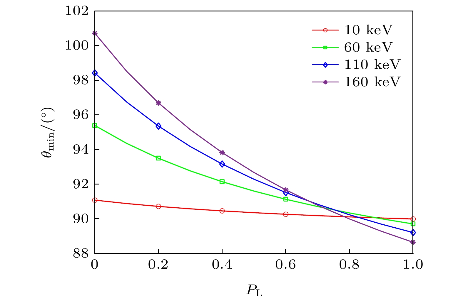
图 3 不同入射能量时,
${\theta _{{\rm{min}}}}$ 随线偏振度变化Fig. 3.
${\theta _{{\rm{min}}}}$ varying with polarization for different incident energy.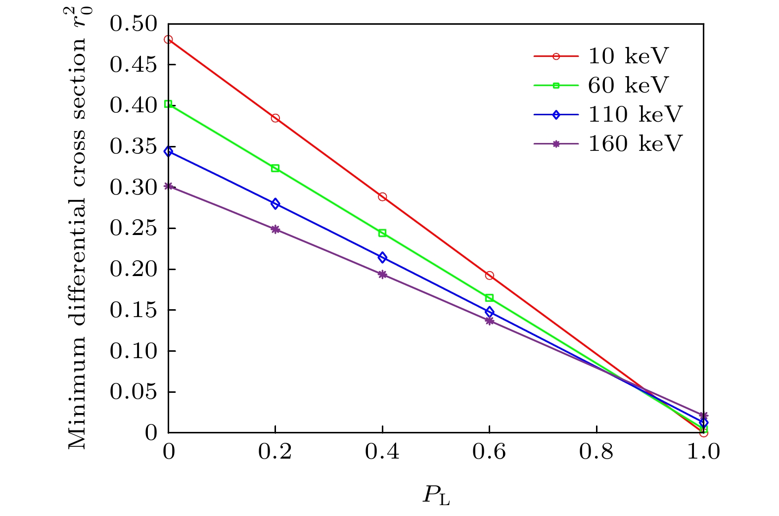
图 4 不同入射能量时最小微分截面随偏振度的变化
Fig. 4. Minimum differential cross section varying with polarization for different incident energy.

图 5
$\varphi = 0{\rm{ , \pi }}$ 处微分截面随散射角变化 (a)${P_{\rm{L}}} = 1$ 时相干、非相干和总微分截面; (b) 不同线偏振度时的总微分截面; (c)${P_{\rm{L}}} = $ $ {\rm{0}}{\rm{.5}}$ 时不同入射能量下的总微分截面Fig. 5. Differential cross section varying with
$\theta $ at$\varphi = 0{\rm{ , \pi }}$ : (a) Incoherent, coherent and total differential cross section at${P_{\rm{L}}} = 1$ ; (b) total differential cross section varying with$\theta $ for different${P_{\rm{L}}}$ ; (c) total differential cross section varying with$\theta $ for different incident energy at${P_{\rm{L}}} = {\rm{0}}{\rm{.5 }}$ 
图 6
$\varphi = 0\;{\rm{ or\; \pi }}$ 处$ \xi _2^{({\rm{f}})} $ 随散射角变化Fig. 6.
$ \xi _2^{({\rm{f}})} $ varying with$ \theta $ at$\varphi = 0,\;{\rm{\pi }}$ .
图 7 不同能量下
$\theta = {\rm{π}}/2{\rm{ , }}\varphi = 0,\;{\rm{\;\pi }}$ 处散射光线偏振度随入射光线偏振度变化Fig. 7. The linear polarization of scattering photons
$ P_{\rm{L}}^{({\rm{f}})} $ varying with$ P_{\rm{L}}^{({\rm{i}})} $ at$\theta = {\rm{π}}/2{\rm{ , }}~\varphi = 0,\;{\rm{ \pi }}$ for different incident energy. -
[1] Boisseau P, Grodzins L 1987 Hyperfine Interact. 33 283
Google Scholar
[2] Cheong S K, Jones B L, Siddiqi A K, Liu F, Manohar N, Cho S H 2010 Phys. Med. Biol. 55 647
Google Scholar
[3] Bazalova M, Kuang Y, Pratx G, Xing L 2012 IEEE Trans. Med. Imaging 31 1620
Google Scholar
[4] Sjölin M, Danielsson M 2014 Phys. Med. Biol. 59 6507
Google Scholar
[5] Li L, Zhang S Y, Li R Z, Chen Z Q 2017 Opt. Eng. 56 043106
Google Scholar
[6] Zhang S Y, Li L, Chen Z Q 2019 IEEE Access 7 113589
Google Scholar
[7] Li L, Li R Z, Zhang S Y, Chen Z Q 2016 Proc. of SPIE 9967 99670F
Google Scholar
[8] Zhang S Y, Li L, Chen J Y, Chen Z Q, Zhang W L, Lu H B 2019 Int. J. Mol. Sci. 20 2315
Google Scholar
[9] Ahmad M, Bazalova M, Xiang L, Xing L 2014 IEEE Trans. Med. Imaging 33 1119
Google Scholar
[10] Sasaya T, Sunaguchi N, Hyodo K, Zeniya T, Takeda T, Yuasa T 2017 Sci. c Rep. 7 44143
Google Scholar
[11] Chi Z, Du Y, Huang W, Tang C 2020 J. Synchrotron Radiat. 27 737
Google Scholar
[12] Vernekohl D, Tzoumas S, Zhao W, Xing L 2018 Med. Phys. 45 3741
Google Scholar
[13] Mcmaster W H 1961 Rev. Mod. Phys. 33 8
Google Scholar
[14] Jones J A, D’Addario A J, Rojec B L, Milione G, Galvez E J 2016 Am. J. Phys. 84 822
Google Scholar
[15] Bambynek W, Crasemann B, Fink R W, Freund H U, Mark H, Swift C D, Price R E, Rao P V 1972 Rev. Mod. Phys. 44 716
Google Scholar
[16] Hubbell JH, Trehan PN, Singh N, Chand B, Mehta D, Garg ML, Garg RR, Singh S, Puri S 1994 J. Phys. Chem. Ref. Data 23 339
Google Scholar
[17] Ertuğral B, Apaydın G, Çevik U, Ertuğrul M, Kobya A İ 2007 Radiat. Phys. Chem. 76 15
Google Scholar
[18] Scofield J H 1974 Phys. Rev. A 9 1041
Google Scholar
[19] Schaart D R 2021 Phys. Med. Biol. 66 09TR01
Google Scholar
[20] Han I, Şahin M, Demir L 2008 Can. J. Phys 86 361
Google Scholar
[21] E G Berezhko, N M Kabachnik 1977 J. Phys. B:At. Mol. Phys. 10 2467
Google Scholar
[22] Kämpfer T, Uschmann I, Wu Z W, Surzhykov A, Fritzsche S, Förster E, Paulus G G 2016 Phys. Rev. A 93 033409
Google Scholar
[23] 柳钰, 徐忠锋, 王兴, 曾利霞, 刘婷 2020 物理学报 69 043201
Google Scholar
Liu Y, Xu Z F, Wang X, Zeng L X, Liu T 2020 Acta Phys. Sin. 69 043201
Google Scholar
[24] Kahlon K S, Aulakh H S, Singh N, Mittal R, Allawadhi K L, Sood B S 1991 Phy. Rev. A 43 1455
Google Scholar
[25] Depaola G O 2003 Nucl. Instrum. Methods Phys. Res., Sect. A 512 619
Google Scholar
[26] Matt G, Feroci M, Rapisarda M, Costa E 1996 Radiat. Phys. Chem. 48 403
Google Scholar
[27] Fano U 1949 JOSA 39 859
Google Scholar
[28] Hamzawy A 2016 Radiat. Phys. Chem. 119 103
Google Scholar
[29] Hubbell J H 1997 Radiat. Phys. Chem. 50 113
Google Scholar
[30] Hubbell J H, Veigele W J, Briggs E A, Brown R T, Cromer D T, Howerton D R 1975 J. Phys. Chem. Ref. Data 4 471
Google Scholar
[31] Hubbell J H, O/verbo/ I 1979 J. Phys. Chem. Ref. Data 8 69
Google Scholar
[32] Chantler C T 1995 J. Phys. Chem. Ref. Data 24 71
Google Scholar
[33] Tartari A, Taibi A, Bonifazzi C, Baraldi C 2002 Phys. Med. Biol. 47 163
Google Scholar
[34] Zhang L, YangDai T Y 2016 Appl. Radiat. Isot. 114 179
Google Scholar

 下载:
下载:


计量
- 文章访问数: 9052
- PDF下载量: 116
- 被引次数: 0
