


-
将InAs/GaAs量子点薄膜样品转移到Ag纳米颗粒覆盖的Si衬底上, 然后将样品放到金刚石对顶砧压力腔室内. 在
$ 1.09—1.98\;\rm{G}\rm{P}\rm{a} $ 的压力范围内, 测量了量子点激子的荧光光谱和时间分辨光谱. 实验结果显示, 随着静水压力的增大, 激子的发光波长蓝移, 激子的发光寿命从$ \left(41\pm 3\right)\times 10\;\rm{n}\rm{s} $ 延长到$ \left(120\pm 4\right)\times 10\;\rm{n}\rm{s} $ , 再减短到$ (7.6\pm 0.2)\;\rm{n}\rm{s} $ , 在激子发光波长为$ 797.49\;\rm{n}\rm{m} $ 时, 寿命达到最长的$ \left(120\pm 4\right)\times 10\;\rm{n}\rm{s} $ . 相比没有Ag纳米颗粒影响的InAs/GaAs量子点中的激子寿命约$ 1\;\rm{n}\rm{s} $ , 激子的寿命延长了约1200倍. 其物理机制为量子点浸润层中激子的辐射场和Ag 纳米颗粒的散射场之间发生相消干涉, 抑制了浸润层中激子的自发辐射, 这些长寿命的浸润层激子将扩散到量子点中, 并辐射复合发光, 从而观察到量子点激子的长寿命衰变曲线. 这一实验结果与基于在散射场下的偶极子辐射模型计算结果一致.-
关键词:
- InAs/GaAs量子点 /
- 自发辐射速率 /
- Ag纳米颗粒 /
- 长寿命激子 /
- 静水压力
In the past few decades, the studies of exciton emissions coupled with the metal nanoparticles have mainly focused on the enhancing exciton radiation and reducing exciton lifetime by near-field coupling interactions between excitons and metal nanoparticles. Only in recent years has the plasmon-field-induced to extend exciton lifetime (inhibition of the exciton emission) been reported. Experimentally, for observing a long-lifetime exciton state it needs to satisfy a condition of$kz\sim1$ , instead of near-field condition of$ kz\ll 1 $ , where$k=2{\pi }n/\lambda$ is the wavevector,$ n $ is the refractive index,$ \lambda $ is the wavelength, and$ z $ is the separation distance between the emitter and metal nanoparticle. Thus, in this paper, we tune the exciton emission wavelength by applying hydrostatic pressure to achieve the condition of$kz\sim1$ in order to in detail investigate the coupling between excitons and metal nanoparticles. The studied InAs/GaAs quantum dot (QD) sample is grown by molecular beam epitaxy on a (001) semi-insulating GaAs substrate. After the AlAs sacrificial layer is etched with hydrofluoric acid, the QD film sample is transferred onto an Si substrate covered with Ag nanoparticles. Then the sample is placed in the diamond anvil cell device combined with a piezoelectric ceramic. In this case we can measure the photoluminescence and time-resolved photoluminescence spectra of the QD sample under different pressures. It is found that the observed longest exciton lifetime is$(120\pm 4)\times 10~\rm{n}\rm{s}$ at a pressure of$ 1.38\;\rm{G}\rm{P}\rm{a} $ , corresponding the exciton emission wavelength of$ 797.49\;\rm{n}\rm{m} $ , which is about$ 1200 $ times longer than the exciton lifetime of$\sim 1\;\rm{n}\rm{s} $ in QDs without the influence of Ag nanoparticles. The experimental results can be understood based on the destructive interference between the quantum dot exciton radiation field and the scattering field of metal nanoparticles. This model proposes a convenient way to increase the emission lifetime of dipoles on a large scale, and is expected to be applied to quantum information processing, optoelectronic applications, fundamental physics researches such as Bose-Einstein condensates.-
Keywords:
- InAs/GaAs quantum dots /
- spontaneous emission rate /
- Ag nanoparticles /
- long-lived excitons /
- hydrostatic pressure
[1] Drexhage K H 1970 J. Lumin. 1–2 693
[2] Ferioli G, Glicenstein A, Henriet L, Ferrier-Barbut I, Browaeys A 2021 Phys. Rev. X 11 021031
[3] Cipris A, Moreira N A, do Espirito Santo T S, Weiss P, Villas-Boas C J, Kaiser R, Guerin W, Bachelard R 2021 Phys. Rev. Lett. 126 103604
 Google Scholar
[4] Pineiro Orioli A, Rey A M 2019 Phys. Rev. Lett. 123 223601
Google Scholar
[5] Heyde K, Sau J 1986 Phys. Rev. C Nucl. Phys. 33 1050
Google Scholar
[6] Zhou Y, Scuri G, Sung J, Gelly R J, Wild D S, De Greve K, Joe A Y, Taniguchi T, Watanabe K, Kim P, Lukin M D, Park H 2020 Phys. Rev. Lett. 124 027401
Google Scholar
[7] Ropp C, Cummins Z, Nah S, Fourkas J T, Shapiro B, Waks E 2015 Nat. Commun. 6 6558
Google Scholar
[8] Bužek V 1990 Z. Phys. D At. Mol. Clust. 17 91
Google Scholar
[9] Gu Y, Wang L, Ren P, Zhang J, Zhang T, Martin O J, Gong Q 2012 Nano Lett. 12 2488
Google Scholar
[10] Anger P, Bharadwaj P, Novotny L 2006 Phys. Rev. Lett. 96 113002
Google Scholar
[11] Delga A, Feist J, Bravo-Abad J, Garcia-Vidal F J 2014 Phys. Rev. Lett. 112 253601
Google Scholar
[12] Gazzano O, Michaelis de Vasconcellos S, Gauthron K, Symonds C, Bloch J, Voisin P, Bellessa J, Lemaitre A, Senellart P 2011 Phys. Rev. Lett. 107 247402
Google Scholar
[13] Evangelou S, Yannopapas V, Paspalakis E 2011 Phys. Rev. A 83 023819
Google Scholar
[14] Felici M, Pettinari G, Biccari F, Boschetti A, Younis S, Birindelli S, Gurioli M, Vinattieri A, Gerardino A, Businaro L, Hopkinson M, Rubini S, Capizzi M, Polimeni A 2020 Phys. Rev. B 101 205403
Google Scholar
[15] 闫晓宏, 牛亦杰, 徐红星, 魏红 2022 物理学报 71 067301
Google Scholar
Yan X H, Niu Y J, Xu H X, Wei H 2022 Acta Phys. Sin. 71 067301
Google Scholar
[16] Kuhn S, Hakanson U, Rogobete L, Sandoghdar V 2006 Phys. Rev. Lett. 97 017402
Google Scholar
[17] Pustovit V N, Shahbazyan T V 2009 Phys. Rev. Lett. 102 077401
Google Scholar
[18] Huang J, Ojambati O S, Chikkaraddy R, Sokolowski K, Wan Q, Durkan C, Scherman O A, Baumberg J J 2021 Phys. Rev. Lett. 126 047402
Google Scholar
[19] Belacel C, Habert B, Bigourdan F, Marquier F, Hugonin J P, de Vasconcellos S M, Lafosse X, Coolen L, Schwob C, Javaux C, Dubertret B, Greffet J J, Senellart P, Maitre A 2013 Nano Lett. 13 1516
Google Scholar
[20] Dey S, Zhou Y, Tian X, Jenkins J A, Chen O, Zou S, Zhao J 2015 Nanoscale 7 6851
Google Scholar
[21] 张炼, 王化雨, 王宁, 陶灿, 翟学琳, 马平准, 钟莹, 刘海涛 2022 物理学报 71 118101
Google Scholar
Zhang L, Wang H Y, Wang N, Tao C, Zhai X L, Ma P Z, Zhong Y, Liu H T 2022 Acta Phys. Sin. 71 118101
Google Scholar
[22] Chen H, Huang J, He X, Ding K, Ni H, Niu Z, Jiang D, Dou X, Sun B 2020 ACS Photon. 7 3228
Google Scholar
[23] Huang J, Chen H, Zhuo Z, Wang J, Li S, Ding K, Ni H, Niu Z, Jiang D, Dou X, Sun B 2021 Chin. Phys. B 30 097805
Google Scholar
[24] Zhuo Z, Chen H, Huang J, Li S, Wang J, Ding K, Ni H, Niu Z, Jiang D, Dou X, Sun B 2021 J. Phys. Chem. Lett. 12 3485
Google Scholar
[25] Carreño F, Antón M A, Arrieta-Yáñez F 2013 Phys. Rev. B 88 195303
Google Scholar
[26] Feldman M A, Dumitrescu E F, Bridges D, Chisholm M F, Davidson R B, Evans P G, Hachtel J A, Hu A, Pooser R C, Haglund R F, Lawrie B J 2018 Phys. Rev. B 97 081404
Google Scholar
[27] Carreño F, Yannopapas V, Antón M A, Paspalakis E 2019 Phys. Rev. A 100 023802
Google Scholar
[28] Hofmann M S, Gluckert J T, Noe J, Bourjau C, Dehmel R, Hogele A 2013 Nat. Nanotechnol. 8 502
Google Scholar
[29] Johansen J, Julsgaard B, Stobbe S, Hvam J M, Lodahl P 2010 Phys. Rev. B 81 081304
Google Scholar
[30] Palummo M, Bernardi M, Grossman J C 2015 Nano Lett. 15 2794
Google Scholar
[31] Butov L V, Lai C W, Ivanov A L, Gossard A C, Chemla D S 2002 Nature 417 47
Google Scholar
[32] Novotny L, Hecht B 2012 Priciples of Nano-Optics (Cambridge: Cambridge University Press) pp335–359
[33] Wu X F, Wei H, Dou X M, Ding K, Yu Y, Ni H Q, Niu Z C, Ji Y, Li S S, Jiang D S, Guo G C, He L X, Sun B Q 2014 Europhys. Lett. 107 27008
Google Scholar
[34] 李元和, 卓志瑶, 王健, 黄君辉, 李叔伦, 倪海桥, 牛智川, 窦秀明, 孙宝权 2022 物理学报 71 067804
Google Scholar
Li Y H, Zhuo Z Y, Wang J, Huang J H, Li S L, Ni H Q, Niu Z C, Dou X M, Sun B Q 2022 Acta Phys. Sin. 71 067804
Google Scholar
[35] Yu Y, Shang X J, Li M F, Zha G W, Xu J X, Wang L J, Wang G W, Ni H Q, Dou X, Sun B, Niu Z C 2013 Appl. Phys. Lett. 102 201103
Google Scholar
[36] 尚向军, 马奔, 陈泽升, 喻颖, 查国伟, 倪海桥, 牛智川 2018 物理学报 67 227801
Google Scholar
Shang X J, Ma B, Chen Z S, Yu Y, Zha G W, Ni H Q, Niu Z C 2018 Acta Phys. Sin. 67 227801
Google Scholar
[37] Wu X, Dou X, Ding K, Zhou P, Ni H, Niu Z, Jiang D, Sun B 2013 Appl. Phys. Lett. 103 252108
Google Scholar
[38] 丁琨, 武雪飞, 窦秀明, 孙宝权 2016 物理学报 65 037701
Google Scholar
Ding K, Wu X F, Dou X M, Sun B Q 2016 Acta Phys. Sin. 65 037701
Google Scholar
[39] 徐章程, 贾国治, 孙亮, 姚江宏, 许京军, Hvam J M, 王占国 2005 物理学报 54 5367
Google Scholar
Xu Z C, Jia G Z, Sun L, Yao J H, Xu J J, Hvam J M, Wang Z G 2005 Acta Phys. Sin. 54 5367
Google Scholar
[40] Itskevich I E, Lyapin S G, Troyan I A, Klipstein P C, Eaves L, Main P C, Henini M 1998 Phys. Rev. B 58 R4250
Google Scholar
[41] Wang G, Fafard S, Leonard D, Bowers J E, Merz J L, Petroff P M 1994 Appl. Phys. Lett. 64 2815
Google Scholar
[42] Seufert J, Bacher G, Schömig H, Forchel A, Hansen L, Schmidt G, Molenkamp L W 2004 Phys. Rev. B 69 035311
Google Scholar
[43] Zhou P Y, Dou X M, Wu X F, Ding K, Luo S, Yang T, Zhu H J, Jiang D S, Sun B Q 2014 J. Appl. Phys. 116 023510
Google Scholar
[44] Eisaman M D, Fan J, Migdall A, Polyakov S V 2011 Rev. Sci. Instrum. 82 071101
Google Scholar
[45] Weber W H, Ford G W 2004 Phys. Rev. B 70 125429
Google Scholar
[46] Johnson P B, Christy R W 1972 Phys. Rev. B 6 4370
Google Scholar
[47] Adachi S 2012 Handbook on Optical Constants of Metals (Singapore: World Scientific Publishing ) pp61–68
[48] Adachi S 2012 The Handbook on Optical Constants of Semiconductors (Singapore: World Scientific Publishing) pp153–162
[49] Saglimbeni F, Bianchi S, Gibson G, Bowman R, Padgett M, Di Leonardo R 2016 Opt. Express 24 27009
Google Scholar
-
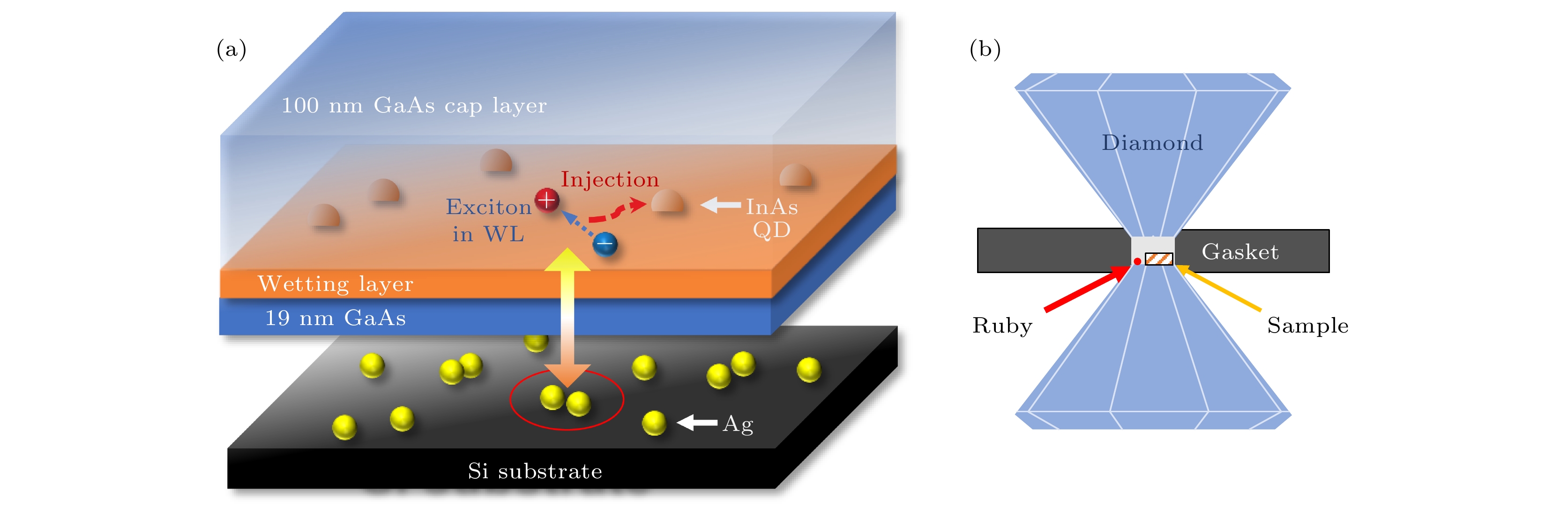
图 1 (a) InAs/GaAs 量子点样品转移到覆盖了Ag纳米颗粒的Si片上示意图, 其中黄色箭头表示量子点浸润层中激子偶极子与金属纳米颗粒偶极子之间的相互作用; (b)金刚石对顶砧设备示意图, 其中金属垫片和金刚石砧面组成样品的压力腔室, 腔室中放置样品和红宝石
Fig. 1. (a) Schematic diagram of the InAs/GaAs QD sample transferred onto a Si substrate covered with Ag nanoparticles. The yellow arrow represents the interaction between exciton dipole in WL and the Ag nanoparticles. (b) Schematic diagram of the diamond anvil cell. The pressure chamber consists of a metal gasket and diamond surfaces. The sample and ruby are placed in the chamber.
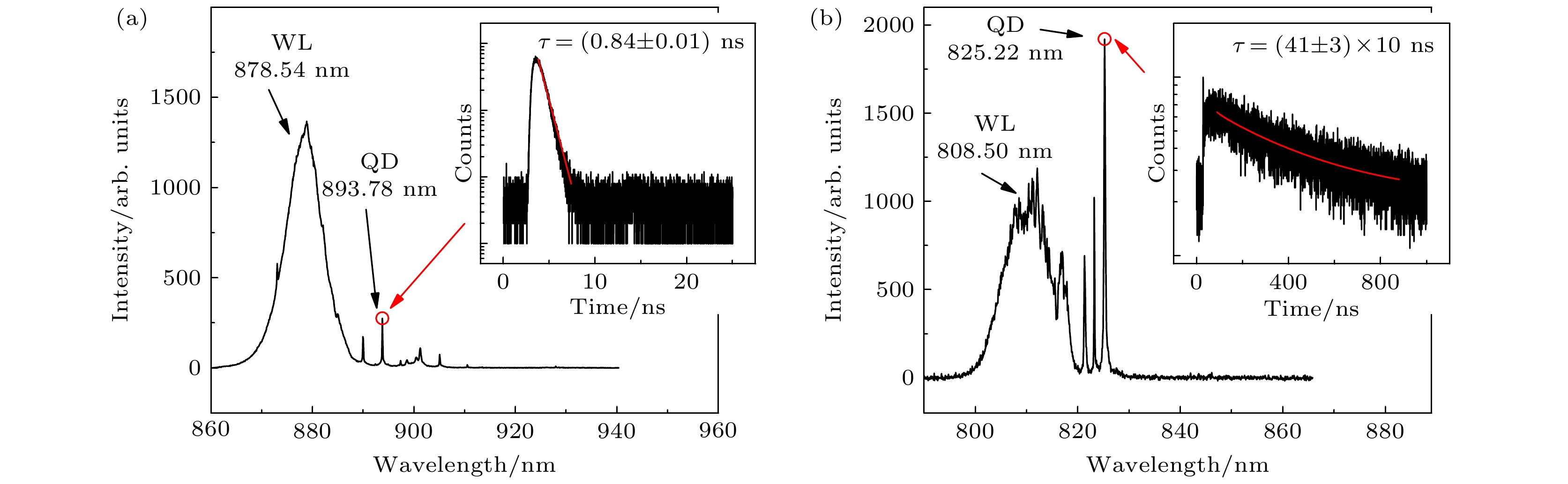
图 2 (a)低温20 K和常压下量子点样品的PL光谱, 激发功率为2.2 μW; 插图为
$ 893.78\;\rm{n}\rm{m} $ 波长的量子点发光谱线的TRPL光谱, 激光为$ 40\;\rm{ }\rm{M}\rm{H}\rm{z} $ 的脉冲光, 激发功率为1.04 μW, 红色实线为使用单指数衰减函数拟合的结果; (b)在低温 20 K和$ 1.09\;\rm{G}\rm{P}\rm{a} $ 压力下, 转移后量子点样品的PL光谱, 激发功率为2.2 μW; 插图为$ 825.22\;\rm{n}\rm{m} $ 波长的发光谱线的TRPL光谱, 激发模式为$ 1\;\rm{M}\rm{H}\rm{z} $ 频率的脉冲光, 激发功率为0.026 μW, 红色实线为类拓展指数衰减函数拟合结果Fig. 2. (a) PL spectrum of QD sample at 20 K and atmospheric pressure, excited by a power of 2.2 μW. Inset: TRPL spectrum of QD emission line of
$ 893.78\;\rm{n}\rm{m} $ at an excitation power of 1.04 μW in pulsed mode of$ 40\;\rm{M}\rm{H}\rm{z} $ . The red solid line represents the single exponential function fitting result. (b) PL spectrum of the transferred QD sample at 20 K and$ 1.09\;\rm{G}\rm{P}\rm{a} $ , excited by a power of 2.2 μW. Inset: TRPL spectrum of QD emission line of$ 825.22\;\rm{n}\rm{m} $ at an excitation power of 0.026 μW in pulsed mode of$ 1\rm{ }\rm{M}\rm{H}\rm{z} $ . The red solid line represents the stretched-like exponential function fitting result.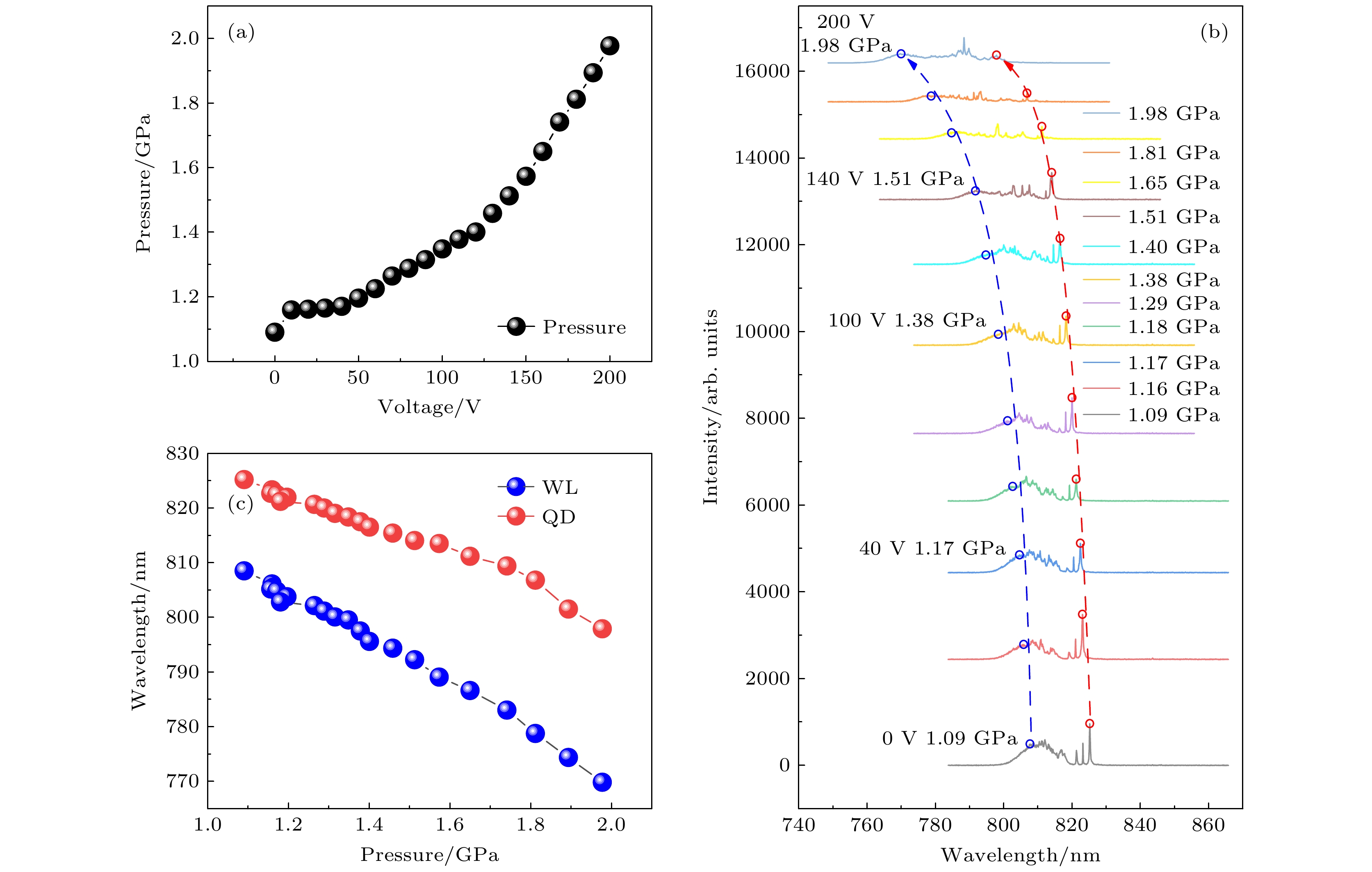
图 3 (a) DAC腔中的压力与PZT电压的函数关系; (b)不同压力下InAs/GaAs量子点样品的PL光谱, 激发功率为2.2 μW, 红色和蓝色虚线箭头分别表示量子点和浸润层发光峰波长蓝移结果; (c)量子点(红色)和浸润层(蓝色)发光峰波长与压力的函数关系
Fig. 3. (a) Hydrostatic pressure in DAC chamber as a function of applied voltage of PZT; (b) PL spectra of the InAs/GaAs QD sample measured under different pressures at an excitation power of 2.2 μW, the red and blue dashed lines indicate the pressure-induced blue shift of QD and WL emission peaks, respectively; (c) pressure dependences of QD (red) and WL (blue) PL peak wavelengths.
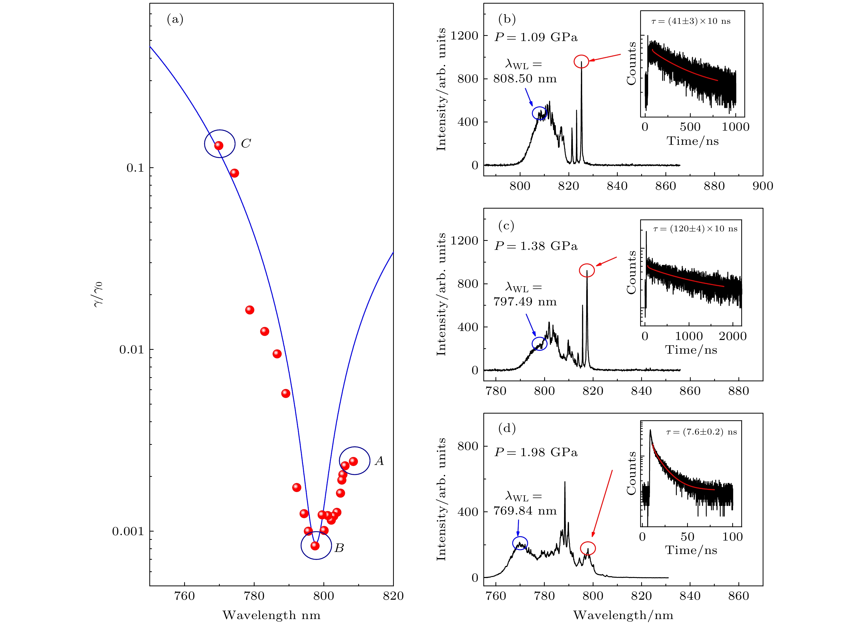
图 4 (a) InAs/GaAs量子点样品中量子点辐射速率与浸润层发光波长的依赖关系, 其中红色点为不同压力下的实验数据, 蓝色实线为(1)式计算结果; (b)—(d)在图(a)中蓝色圆圈A, B和C三个浸润层发光波长 (
$ 808.50\;\rm{n}\rm{m} $ ,$ 797.49\;\rm{ }\rm{n}\rm{m} $ 和$ 769.84\;\rm{ }\rm{n}\rm{m} $ ) 位置对应的量子点PL 和TRPL光谱(插图), 对应激发光的脉冲频率分别为$ 1\;\rm{ }\rm{M}\rm{H}\rm{z} $ ,$ 0.25\;\rm{ }\rm{M}\rm{H}\rm{z} $ 和$ 10\;\rm{ }\rm{M}\rm{H}\rm{z} $ , 其中插图内红色实线表示类拓展指数函数拟合结果Fig. 4. (a) Dependence of QD radiation rate and WL emission wavelength for the transferred InAs/GaAs QD sample, the red dots are experimental data under different hydrostatic pressures and the blue solid line represents the calculated result based on Eq. (1); (b)–(d) PL and TRPL spectra (Inset) of QD for the experimental condition of WL wavelengths at
$ 808.50 $ ,$ 797.49 $ and$ 769.84\;\rm{n}\rm{m} $ , respectively, corresponding to the data points A, B and C in Fig. 4(a), with a laser excitation repetition rate of$ 1\;\rm{M}\rm{H}\rm{z} $ ,$ 0.25\;\rm{M}\rm{H}\rm{z} $ , and$ 10\;\rm{ }\rm{M}\rm{H}\rm{z} $ respectively. The red solid lines in inset represent the stretched-like exponential function fitting results. -
[1] Drexhage K H 1970 J. Lumin. 1–2 693
[2] Ferioli G, Glicenstein A, Henriet L, Ferrier-Barbut I, Browaeys A 2021 Phys. Rev. X 11 021031
[3] Cipris A, Moreira N A, do Espirito Santo T S, Weiss P, Villas-Boas C J, Kaiser R, Guerin W, Bachelard R 2021 Phys. Rev. Lett. 126 103604
Google Scholar
[4] Pineiro Orioli A, Rey A M 2019 Phys. Rev. Lett. 123 223601
Google Scholar
[5] Heyde K, Sau J 1986 Phys. Rev. C Nucl. Phys. 33 1050
Google Scholar
[6] Zhou Y, Scuri G, Sung J, Gelly R J, Wild D S, De Greve K, Joe A Y, Taniguchi T, Watanabe K, Kim P, Lukin M D, Park H 2020 Phys. Rev. Lett. 124 027401
Google Scholar
[7] Ropp C, Cummins Z, Nah S, Fourkas J T, Shapiro B, Waks E 2015 Nat. Commun. 6 6558
Google Scholar
[8] Bužek V 1990 Z. Phys. D At. Mol. Clust. 17 91
Google Scholar
[9] Gu Y, Wang L, Ren P, Zhang J, Zhang T, Martin O J, Gong Q 2012 Nano Lett. 12 2488
Google Scholar
[10] Anger P, Bharadwaj P, Novotny L 2006 Phys. Rev. Lett. 96 113002
Google Scholar
[11] Delga A, Feist J, Bravo-Abad J, Garcia-Vidal F J 2014 Phys. Rev. Lett. 112 253601
Google Scholar
[12] Gazzano O, Michaelis de Vasconcellos S, Gauthron K, Symonds C, Bloch J, Voisin P, Bellessa J, Lemaitre A, Senellart P 2011 Phys. Rev. Lett. 107 247402
Google Scholar
[13] Evangelou S, Yannopapas V, Paspalakis E 2011 Phys. Rev. A 83 023819
Google Scholar
[14] Felici M, Pettinari G, Biccari F, Boschetti A, Younis S, Birindelli S, Gurioli M, Vinattieri A, Gerardino A, Businaro L, Hopkinson M, Rubini S, Capizzi M, Polimeni A 2020 Phys. Rev. B 101 205403
Google Scholar
[15] 闫晓宏, 牛亦杰, 徐红星, 魏红 2022 物理学报 71 067301
Google Scholar
Yan X H, Niu Y J, Xu H X, Wei H 2022 Acta Phys. Sin. 71 067301
Google Scholar
[16] Kuhn S, Hakanson U, Rogobete L, Sandoghdar V 2006 Phys. Rev. Lett. 97 017402
Google Scholar
[17] Pustovit V N, Shahbazyan T V 2009 Phys. Rev. Lett. 102 077401
Google Scholar
[18] Huang J, Ojambati O S, Chikkaraddy R, Sokolowski K, Wan Q, Durkan C, Scherman O A, Baumberg J J 2021 Phys. Rev. Lett. 126 047402
Google Scholar
[19] Belacel C, Habert B, Bigourdan F, Marquier F, Hugonin J P, de Vasconcellos S M, Lafosse X, Coolen L, Schwob C, Javaux C, Dubertret B, Greffet J J, Senellart P, Maitre A 2013 Nano Lett. 13 1516
Google Scholar
[20] Dey S, Zhou Y, Tian X, Jenkins J A, Chen O, Zou S, Zhao J 2015 Nanoscale 7 6851
Google Scholar
[21] 张炼, 王化雨, 王宁, 陶灿, 翟学琳, 马平准, 钟莹, 刘海涛 2022 物理学报 71 118101
Google Scholar
Zhang L, Wang H Y, Wang N, Tao C, Zhai X L, Ma P Z, Zhong Y, Liu H T 2022 Acta Phys. Sin. 71 118101
Google Scholar
[22] Chen H, Huang J, He X, Ding K, Ni H, Niu Z, Jiang D, Dou X, Sun B 2020 ACS Photon. 7 3228
Google Scholar
[23] Huang J, Chen H, Zhuo Z, Wang J, Li S, Ding K, Ni H, Niu Z, Jiang D, Dou X, Sun B 2021 Chin. Phys. B 30 097805
Google Scholar
[24] Zhuo Z, Chen H, Huang J, Li S, Wang J, Ding K, Ni H, Niu Z, Jiang D, Dou X, Sun B 2021 J. Phys. Chem. Lett. 12 3485
Google Scholar
[25] Carreño F, Antón M A, Arrieta-Yáñez F 2013 Phys. Rev. B 88 195303
Google Scholar
[26] Feldman M A, Dumitrescu E F, Bridges D, Chisholm M F, Davidson R B, Evans P G, Hachtel J A, Hu A, Pooser R C, Haglund R F, Lawrie B J 2018 Phys. Rev. B 97 081404
Google Scholar
[27] Carreño F, Yannopapas V, Antón M A, Paspalakis E 2019 Phys. Rev. A 100 023802
Google Scholar
[28] Hofmann M S, Gluckert J T, Noe J, Bourjau C, Dehmel R, Hogele A 2013 Nat. Nanotechnol. 8 502
Google Scholar
[29] Johansen J, Julsgaard B, Stobbe S, Hvam J M, Lodahl P 2010 Phys. Rev. B 81 081304
Google Scholar
[30] Palummo M, Bernardi M, Grossman J C 2015 Nano Lett. 15 2794
Google Scholar
[31] Butov L V, Lai C W, Ivanov A L, Gossard A C, Chemla D S 2002 Nature 417 47
Google Scholar
[32] Novotny L, Hecht B 2012 Priciples of Nano-Optics (Cambridge: Cambridge University Press) pp335–359
[33] Wu X F, Wei H, Dou X M, Ding K, Yu Y, Ni H Q, Niu Z C, Ji Y, Li S S, Jiang D S, Guo G C, He L X, Sun B Q 2014 Europhys. Lett. 107 27008
Google Scholar
[34] 李元和, 卓志瑶, 王健, 黄君辉, 李叔伦, 倪海桥, 牛智川, 窦秀明, 孙宝权 2022 物理学报 71 067804
Google Scholar
Li Y H, Zhuo Z Y, Wang J, Huang J H, Li S L, Ni H Q, Niu Z C, Dou X M, Sun B Q 2022 Acta Phys. Sin. 71 067804
Google Scholar
[35] Yu Y, Shang X J, Li M F, Zha G W, Xu J X, Wang L J, Wang G W, Ni H Q, Dou X, Sun B, Niu Z C 2013 Appl. Phys. Lett. 102 201103
Google Scholar
[36] 尚向军, 马奔, 陈泽升, 喻颖, 查国伟, 倪海桥, 牛智川 2018 物理学报 67 227801
Google Scholar
Shang X J, Ma B, Chen Z S, Yu Y, Zha G W, Ni H Q, Niu Z C 2018 Acta Phys. Sin. 67 227801
Google Scholar
[37] Wu X, Dou X, Ding K, Zhou P, Ni H, Niu Z, Jiang D, Sun B 2013 Appl. Phys. Lett. 103 252108
Google Scholar
[38] 丁琨, 武雪飞, 窦秀明, 孙宝权 2016 物理学报 65 037701
Google Scholar
Ding K, Wu X F, Dou X M, Sun B Q 2016 Acta Phys. Sin. 65 037701
Google Scholar
[39] 徐章程, 贾国治, 孙亮, 姚江宏, 许京军, Hvam J M, 王占国 2005 物理学报 54 5367
Google Scholar
Xu Z C, Jia G Z, Sun L, Yao J H, Xu J J, Hvam J M, Wang Z G 2005 Acta Phys. Sin. 54 5367
Google Scholar
[40] Itskevich I E, Lyapin S G, Troyan I A, Klipstein P C, Eaves L, Main P C, Henini M 1998 Phys. Rev. B 58 R4250
Google Scholar
[41] Wang G, Fafard S, Leonard D, Bowers J E, Merz J L, Petroff P M 1994 Appl. Phys. Lett. 64 2815
Google Scholar
[42] Seufert J, Bacher G, Schömig H, Forchel A, Hansen L, Schmidt G, Molenkamp L W 2004 Phys. Rev. B 69 035311
Google Scholar
[43] Zhou P Y, Dou X M, Wu X F, Ding K, Luo S, Yang T, Zhu H J, Jiang D S, Sun B Q 2014 J. Appl. Phys. 116 023510
Google Scholar
[44] Eisaman M D, Fan J, Migdall A, Polyakov S V 2011 Rev. Sci. Instrum. 82 071101
Google Scholar
[45] Weber W H, Ford G W 2004 Phys. Rev. B 70 125429
Google Scholar
[46] Johnson P B, Christy R W 1972 Phys. Rev. B 6 4370
Google Scholar
[47] Adachi S 2012 Handbook on Optical Constants of Metals (Singapore: World Scientific Publishing ) pp61–68
[48] Adachi S 2012 The Handbook on Optical Constants of Semiconductors (Singapore: World Scientific Publishing) pp153–162
[49] Saglimbeni F, Bianchi S, Gibson G, Bowman R, Padgett M, Di Leonardo R 2016 Opt. Express 24 27009
Google Scholar


 下载:
下载:


计量
- 文章访问数: 6851
- PDF下载量: 68
- 被引次数: 0
