


-
上海软X射线自由电子激光装置(SXFEL)是我国首台X射线自由电子激光用户装置, 目前建有2条波荡器线、2条光束线以及5个实验站. 装置可提供2—15 nm波长(80—620 eV )的X射线脉冲, 用于高时空分辨的前沿科学研究. 利用XFEL高亮度、短脉冲和全相干的特性实现单脉冲相干衍射成像, 可以有效地减轻辐射损伤, 提高图像的空间分辨率. SXFEL设计重复频率为50 Hz, 实现单脉冲成像的关键在于通过定时系统能够精确地控制X射线脉冲到达样品点的时间, 以确保只有一个脉冲被选中用于成像. 同时, 还需要与成像系统的触发进行同步, 以确保成像系统在正确的时间采集X射线脉冲与样品作用后的图像. 本文介绍了SXFEL单脉冲成像定时的设计与实现. 通过单脉冲成像的结果表明该定时方案能满足在50 Hz的SXFEL开展单脉冲成像的需求.
-
关键词:
- 相干 X 射线衍射成像 /
- X射线自由电子激光 /
- 单脉冲成像 /
- 定时系统
X-ray free-electron laser (XFEL), as a novel advanced X-ray light source, has excellent properties such as ultra-high brightness, ultra-shot pulse duration, and full coherence. The coherent X-ray diffraction imaging (CDI) has a lot of advantages at high resolution and quantitative imaging compared with the traditional lens based X-ray imaging methods. By combining the excellent properties of XFEL and advantages of CDI, the single-shot imaging has been realized, based on the concept of “diffraction before destruction”. Shanghai soft X-ray free-electron laser facility (SXFEL) is the first XFEL facility operated at the X-ray wavelength in China. The coherent scattering and imaging (CSI) endstation is the first commissioned endstation at SXFEL, focusing on the high spatiotemporal imaging for nano materials and micro materials by using a single-shot imaging method. To realize the single-shot experiment at XFEL, especially for single-shot imaging, the timing system plays a crucial role in ensuring the operation of the equipment in sequence. This paper introduces the design and implementation process of SXFEL single-shot imaging timing. The timing system is implemented with White Rabbit (WR) and digital delay and pulse generator (BNC505). Single-shot imaging is realized by synchronously moving the sample scanning stages and X-ray shutter to select a single pulse to illuminate the sample. At the same time, the X-ray detector is triggered with the timing system to record the single-shot diffraction pattern. During debugging, a gold nanodisks each with a side length of approximately 300 nm and a thickness of about 30 nm, as test samples, are imaged at the CSI endstation. The nanodisks are uniformly dispersed on Si3N4 membranes for single-shot imaging. Because of the ultra-high peak intensity at the focus spot, the samples and membrane are ionized for each XFEL pulse shot. A raster scan is performed on the membranes at intervals of 50 μm to update the sample. With the timing system and X-ray shutter, single-shot diffraction patterns can be recorded by using an X-ray detector. From the image of the Si3N4 membrane after raster scanning, the ionized holes with an interval of 50 μm can be recognized. Finally, phase retrieval is applied to the single-shot diffraction pattern to obtain a real-space image of the sample. The resolution of the reconstructed image is estimated by calculating the phase-retrieval transfer function (PRTF). With a citation of the PRTF curve dropping below$ 1/{\mathrm{e}} $ , the spatial frequency cutoff is determined to be 22.6 μm–1, corresponding to a half period resolution of 22.1 nm. The results show that the designed timing system can accurately control the time sequence of the imaging process, meeting the requirement for single-shot imaging within 50 Hz at SXFEL.-
Keywords:
- coherent X-ray diffraction imaging /
- X-ray free electron laser /
- single shot imaging /
- timing system
[1] 范家东, 江怀东 2012 物理学报 61 218702
 Google Scholar
Fan J D, Jiang H D 2012 Acta Phys. Sin. 61 218702
Google Scholar
[2] Sakdinawat A, Attwood D 2010 Nat. Photonics 4 840
Google Scholar
[3] 周光照, 佟亚军, 陈灿, 任玉琦, 王玉丹, 肖体乔 2011 物理学报 60 028701
Google Scholar
Zhou G Z, Tong Y J, Chen C, Ren Y Q, Wang Y D, Xiao T Q 2011 Acta Phys. Sin. 60 028701
Google Scholar
[4] 周光照, 王玉丹, 任玉琦, 陈灿, 叶琳琳, 肖体乔 2012 物理学报 61 018701
Google Scholar
Zhou G Z, Wang Y D, Ren Y Q, Chen C, Ye L L, Xiao T Q 2012 Acta Phys. Sin. 61 018701
Google Scholar
[5] Thibault P, Dierolf M, Menzel A, Bunk O, David C, Pfeiffer F 2008 Science 321 379
Google Scholar
[6] Song C Y, Jiang H D, Mancuso A, Amirbekian B, Peng L, Sun R, Shah S S, Zhou Z H, Ishikawa T, Miao J W 2008 Phys. Rev. Lett. 101 158101
Google Scholar
[7] Jiang H D, Song C Y, Chen C C, Xu R, Raines K S, Fahimian B P, Lu C H, Lee T K, Nakashima A, Urano J, Ishikawa T, Tamano F, Miao J W 2010 Proc. Natl. Acad. Sci. U. S. A. 107 11234
Google Scholar
[8] Chapman H N, Barty A, Bogan M J, Boutet S, Frank M, Hau-Riege S P, Marchesini S, Woods B W, Bajt S, Benner W H, London R A, Plönjes E, Kuhlmann M, Treusch R, Düsterer S, Tschentscher T, Schneider J R, Spiller E, Möller T, Bostedt C, Hoener M, Shapiro D A, Hodgson K O, van der Spoel D, Burmeister F, Bergh M, Caleman C, Huldt G, Seibert M M, Maia F R N C, Lee R W, Szöke A, Timneanu N, Hajdu J 2006 Nat. Phys. 2 839
Google Scholar
[9] Neutze R, Wouts R, van der Spoel D, Weckert E, Hajdu J 2000 Nature 406 752
Google Scholar
[10] Ihm Y, Cho D H, Sung D, Nam D, Jung C, Sato T, Kim S, Park J, Kim S, Gallagher-Jones M, Kim Y, Xu R, Owada S, Shim J H, Tono K, Yabashi M, Ishikawa T, Miao J, Noh D Y, Song C 2019 Nat. Commun. 10 2411
Google Scholar
[11] Gaffney K J, Chapman H N 2007 Science 316 1444
Google Scholar
[12] Miao J, Ishikawa T, Robinson I K, Murnane M M 2015 Science 348 530
Google Scholar
[13] Hidvegi A, Gessler P, Rehlich K, Bohm C 2011 IEEE Trans. Nucl. Sci. 58 1852
Google Scholar
[14] Ye Y, Li H, Li J, Yan Y, Yu P, Gong G 2022 J. Instrum. 17 T09009
Google Scholar
[15] Fan J D, Tong Y J, Nie Y G, Gao Z C, He B, Luan H, Lu D H, Zhang J H, Zhang D F, Yuan X Y, Chen J H, Guo Z, Liu T, Zhang M, Feng C, Deng H X, Liu B, Zhao Z T, Liu Z, Jiang H D 2022 Nucl. Sci. Tech. 33 114
Google Scholar
[16] MFR homepage, http://www.mrf.fi/ [2024-4-11]
[17] Kim C, Baek S Y, Kang H S, Kim J, Kim K W, Ko I S, Mun G, Park B R 2015 Proceedings of the 15th International Conference on Accelerator and Large Experimental Physics Control Systems Melbourne, Australia, October 17–23, 2015 p79
[18] Kalantari B, Biffiger R 2017 Proceedings of 16th International Conference on Accelerator and Large Experimental Physics Control Systems Barcelona, Spain, October 8–13, 2017 p232
[19] Krejcik P, Akre R A, Allison S, Browne M, Dalesio L R, Dusatko J E, Frisch J C, Fuller R, Gromme A E, Kotturi K D, Norum S, Rogind D, White W E, Zelazny M 2007 Proceedings of the 8th European Workshop on Beam Diagnostics and Instrumentation for Particle Accelerators Venice, Italy, May 20–23, 2007 p373
[20] WR homepage, https://white-rabbit.web.cern.ch/ [2024-4-11]
[21] 余鹏翔, 阎映炳 2023 核电子学与探测技术 43 923
Google Scholar
Yu P G, Yan Y B 2023 Nucl. Electron. Detect. Technol. 43 923
Google Scholar
[22] Yan Y B, Chen G H, Gong G H, Gu J L, Jiang Z Y, Xiao Q W, Ye Y M, Yu P X, Zhao L 2022 Proceedings of the 13th International Particle Accelerator Conference Bangkok, Thailand, June 12-17, 2022 p2415
[23] 于春蕾, 赵欢, 胡守明, 丁建国 2019 核技术 42 040102
Google Scholar
Yu C L, Zhao H, Hu S M, Ding J G 2019 Nucl. Tech. 42 040102
Google Scholar
[24] Tong Y J, Fan J D, Nie Y G, Guo Z, Gao Z C, Yuan X Y, He B, Chen J H, Zhang D F, Luan H, Zhang J H, Lu D H, Xie M H, Cheng P, Feng C, Liu T, Deng H X, Liu B, Liu Z, Jiang H D 2022 Front. Phys. 10 977957
Google Scholar
[25] Park J, Eom I, Kang T H, Rah S, Nam K H, Park J, Kim S, Kwon S, Park S H, Kim K S, Hyun H, Kim S N, Lee E H, Shin H, Kim S, Kim M J, Shin H J, Ahn D, Lim J, Yu C J, Song C, Kim H, Noh D Y, Kang H S, Kim B, Kim K W, Ko I S, Cho M H, Kim S 2016 Nucl. Instrum. Methods Phys. Res. , Sect. A 810 74
Google Scholar
[26] Rodriguez J A, Xu R, Chen C C, Zou Y, Miao J 2013 J. Appl. Crystallogr. 46 312
Google Scholar
[27] Shapiro D, Thibault P, Beetz T, Elser V, Howells M, Jacobsen C, Kirz J, Lima E, Miao H, Neiman A M, Sayre D 2005 Proc. Natl. Acad. Sci. U. S. A. 102 15343
Google Scholar
[28] Chapman H N, Barty A, Marchesini S, Noy A, Hau-Riege S P, Cui C, Howells M R, Rosen R, He H, Spence J C H, Weierstall U, Beetz T, Jacobsen C, Shapiro D 2006 J. Opt. Soc. Am. A 23 1179
Google Scholar
-
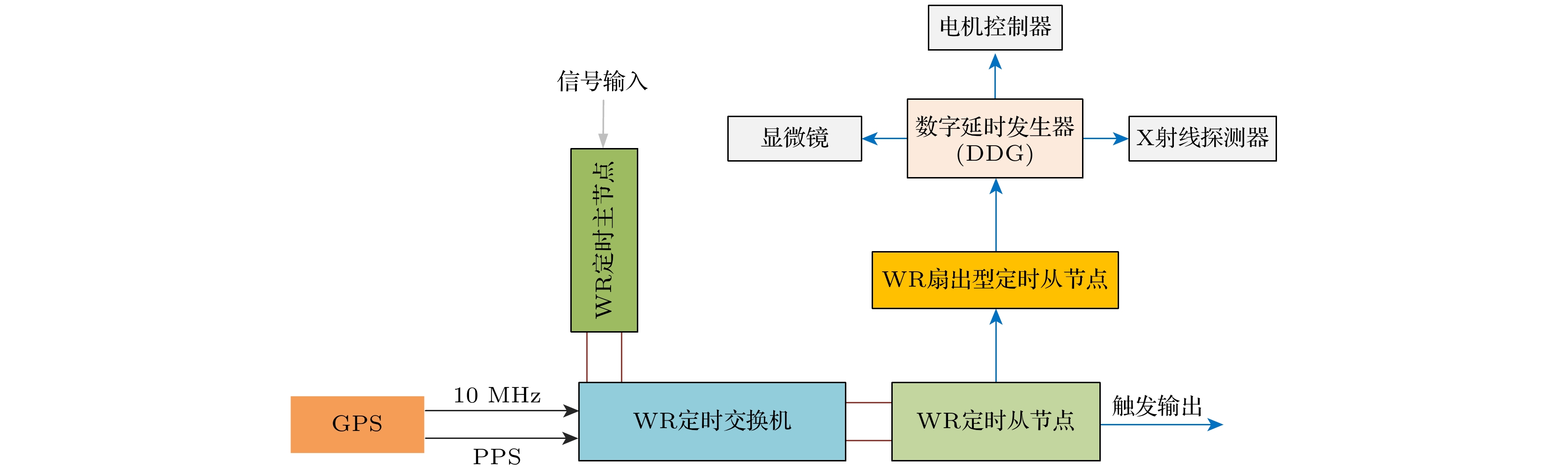
图 1 定时系统硬件结构图: 该定时系统包括两部分——WR定时系统和数字脉冲延时发生器, DDG接收WR定时信号, 经DDG后将特定时序关系的触发信号分发给需要定时的设备(GPS, 全球定位系统; PPS, 秒脉冲)
Fig. 1. Hardware structure diagram of timing system: This timing system consists of two parts, namely WR timing system and digital delay pulse generator. The DDG receives signals from the WR timing system, and then distributes trigger signals with specific timing relationships to the devices that require timing. GPS, global positioning system; PPS, pulse per second.
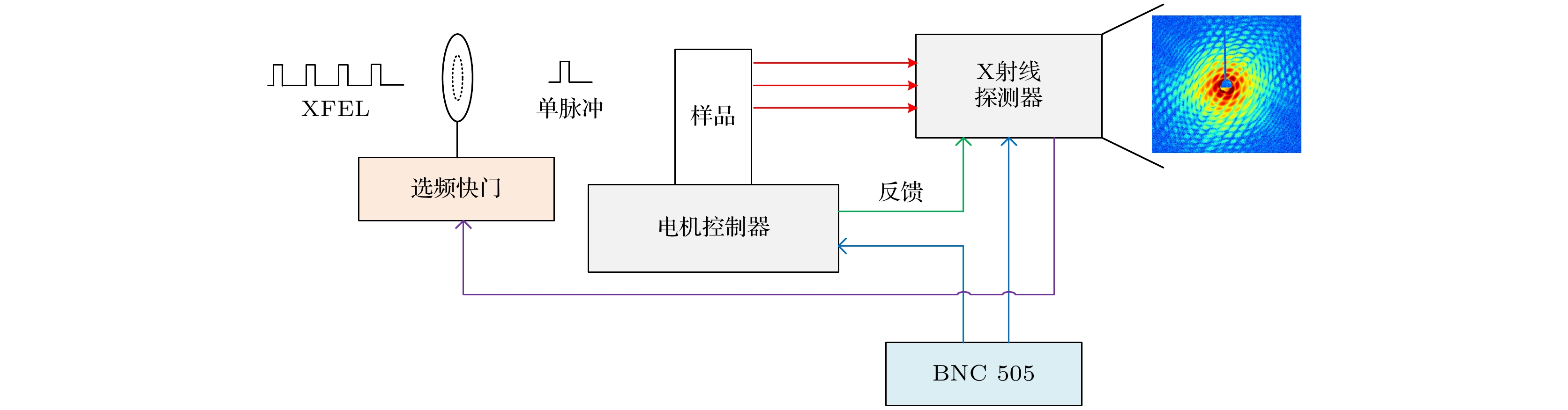
图 2 XFEL单脉冲成像设备连接示意图: 特定时序关系的触发信号经BNC505分发给X射线探测器和电机控制器, 用于同步XFEL脉冲. X射线探测器的触发输出信号用于控制X射线快门的开关时间
Fig. 2. XFEL single-shot imaging devices connection schematic: The trigger signal with specific timing relationships is distributed to the X-ray detector and the motor controller via the BNC505, used to synchronize the XFEL pulses. The trigger output signal from the X-ray detector is used to control the switching time of the X-ray shutter.
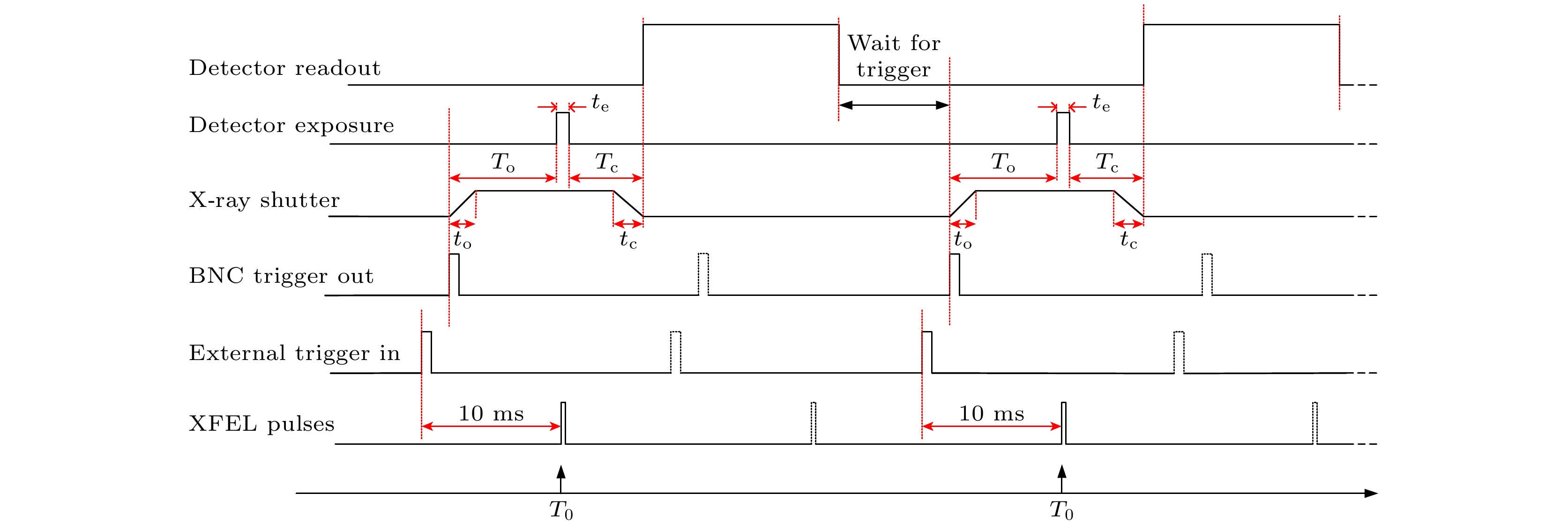
图 3 XFEL单脉冲成像工作时序图: 外部触发输入和BNC505触发输出的时间延迟由快门的固有开关时间决定. 对X射线快门的操作, 保证只有一个XFEL脉冲与样品的交互发生在探测器曝光时间内, 避免多余脉冲损害样品和探测器
Fig. 3. XFEL single-shot imaging working sequence diagram: The time delay between the external trigger input and the BNC505 trigger output is determined by the inherent switching time of the shutter. The operation of the X-ray shutter ensures that only one XFEL pulse interacts with the sample during the detector’s exposure time, preventing additional pulses from damaging the sample and the detector.
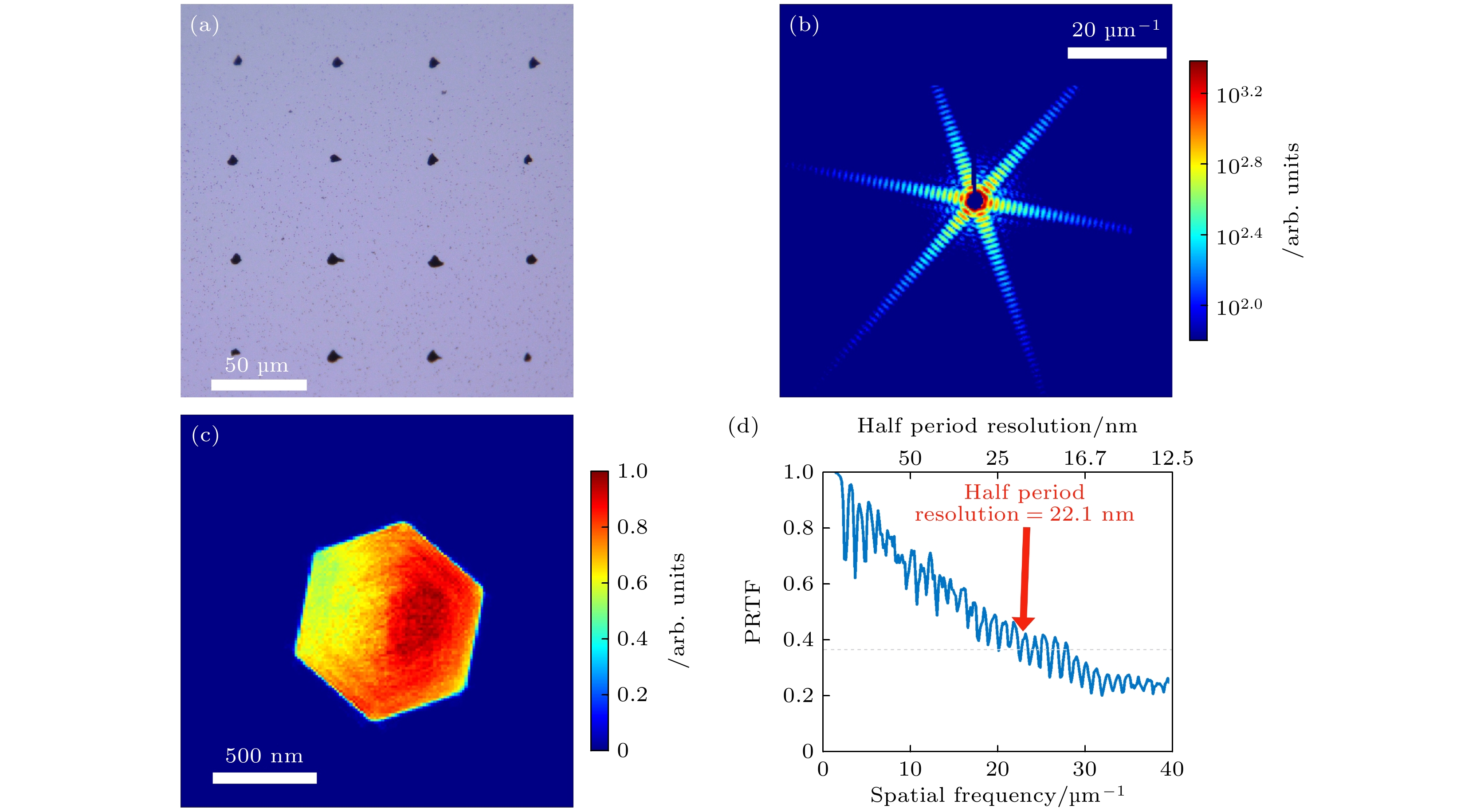
图 4 单脉冲相干衍射成像结果与分析 (a) XFEL脉冲逐点扫描沉积有六边金纳米盘样品的Si3N4窗口, 其中孔洞是XFEL的高亮度、飞秒脉冲将Si3N4窗口与样品一同离子化的结果; (b) 六边金纳米盘的单脉冲衍射图样; (c) 1000次独立重构, 选取误差最小的100组重构的平均结果; (d) 重构图像的PRTF, 以灰色虚线所示的1/e为标准, 样品成像的半周期分辨率为22.1 nm
Fig. 4. Results and analysis of single-shot coherent diffraction imaging. (a) A membrane with holes after the single-shot experiment. The hole is caused by ionization of gold nanoparticles Si3N4 window deposited by a single XFEL pulse hit. Scanning interval of 50 μm is used. (b) Single-shot diffraction pattern of a hexagonal gold nanodisks. (c) Reconstructed image from (b), from 1000 independent reconstructions, the 100 groups with the smallest error were chosen to calculate the average value. (d) The PRTF curve dropping below 1/e as the grey dashed line shows, the half-period resolution is 22.1 nm.
-
[1] 范家东, 江怀东 2012 物理学报 61 218702
Google Scholar
Fan J D, Jiang H D 2012 Acta Phys. Sin. 61 218702
Google Scholar
[2] Sakdinawat A, Attwood D 2010 Nat. Photonics 4 840
Google Scholar
[3] 周光照, 佟亚军, 陈灿, 任玉琦, 王玉丹, 肖体乔 2011 物理学报 60 028701
Google Scholar
Zhou G Z, Tong Y J, Chen C, Ren Y Q, Wang Y D, Xiao T Q 2011 Acta Phys. Sin. 60 028701
Google Scholar
[4] 周光照, 王玉丹, 任玉琦, 陈灿, 叶琳琳, 肖体乔 2012 物理学报 61 018701
Google Scholar
Zhou G Z, Wang Y D, Ren Y Q, Chen C, Ye L L, Xiao T Q 2012 Acta Phys. Sin. 61 018701
Google Scholar
[5] Thibault P, Dierolf M, Menzel A, Bunk O, David C, Pfeiffer F 2008 Science 321 379
Google Scholar
[6] Song C Y, Jiang H D, Mancuso A, Amirbekian B, Peng L, Sun R, Shah S S, Zhou Z H, Ishikawa T, Miao J W 2008 Phys. Rev. Lett. 101 158101
Google Scholar
[7] Jiang H D, Song C Y, Chen C C, Xu R, Raines K S, Fahimian B P, Lu C H, Lee T K, Nakashima A, Urano J, Ishikawa T, Tamano F, Miao J W 2010 Proc. Natl. Acad. Sci. U. S. A. 107 11234
Google Scholar
[8] Chapman H N, Barty A, Bogan M J, Boutet S, Frank M, Hau-Riege S P, Marchesini S, Woods B W, Bajt S, Benner W H, London R A, Plönjes E, Kuhlmann M, Treusch R, Düsterer S, Tschentscher T, Schneider J R, Spiller E, Möller T, Bostedt C, Hoener M, Shapiro D A, Hodgson K O, van der Spoel D, Burmeister F, Bergh M, Caleman C, Huldt G, Seibert M M, Maia F R N C, Lee R W, Szöke A, Timneanu N, Hajdu J 2006 Nat. Phys. 2 839
Google Scholar
[9] Neutze R, Wouts R, van der Spoel D, Weckert E, Hajdu J 2000 Nature 406 752
Google Scholar
[10] Ihm Y, Cho D H, Sung D, Nam D, Jung C, Sato T, Kim S, Park J, Kim S, Gallagher-Jones M, Kim Y, Xu R, Owada S, Shim J H, Tono K, Yabashi M, Ishikawa T, Miao J, Noh D Y, Song C 2019 Nat. Commun. 10 2411
Google Scholar
[11] Gaffney K J, Chapman H N 2007 Science 316 1444
Google Scholar
[12] Miao J, Ishikawa T, Robinson I K, Murnane M M 2015 Science 348 530
Google Scholar
[13] Hidvegi A, Gessler P, Rehlich K, Bohm C 2011 IEEE Trans. Nucl. Sci. 58 1852
Google Scholar
[14] Ye Y, Li H, Li J, Yan Y, Yu P, Gong G 2022 J. Instrum. 17 T09009
Google Scholar
[15] Fan J D, Tong Y J, Nie Y G, Gao Z C, He B, Luan H, Lu D H, Zhang J H, Zhang D F, Yuan X Y, Chen J H, Guo Z, Liu T, Zhang M, Feng C, Deng H X, Liu B, Zhao Z T, Liu Z, Jiang H D 2022 Nucl. Sci. Tech. 33 114
Google Scholar
[16] MFR homepage, http://www.mrf.fi/ [2024-4-11]
[17] Kim C, Baek S Y, Kang H S, Kim J, Kim K W, Ko I S, Mun G, Park B R 2015 Proceedings of the 15th International Conference on Accelerator and Large Experimental Physics Control Systems Melbourne, Australia, October 17–23, 2015 p79
[18] Kalantari B, Biffiger R 2017 Proceedings of 16th International Conference on Accelerator and Large Experimental Physics Control Systems Barcelona, Spain, October 8–13, 2017 p232
[19] Krejcik P, Akre R A, Allison S, Browne M, Dalesio L R, Dusatko J E, Frisch J C, Fuller R, Gromme A E, Kotturi K D, Norum S, Rogind D, White W E, Zelazny M 2007 Proceedings of the 8th European Workshop on Beam Diagnostics and Instrumentation for Particle Accelerators Venice, Italy, May 20–23, 2007 p373
[20] WR homepage, https://white-rabbit.web.cern.ch/ [2024-4-11]
[21] 余鹏翔, 阎映炳 2023 核电子学与探测技术 43 923
Google Scholar
Yu P G, Yan Y B 2023 Nucl. Electron. Detect. Technol. 43 923
Google Scholar
[22] Yan Y B, Chen G H, Gong G H, Gu J L, Jiang Z Y, Xiao Q W, Ye Y M, Yu P X, Zhao L 2022 Proceedings of the 13th International Particle Accelerator Conference Bangkok, Thailand, June 12-17, 2022 p2415
[23] 于春蕾, 赵欢, 胡守明, 丁建国 2019 核技术 42 040102
Google Scholar
Yu C L, Zhao H, Hu S M, Ding J G 2019 Nucl. Tech. 42 040102
Google Scholar
[24] Tong Y J, Fan J D, Nie Y G, Guo Z, Gao Z C, Yuan X Y, He B, Chen J H, Zhang D F, Luan H, Zhang J H, Lu D H, Xie M H, Cheng P, Feng C, Liu T, Deng H X, Liu B, Liu Z, Jiang H D 2022 Front. Phys. 10 977957
Google Scholar
[25] Park J, Eom I, Kang T H, Rah S, Nam K H, Park J, Kim S, Kwon S, Park S H, Kim K S, Hyun H, Kim S N, Lee E H, Shin H, Kim S, Kim M J, Shin H J, Ahn D, Lim J, Yu C J, Song C, Kim H, Noh D Y, Kang H S, Kim B, Kim K W, Ko I S, Cho M H, Kim S 2016 Nucl. Instrum. Methods Phys. Res. , Sect. A 810 74
Google Scholar
[26] Rodriguez J A, Xu R, Chen C C, Zou Y, Miao J 2013 J. Appl. Crystallogr. 46 312
Google Scholar
[27] Shapiro D, Thibault P, Beetz T, Elser V, Howells M, Jacobsen C, Kirz J, Lima E, Miao H, Neiman A M, Sayre D 2005 Proc. Natl. Acad. Sci. U. S. A. 102 15343
Google Scholar
[28] Chapman H N, Barty A, Marchesini S, Noy A, Hau-Riege S P, Cui C, Howells M R, Rosen R, He H, Spence J C H, Weierstall U, Beetz T, Jacobsen C, Shapiro D 2006 J. Opt. Soc. Am. A 23 1179
Google Scholar


 下载:
下载:
计量
- 文章访问数: 5212
- PDF下载量: 147
- 被引次数: 0
