


-
多焦点结构光照明显微技术(multifocal structured illumination microscopy, MSIM)能在50 μm的成像深度内和1Hz的成像速度下实现两倍于衍射极限分辨率的提升, 相比传统的宽场结构光照明显微技术, 具有较大的成像深度和层析能力, 更适合应用于厚样品的长时程三维超分辨成像. 然而, MSIM存在成像速度慢、图像处理过程复杂等问题. 本文提出了一种基于平场复用多焦点结构光照明的快速超分辨显微成像方法和系统(flat-field multiplexed MSIM, FM-MSIM), 通过在照明光路中插入光束整形器件, 将高斯光束转变为均为分布的平顶光束, 提高激发点阵的强度均匀性和扩大视场; 通过将每个衍射受限的激发点沿y方向延长, 形成新的多路复用多焦点阵照明图案, 提高能量利用率, 减少扫描步数, 进而提高成像速度和信噪比; 结合基于多重测量矢量模型的稀疏贝叶斯学习图像重构算法, 简化图像重构步骤, 在保证空间分辨率的同时实现至少4倍于传统MSIM的成像速度. 在此基础上, 利用搭建的FM-MSIM系统进行了BSC细胞微管样片和小鼠肾切片标准样片的超分辨成像实验, 实验结果证明了该系统的快速三维超分辨成像能力, 对于MSIM的发展具有重要的意义.
-
关键词:
- 多焦点结构光照明显微技术 /
- 超分辨成像 /
- 平场照明 /
- 贝叶斯学习算法
Multifocal structured illumination microscopy (MSIM) can achieve optically sectioned images with twice the diffraction limited resolution at an imaging speed of 1 Hz and an imaging depth of up to 50 μm. Compared with the traditional wide-field SIM, the MSIM has greater imaging depth and optical sectionning ability, and it is more suitable for long-term three-dimensional (3D) super-resolution imaging of living thick samples. However, the MSIM has some problems, such as slow imaging speed and complex image post-processing process. In this work, a fast super-resolution imaging method and system based on the flat-field multiplexed MSIM (FM-MSIM) is proposed. By inserting a beam shaping device into the illumination light path, the Gaussian beam is reshaped into a uniform flat-top profile, thereby improving the intensity uniformity of excitation multi-spot focal array and expanding the field of view. By elongating each diffraction limited excitation focal point four times along the Y direction to form a new multiplexed multifocal array pattern, the number of scanning steps is reduced, the energy utilization is improved, and then the imaging speed and signal-to-noise ratio are improved. Combined with the sparse Bayesian learning image reconstruction algorithm based on multiple measurement vector model, the image reconstruction steps are simplified, the imaging speed can be improved at least 4 times while ensuring the spatial resolution of MSIM. On this basis, the established FM-MSIM system is used to carry out the super-resolution imaging experiments on the BSC cell microtubule samples and mouse kidney slices. The experimental results prove the fast three-dimensional super-resolution imaging ability of the system, which is of great significance in developing the fast MSIM.-
Keywords:
- multifocal structured illumination microscopy /
- super-resolution imaging /
- flat-field illumination /
- Bayesian learning algorithm
[1] Hell S W, Wichmann J 1994 Opt. Lett. 19 780
 Google Scholar
[2] Klar T A, Jakobs S, Dyba M, Egner A, Hell S W 2000 Proc. Natl. Acad. Sci. U. S. A. 97 8206
Google Scholar
[3] Willig K I, Harke B, Medda R, Hell S W 2007 Nat. Methods 4 915
Google Scholar
[4] Rust M J, Bates M, Zhuang X 2006 Nat. Methods 3 793
Google Scholar
[5] Betzig E, Patterson G H, Sougrat R, Lindwasser O W, Olenych S, Bonifacino J S, Davidson M W, Lippincott-Schwartz J, Hess H F 2006 Science 313 1642
Google Scholar
[6] Gustafsson M G L 2000 J. Microsc. 198 82
Google Scholar
[7] Gustafsson M G L 2005 Proc. Natl. Acad. Sci. U. S. A. 102 13081
Google Scholar
[8] Gustafsson M G L, Shao L, Carlton P M, Wang C J, Golubovskaya I N, Cande W Z, Agard D A, Sedat J W 2008 Biophys. J. 94 4957
Google Scholar
[9] Huang X, Fan J, Li L, Liu H, Wu R, Wu Y, Wei L, Mao H, Lal A, Xi P, Tang L, Zhang Y, Liu Y, Tan S, Chen L 2018 Nat. Biotechnol. 36 451
Google Scholar
[10] Guo Y, Li D, Zhang S, Yang Y, Liu J J, Wang X, Liu C, Milkie D E, Moore R P, Tulu U S, Kiehart D P, Hu J, Lippincott-Schwartz J, Betzig E, Li D 2018 Cell 175 1430
Google Scholar
[11] Muller C B, Enderlein J 2010 Phys. Rev. Lett. 104 198101
Google Scholar
[12] York A G, Parekh S H, Dalle Nogare D, Fischer R S, Temprine K, Mione M, Chitnis A B, Combs C A, Shroff H 2012 Nat. Methods 9 749
Google Scholar
[13] Schulz O, Pieper C, Clever M, Pfaff J, Ruhlandt A, Kehlenbach R H, Wouters F S, Grosshans J, Bunt G, Enderlein J 2013 Proc. Natl. Acad. Sci. U. S. A. 110 21000
Google Scholar
[14] York A G, Chandris P, Nogare D D, Head J, Wawrzusin P, Fischer R S, Chitnis A, Shroff H 2013 Nat. Methods 10 1122
Google Scholar
[15] De Luca G M, Breedijk R M, Brandt R A, Zeelenberg C H, de Jong B E, Timmermans W, Azar L N, Hoebe R A, Stallinga S, Manders E M 2013 Biomed. Opt. Express 4 2644
Google Scholar
[16] Roth S, Heintzmann R 2016 Methods Appl. Fluoresc. 4 045005
Google Scholar
[17] Wu J J, Li S W, Cao H Q, Lin D Y, Yu B, Qu J L 2018 Opt. Express 26 31430
Google Scholar
-
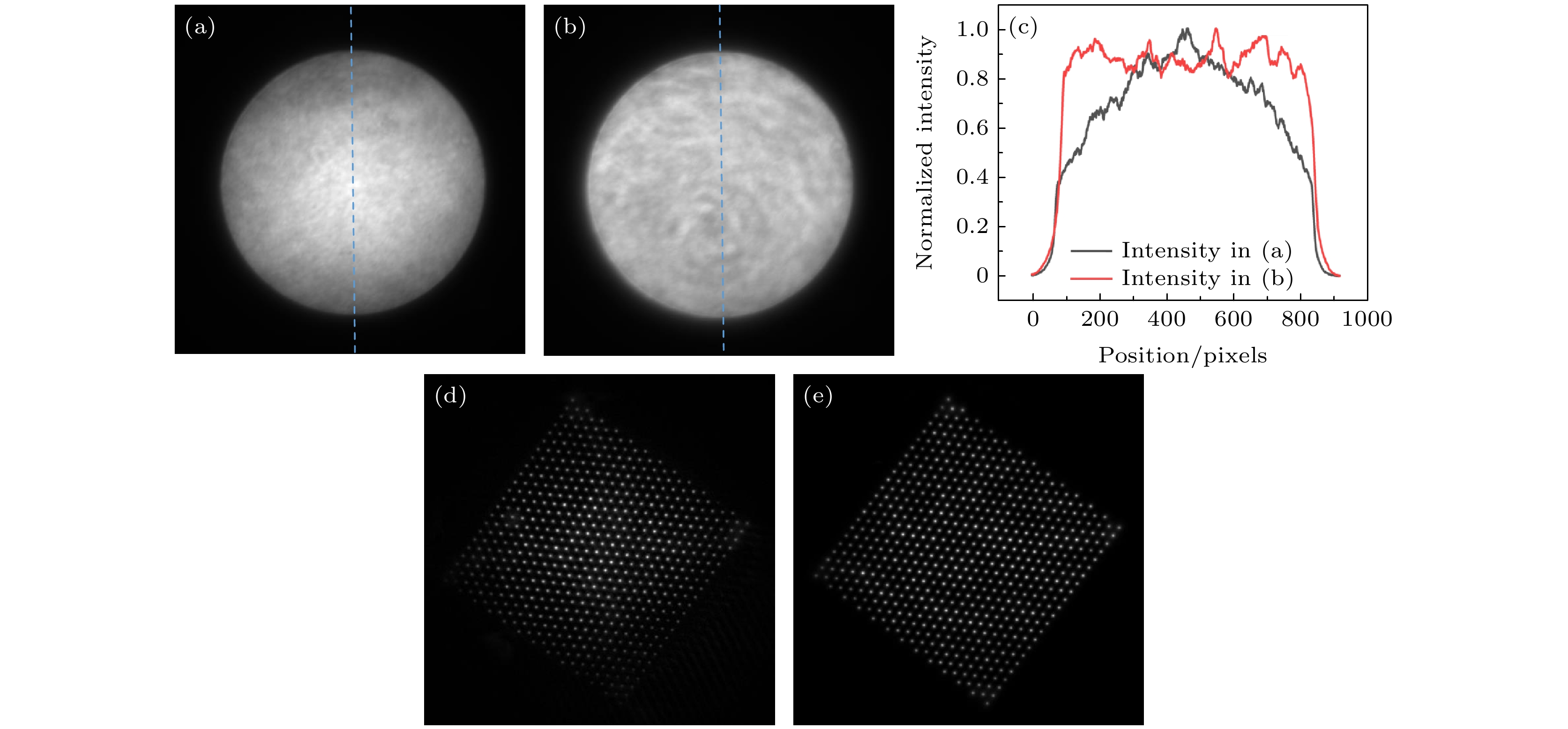
图 2 平场照明实验表征 (a)无光束整形器的罗丹明均匀溶液宽场成像; (b)有光束整形器的罗丹明均匀溶液宽场成像; (c)图(a)和图(b)中蓝色虚线部分布归一化强度轮廓图; (d)无光束整形器的激发点阵; (e)有光束整形器的激发点阵
Fig. 2. Experimental characterization of flat-field illumination: (a) Wide field imaging of uniform Rhodamine 6 G solution without a beam shaper; (b) wide field imaging of uniform Rhodamine 6 G solution with a beam shaper; (c) normalized intensity fitting profiles along blue dotted line in panel (a) and panel (b); (d) a multifocal excitation pattern without a beam shaper; (e) a multifocal excitation pattern with a beam shaper.

图 3 复用多焦点照明原理图 (a) 4 × 1激发点阵扫描原理图; (b) 1 × 1激发点阵扫描原理图; (c)复用的多焦点激发罗丹明均匀染料样品荧光图像
Fig. 3. Schematic diagram of multiplexed multifocal excitation illumination: (a) Schematic diagram of 4 × 1 excitation spot array scanning; (b) schematic diagram of 1 × 1 excitation spot array scanning; (c) fluorescece image of the excitation foci in a uniform solution of Rhodamine 6G at the sample plane.
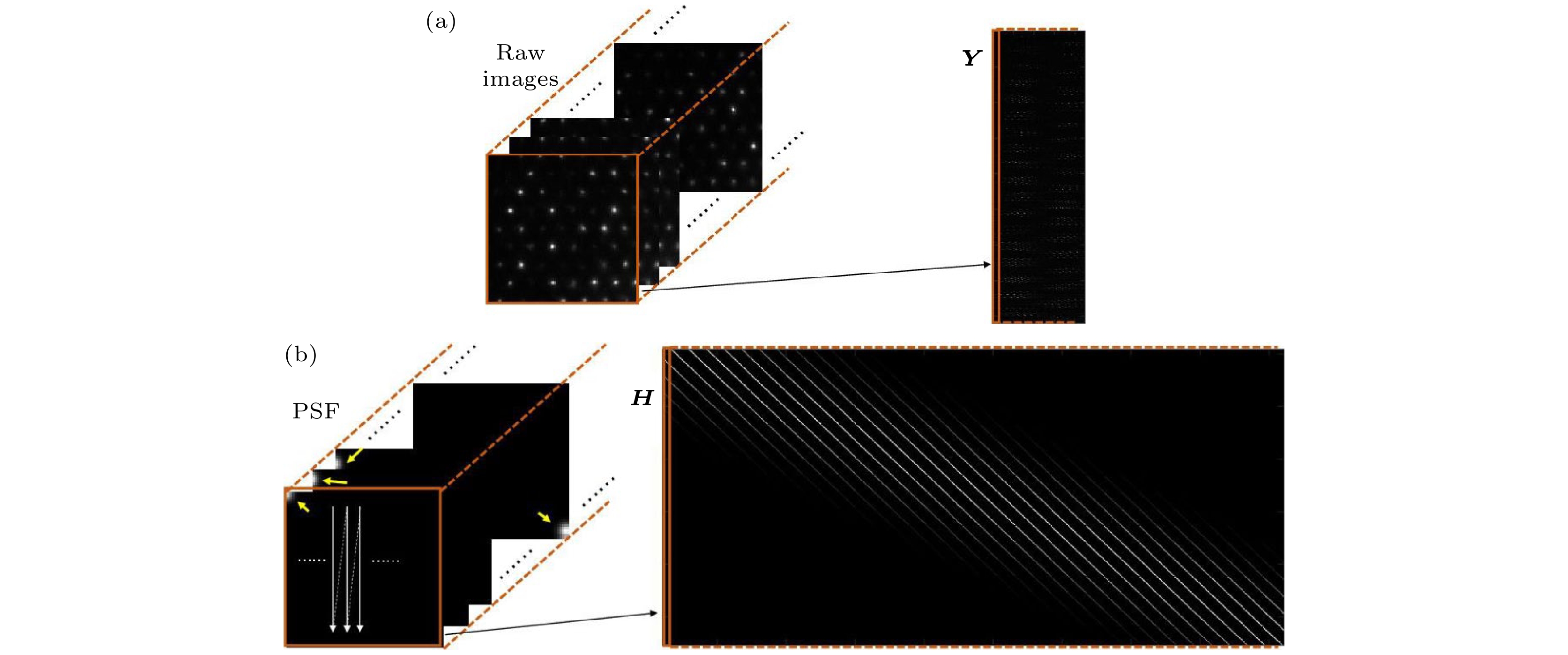
图 4 原始数据与系统PSF转换为矩阵形式的过程 (a) FM-MSIM点阵数据转换为矩阵Y; (b)系统PSF转换为矩阵H
Fig. 4. Process of converting raw images and system PSF into matrix form: (a) FM-MSIM multifocal patterns data is converted to matrix Y; (b) System PSF is converted to matrix H.
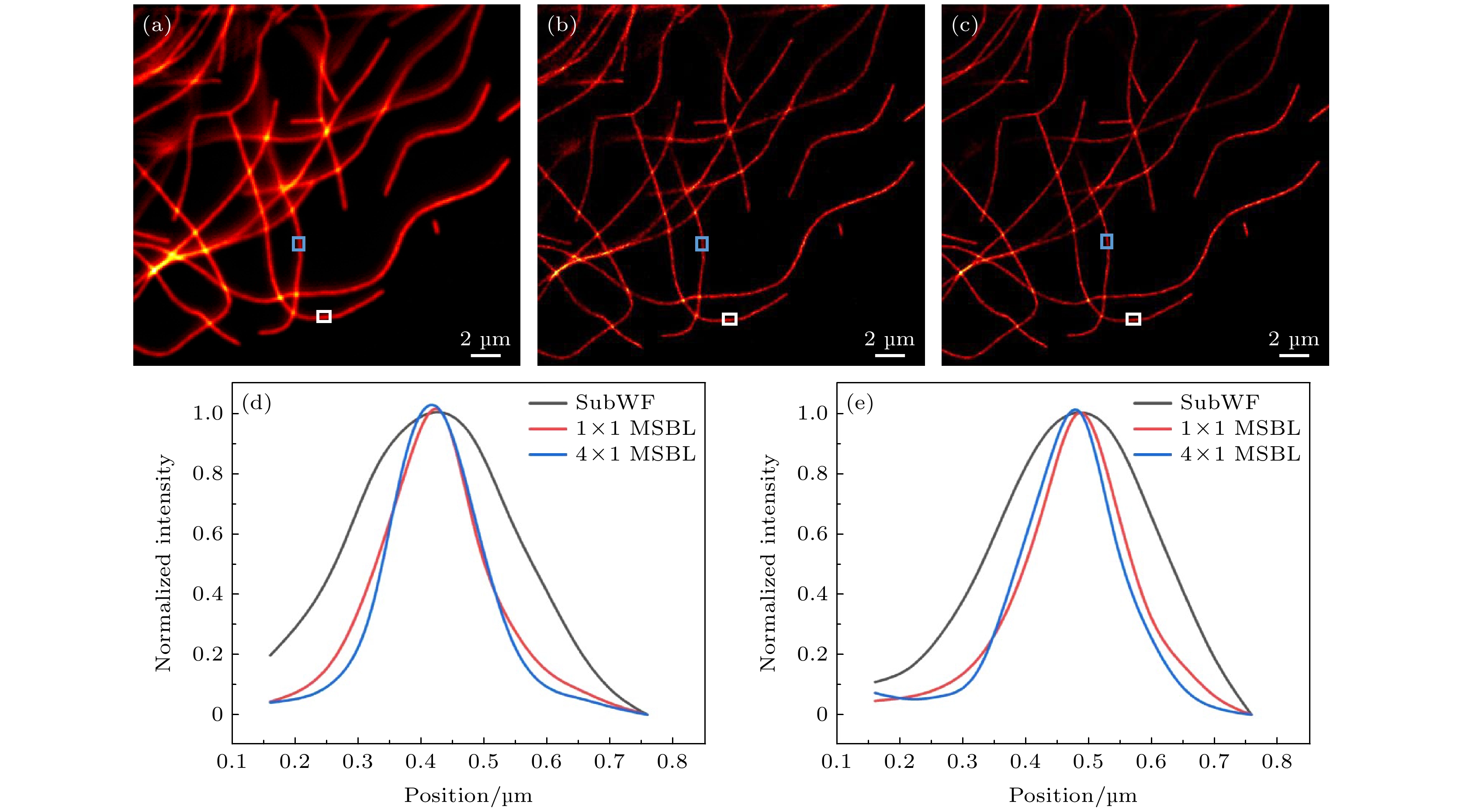
图 5 FM-MSIM 分辨率标定 (a)减去背景噪声的宽场微管图像; (b) 1 × 1点扫模板的MSBL重构图像; (c) 4 × 1点扫模板的MSBL重构图像; (d)蓝色实线处微管强度轮廓线; (e) 白色实线处微管强度轮廓线
Fig. 5. Resolution in FM-MSIM: (a) Wide field image of microtubule in BSCs labeled with Alexa Fluor 488 phalloidin by subtracting background noise; (b) MSBL reconstructed image of 1 × 1 point scan mode; (c) MSBL reconstructed image of 4 × 1 point scan mode; (d) plots of intensity along blue solid line in panels (a)–(c); (e) plots of intensity along white solid line in panels (a)–(c).
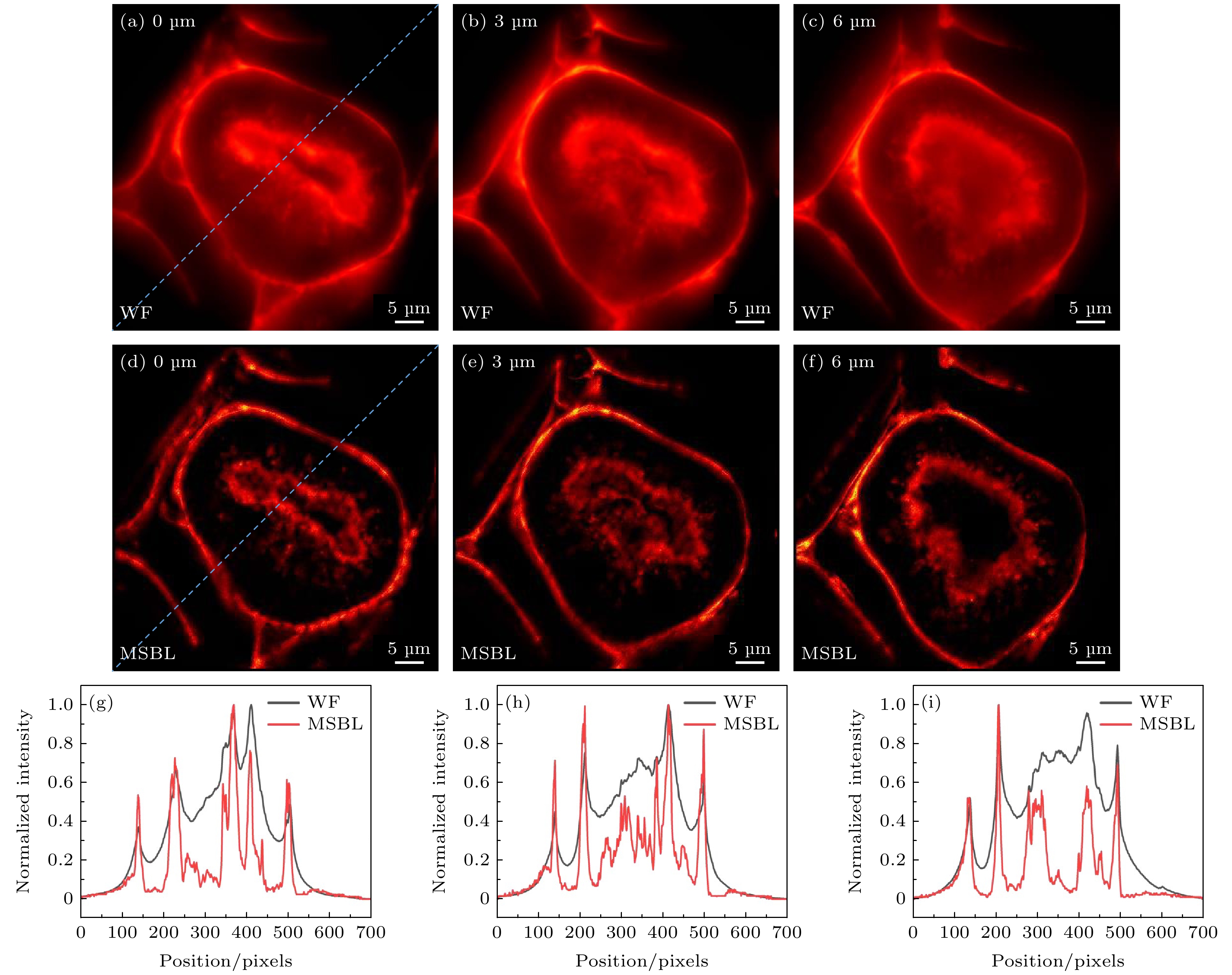
图 6 小鼠肾切片不同轴向位置(z = 0, 3 , 6 μm)处的图像: (a)—(c)宽场图像; (d)—(f)MSBL重构图像; (g)图(a)和图(d)中蓝色虚线处的归一化强度分布轮廓曲线; (h) 图(b)和图(e)相同位置处归一化强度分布轮廓曲线; (i)图(c)和图(f)相同位置处归一化强度分布轮廓曲线
Fig. 6. Images of mouse kidney section at different axial positions(z = 0, 3 and 6 μm): (a)–(c) Wide field images; (d)–(f) MSBL reconstruction images; (g) normalized intensity distribution profiles along the blue dotted line in panel (a) and panel (d); (h) normalized intensity distribution profiles along the same position in panel (b) and panel (e); (i) normalized intensity distribution profiles along the same position in panel (c) and panel (f).
-
[1] Hell S W, Wichmann J 1994 Opt. Lett. 19 780
Google Scholar
[2] Klar T A, Jakobs S, Dyba M, Egner A, Hell S W 2000 Proc. Natl. Acad. Sci. U. S. A. 97 8206
Google Scholar
[3] Willig K I, Harke B, Medda R, Hell S W 2007 Nat. Methods 4 915
Google Scholar
[4] Rust M J, Bates M, Zhuang X 2006 Nat. Methods 3 793
Google Scholar
[5] Betzig E, Patterson G H, Sougrat R, Lindwasser O W, Olenych S, Bonifacino J S, Davidson M W, Lippincott-Schwartz J, Hess H F 2006 Science 313 1642
Google Scholar
[6] Gustafsson M G L 2000 J. Microsc. 198 82
Google Scholar
[7] Gustafsson M G L 2005 Proc. Natl. Acad. Sci. U. S. A. 102 13081
Google Scholar
[8] Gustafsson M G L, Shao L, Carlton P M, Wang C J, Golubovskaya I N, Cande W Z, Agard D A, Sedat J W 2008 Biophys. J. 94 4957
Google Scholar
[9] Huang X, Fan J, Li L, Liu H, Wu R, Wu Y, Wei L, Mao H, Lal A, Xi P, Tang L, Zhang Y, Liu Y, Tan S, Chen L 2018 Nat. Biotechnol. 36 451
Google Scholar
[10] Guo Y, Li D, Zhang S, Yang Y, Liu J J, Wang X, Liu C, Milkie D E, Moore R P, Tulu U S, Kiehart D P, Hu J, Lippincott-Schwartz J, Betzig E, Li D 2018 Cell 175 1430
Google Scholar
[11] Muller C B, Enderlein J 2010 Phys. Rev. Lett. 104 198101
Google Scholar
[12] York A G, Parekh S H, Dalle Nogare D, Fischer R S, Temprine K, Mione M, Chitnis A B, Combs C A, Shroff H 2012 Nat. Methods 9 749
Google Scholar
[13] Schulz O, Pieper C, Clever M, Pfaff J, Ruhlandt A, Kehlenbach R H, Wouters F S, Grosshans J, Bunt G, Enderlein J 2013 Proc. Natl. Acad. Sci. U. S. A. 110 21000
Google Scholar
[14] York A G, Chandris P, Nogare D D, Head J, Wawrzusin P, Fischer R S, Chitnis A, Shroff H 2013 Nat. Methods 10 1122
Google Scholar
[15] De Luca G M, Breedijk R M, Brandt R A, Zeelenberg C H, de Jong B E, Timmermans W, Azar L N, Hoebe R A, Stallinga S, Manders E M 2013 Biomed. Opt. Express 4 2644
Google Scholar
[16] Roth S, Heintzmann R 2016 Methods Appl. Fluoresc. 4 045005
Google Scholar
[17] Wu J J, Li S W, Cao H Q, Lin D Y, Yu B, Qu J L 2018 Opt. Express 26 31430
Google Scholar

 下载:
下载:
计量
- 文章访问数: 9901
- PDF下载量: 187
- 被引次数: 0
