


-
活体小动物成像系统是疾病研究、新药开发的重要组成部分. 其中, X射线微型锥束计算机断层成像(X-ray micro cone-beam computed tomography, Micro-CBCT)能实现数十至数百微米空间分辨率的解剖结构成像研究. Micro-CBCT成像仪的一个关键挑战是其空间和对比度分辨率主要取决于X射线源焦斑大小、探测器分辨率和系统几何结构等因素. 为提高Micro-CBCT的空间分辨率、对比度分辨率和成像均一性, 本文基于毛细管X光透镜研制了一款能够调控照射X射线束斑孔径的Micro-CBCT扫描仪, 用于小动物成像研究. 此系统由微焦斑X射线源、非晶硅平板探测器、旋转工作台和控制电脑组成, 并采用Feldkamp-Daivs-Kress算法重建投影图像. 对该系统的性能进行了测试, 结果表明, 系统在10%调制传递函数下的空间分辨率为9.1 lp/mm, 提高了1.35倍. 同时, 由于毛细管X光透镜对低能X射线的吸收和散射抑制作用, 实现了2倍以上的对比度增强, 减轻了多色X射线束硬化效应引起的图像均一性恶化问题. 应用该Micro-CBCT系统对麻醉小鼠进行了活体成像, 验证了该系统在小动物成像研究中的实用性.In-vivo small animal imaging system is an important part of disease research and new drug development. It is essential for living small animal imaging system to be able to provide the anatomical structure, molecular and functional information. The X-ray micro cone-beam computed tomography (micro-CBCT) can perform longitudinal study with a resolution of tens-to-hundreds of microns in a short imaging time at a relatively low cost. Furthermore, it is easy to combine with other modalities to provide abundant information about small animals. A key challenge to the micro-CBCT scanner is that its spatial and contrast resolution determined primarily by the X-ray focal spot size, the detector element size, and the system geometry. Aiming to improve the spatial resolution, contrast resolution, and imaging uniformity of the micro-CBCT system, we use the X-ray polycapillary optics for adjusting the X-ray source. A micro-CBCT based on X-ray polycapillary optics with a large field of view is constructed for the small animal imaging study. The micro-CBCT system is composed of microfocus X-ray tube with an attached polycapillary focusing X-ray lens, amorphous silicon-based flat panel detector, rotation stage, and controlling PC. The Feldkamp-Daivs-Kress (FDK) algorithm is adopted to reconstruct the image. The system performances are evaluated. The magnification of this micro-CBCT system is 1.97. The results show that the spatial resolution of the system at 10% modulation transfer function (MTF) is 9.1 lp/mm, which is 1.35 times higher than that in the case of no optics. The image uniformity deterioration caused by hardening effect is effectively alleviated by filtrating the low energy X-rays with the X-ray polycapillary optics and the contrast enhancement is more than twice. The anesthetic rats are imaged with this micro-CBCT system in vivo and the practicability of the system in small animal imaging research is verified.
-
Keywords:
- X-ray polycapillary optics /
- micro cone-beam CT /
- X-ray imaging /
- modulation transfer function
[1] Gregory S G, Sekhon M, Schein J, et al. 2002 Nature 418 743
 Google Scholar
[2] Ntziachristos V, Ripoll J, Wang L V, Weissleder R 2005 Nat. Biotechnol. 23 313
Google Scholar
[3] Guerra A D, Belcari N 2007 Nucl. Instrum. Meth. Phys. Res. A 583 119
Google Scholar
[4] Badea C T, Drangova M, Holdsworth D W, Johnson G A 2008 Phys. Med. Biol. 53 319
Google Scholar
[5] Jan M L, Ni Y C, Chen K W, Ching H 2006 Nucl. Instrum. Meth. Phys. Res. A 569 314
Google Scholar
[6] Biederer J, Mirsadraee S, Beer M, Molinari F, Puderbach M 2012 Insights Into Imaging 3 373
Google Scholar
[7] Hoyer C, Gass N, Fahr W W, Sartorius A 2014 Neuropsychobiology 69 187
Google Scholar
[8] Kunjachan S, Ehling J, Storm G, Kiessling F, Lammers T 2015 Chem. Rev. 115 10907
Google Scholar
[9] Eghtedari M, Oraevsky A, Copland J A, Kotov N A, Conjusteau A, Motamedi M 2007 Nano Lett. 7 1914
Google Scholar
[10] Taruttis A, Ntziachristos V 2015 Nat. Photonics 9 219
Google Scholar
[11] Paulus M J, Gleason S S, Kennel S J, Hunsicker P R, Johnson D K 2000 Neoplasia 2 62
Google Scholar
[12] 罗召洋, 杨孝全, 孟远征, 邓勇 2010 物理学报 58 8237
Google Scholar
Luo Z Y, Yang X Q, Meng Y Z, Deng Y 2010 Acta Phys. Sin. 58 8237
Google Scholar
[13] 魏星, 闫镔, 张峰, 李永丽, 席晓琦, 李磊 2014 物理学报 63 058702
Google Scholar
Wei X, Yan B, Zhang F, Li Y L, Xi X Q, Li L 2014 Acta Phys. Sin. 63 058702
Google Scholar
[14] Mazel V, Reiche I, Busignies V, Walter P, Tchoreloff P 2011 Talanta 85 556
Google Scholar
[15] Sun T, Liu Z, Li Y, Lin X, Wang G, Zhu G, Xu Q, Luo P, Pan Q, Liu H 2010 Nucl. Instrum. Meth. Phys. Res. A 622 295
Google Scholar
[16] Macdonald C A, Gibson W M 2003 X-Ray Spectrom. 32 258
Google Scholar
[17] Albertini V R, Paci B, Generosi A, Dabagov S B, Kumakhov M A 2007 Spectrochim. Acta B 62 1203
Google Scholar
[18] Huang R, Bilderback D H 2006 J. Synchrotron Radiat. 13 74
Google Scholar
[19] Balaic D X, Barnea Z, Nugent K A, Garrett R F, Wilkins S W 1996 J. Synchrotron Radiat. 3 289
Google Scholar
[20] MacDonald C A, Owens S M, Gibson W M 1999 J. Appl. Crystallogr. 32 160
Google Scholar
[21] Bjeoumikhov A, Bjeoumikhova S, Langhoff N, Wedell R 2005 Appl. Phys. Lett. 86 144102
Google Scholar
[22] Sun T, Liu Z, Ding X 2007 Nucl. Instrum. Meth. Phys. Res. B 262 153
Google Scholar
[23] Sun T, Peng S, Liu Z, Sun W, Ma Y, Ding X 2013 J. Appl. Crystallogr. 46 1880
Google Scholar
[24] Sun T, Macdonald C A 2013 J. Appl. Phys. 113 053104
Google Scholar
[25] Lamb J S, Bilderback D H, Pollack L, Kwok L, Smilgies D M 2007 J. Appl. Crystallogr. 40 193
Google Scholar
[26] Barrea R A, Huang R, Cornaby S, Bilderback D H, Irving T C 2009 J. Synchrotron Radiat. 16 76
Google Scholar
[27] Zeng X, Duewer F, Feser M, Huang C, Lyon A, Tkachuk A, Yun W 2008 Appl. Opt. 47 2376
Google Scholar
[28] Li F, Liu Z, Sun T, Jiang B, Zhu Y 2016 J. Chem. Phys. 144 104201
Google Scholar
[29] Li F, Liu Z, Sun T 2016 J. Appl. Crystallogr. 49 627
Google Scholar
[30] Li F, Liu Z, Sun T 2016 Rev. Sci. Instrum. 87 093106
Google Scholar
[31] Li F, Liu Z, Sun T 2016 Food Chem. 210 435
Google Scholar
[32] Li F, Liu Z, Sun T, Ma Y, Ding X 2015 Food Control 54 120
Google Scholar
[33] Abreu C C, Kruger D G, MacDonald C A, Mistretta C A, Peppler W W, Xiao Q F 1995 Med. Phys. 22 1793
Google Scholar
[34] Goertzen A L, Nagarkar V, Street R A, Paulus M J, Boone J M, Cherry S R 2004 Phys. Med. Biol. 49 5251
Google Scholar
[35] Kim H K, Min K C, Achterkirchen T, Lee W 2009 IEEE Trans. Nucl. Sci. 56 1179
Google Scholar
[36] Feldkamp L A, Davis L C, Kress J W 1984 J. Opt. Soc. Am. A 1 612
Google Scholar
[37] Flannery B P, Deckman H W, Roberge W G, D'Amico K L 1987 Science 237 1439
Google Scholar
[38] Sun T, Ding X 2005 J. Appl. Phys. 97 124904
Google Scholar
[39] Kai Y, Kwan A L C, Miller D W F, Boone J M 2006 Med. Phys. 33 1695
Google Scholar
[40] Kwan A L C, Boone J M, Yang K, Huang S Y 2007 Med. Phys. 34 275
[41] 余晓锷, 占杰, 李萍, 李婵娟 2006 第四军医大学学报 27 978
Google Scholar
Yu X E, Zhan J, Li P, Li C J 2006 J. Fourth Mil. Med. Univ. 27 978
Google Scholar
-
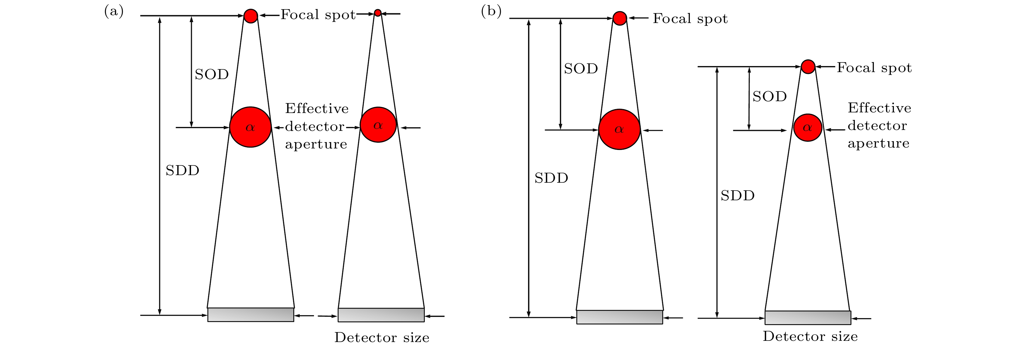
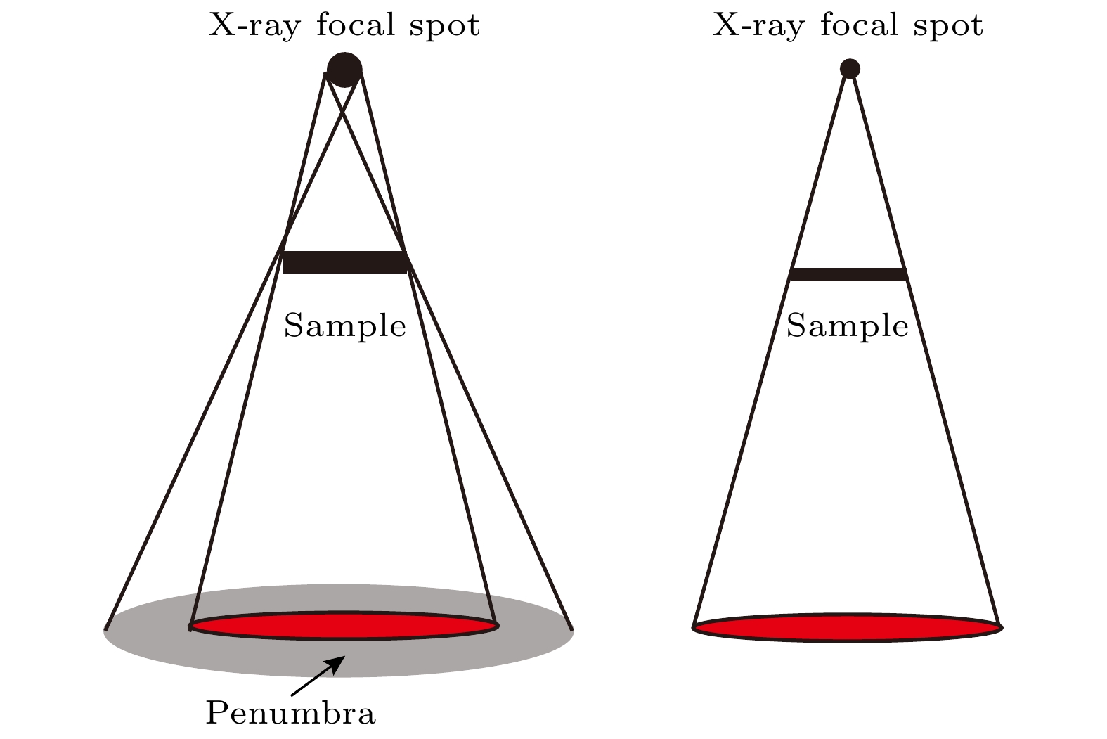
图 1 X射线源焦斑大小、SOD和SDD共同决定了Micro-CBCT的有效探测器孔径大小 (a) 焦点大小与有效探测器孔径α成正比; (b) SOD/SDD比值与有效探测器孔径α成正比
Fig. 1. X-ray tube focal spot size, SOD and SDD jointly determine the effective detector aperture size of the micro-CT system: (a) Focal spot size is proportional to the effective detector aperture (α); (b) ratio of SOD/SDD is proportional to the effective detector aperture (α).
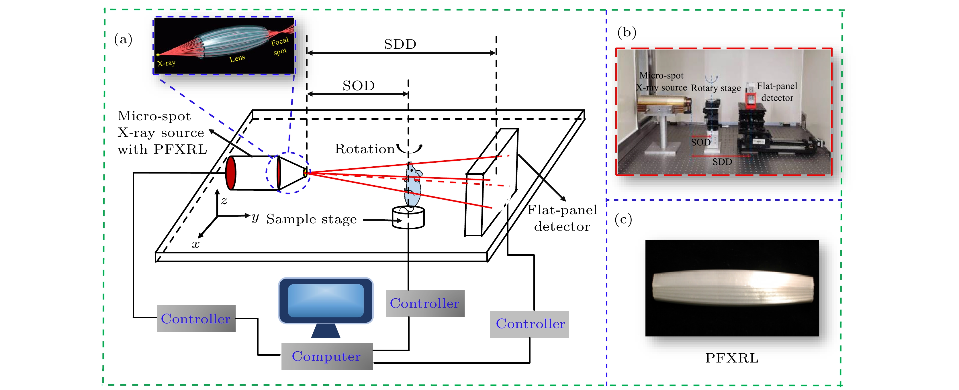
图 3 Micro-CBCT系统, 该系统由一个结合PFXRL的微聚焦X射线源、一个旋转样品台和一个非晶硅平板探测器组成 (a) Micro-CBCT原理图; (b) Micro-CBCT实物图; (c) 采用的PFXRL实物图
Fig. 3. Micro-CBCT system. The system consists of a microfocus X-ray source combined with a PFXRL, a rotating sample stage and an amorphous silicon-based FPD: (a) Micro-CBCT schematic diagram; (b) desktop micro-CBCT system; (c) the PFXRL.
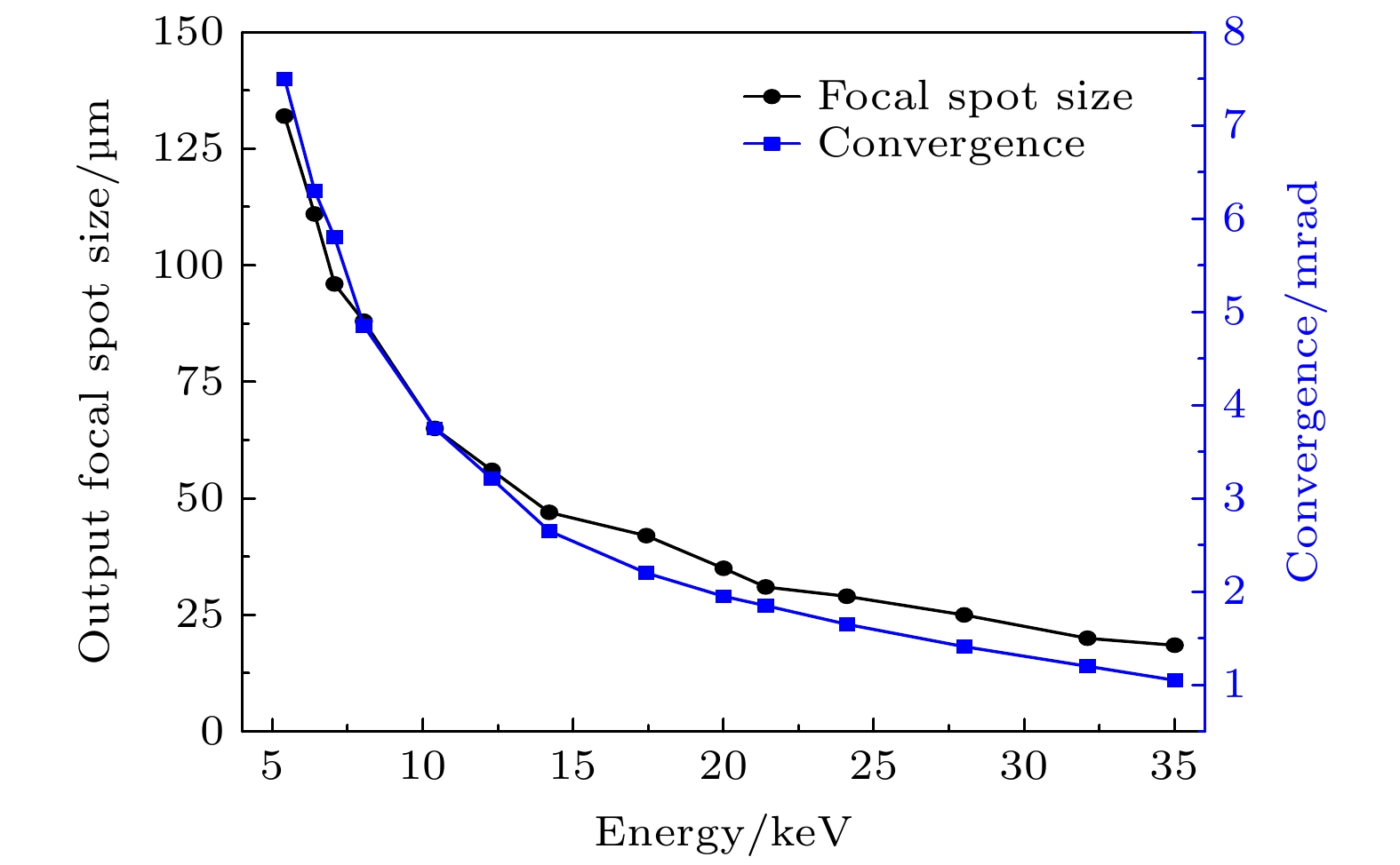
图 4 PFXRL的出口焦斑尺寸和收敛度随X射线能量的依赖关系
Fig. 4. Energy dependence of the output focal spot size and convergence for the PFXRL, respectively.
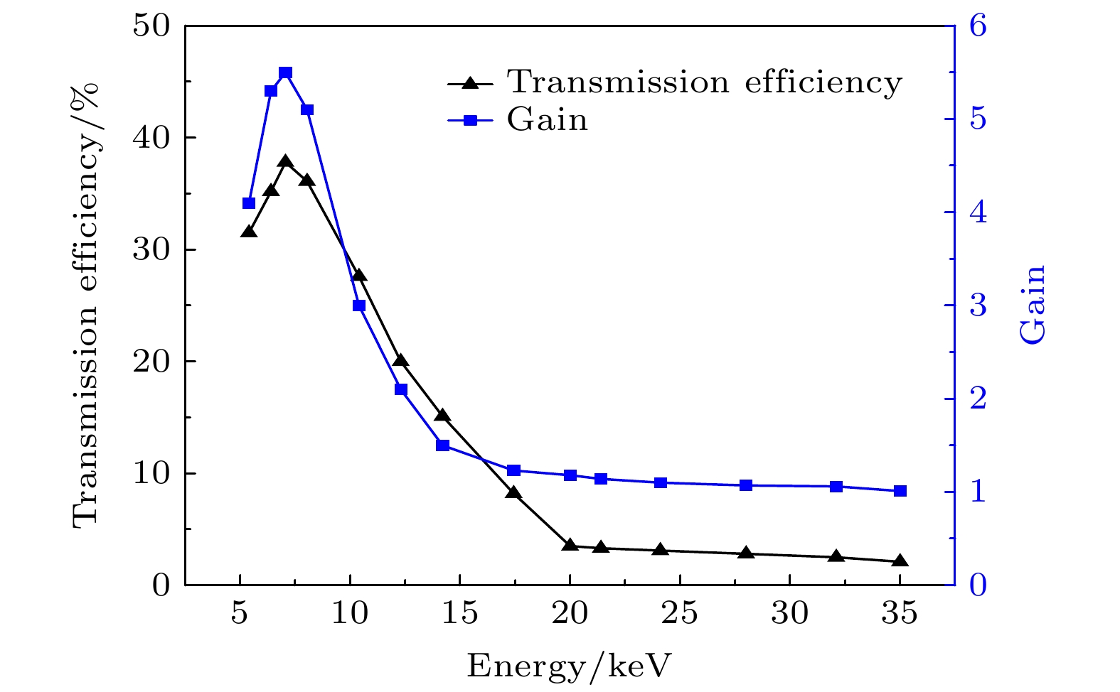
图 5 PFXRL的传输效率和能量密度增益随X射线能量的依赖关系
Fig. 5. Energy dependence of transmission efficiency and gain in power density of the PFXRL, respectively.

图 6 使用和不使用PFXRL两种条件下的Micro-CBCT图像和对应的MTF (a)使用PFXRL; (b)不使用PFXRL; (c) MTF曲线
Fig. 6. Measured images and corresponding MTF of the micro-CBCT system with and without PFXRL: (a) With PFXRL; (b) without PFXRL; (c) MTF curves.
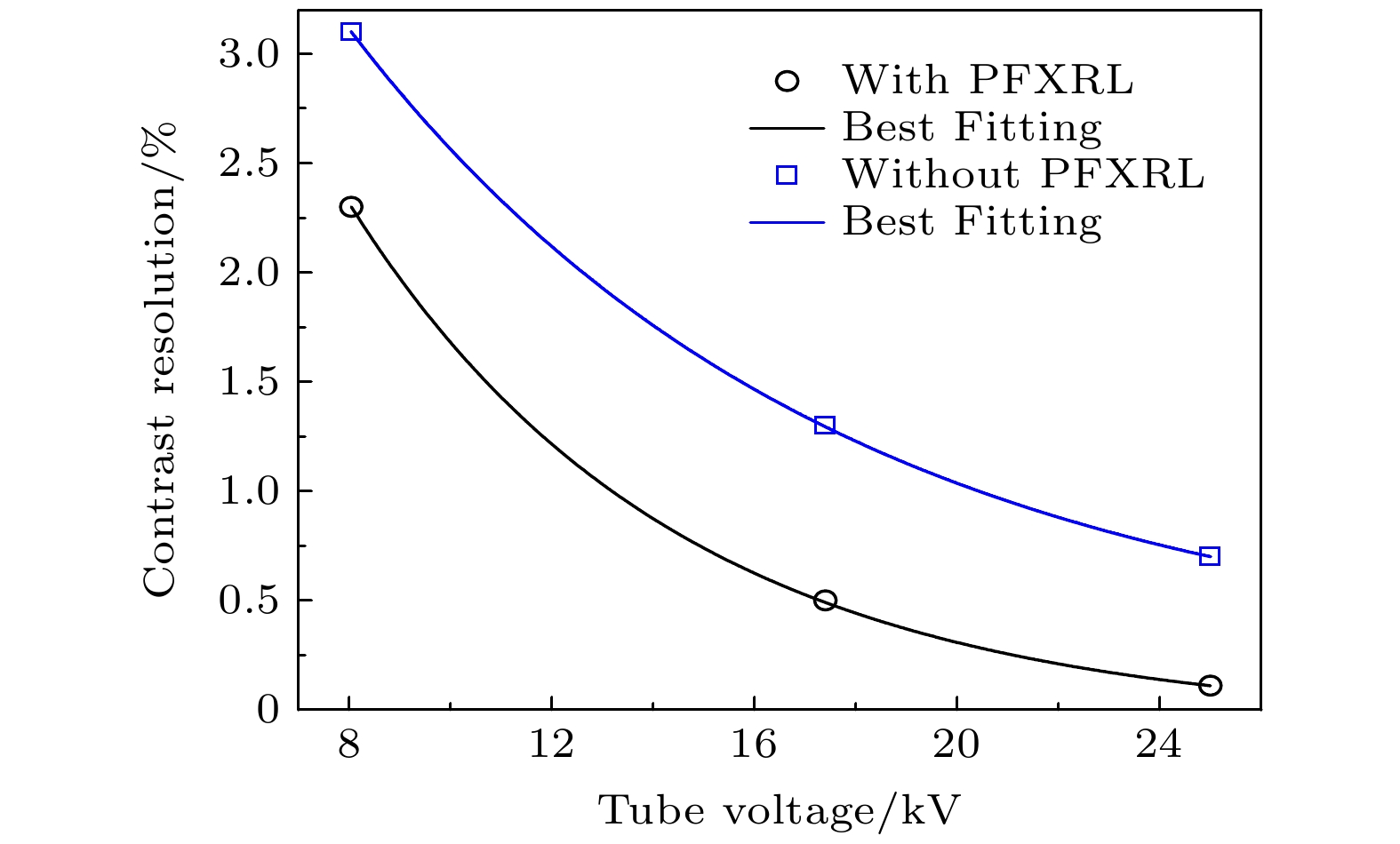
图 7 对于测量的水模, 在使用和不使用PFXRL下的Micro-CBCT系统对比度分辨率与管电压的关系
Fig. 7. Measured contrast resolution of Micro-CBCT system as a function of tube voltages for water phantom.

图 8 水模的均匀性响应 (a) 不使用PFXRL, 重建的水模中平横断面图像及绿线对应的CT值; (b) 使用PFXRL, 重建的水模中平横断面图像及绿线对应的CT值; (c) 采用0.5 mm厚的铝片附加滤过, 重建的水模中平横断面图像及绿线对应的CT值. 空气和水的CT值分别归一化为0和50
Fig. 8. Uniformity response of the water phantom: (a) Reconstructed transaxial image of the uniformity phantom without using PFXRL and radial signal profile taken from the green line; (b) reconstructed transaxial image of the uniformity phantom with using PFXRL and radial signal profile taken from the green line; (c) reconstructed transaxial image of the uniformity phantom with a 0.5 mm thick aluminum sheet as filter and radial signal profile taken from the green line. The CT values of air and water are normalized to 0 and 50, respectively.
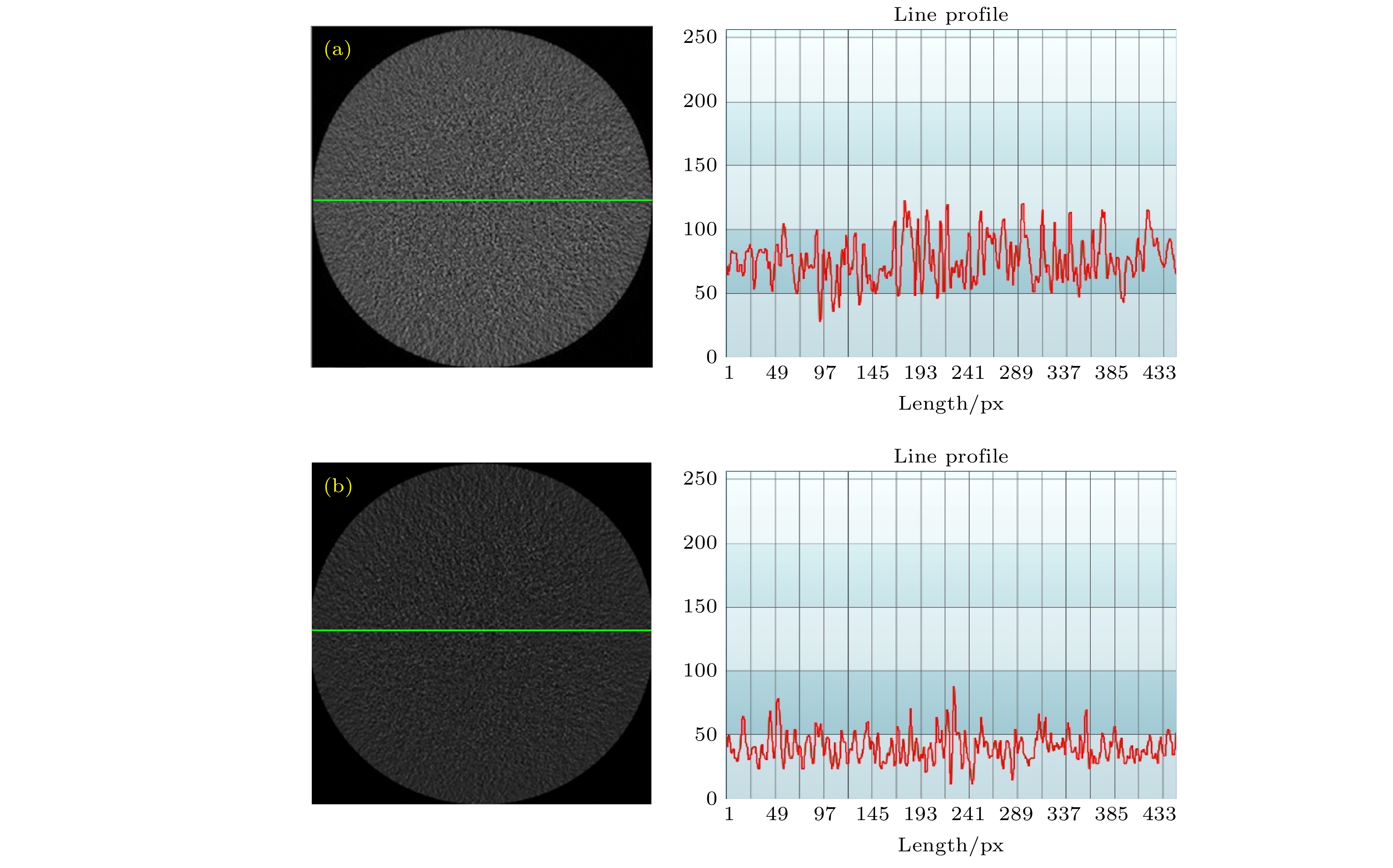
图 9 水模的噪声响应 (a)不使用PFXRL (附加滤过: 0.5 mm Al); (b)使用PFXRL
Fig. 9. Noise response of the water phantom: (a) Without PFXRL (additional filtration: 0.5 mm Al); (b) with PFXRL.
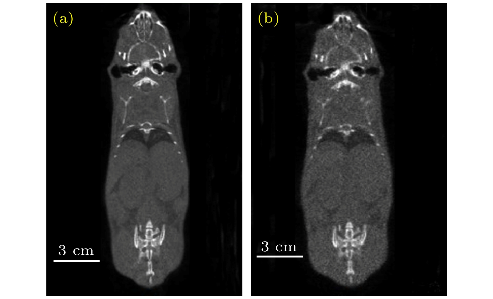
图 10 小鼠Micro-CBCT成像 (a)使用PFXRL; (b)不使用PFXRL
Fig. 10. CT images of rats: (a) With PFXRL; (b) without PFXRL.
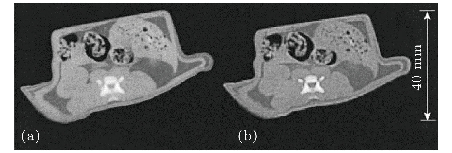
图 11 麻醉小鼠横断面图像比较 (a)使用PFXRL; (b)不使用PFXRL
Fig. 11. Comparison of axial images of the anesthetized mice: (a) With PFXRL; (b) without PFXRL.
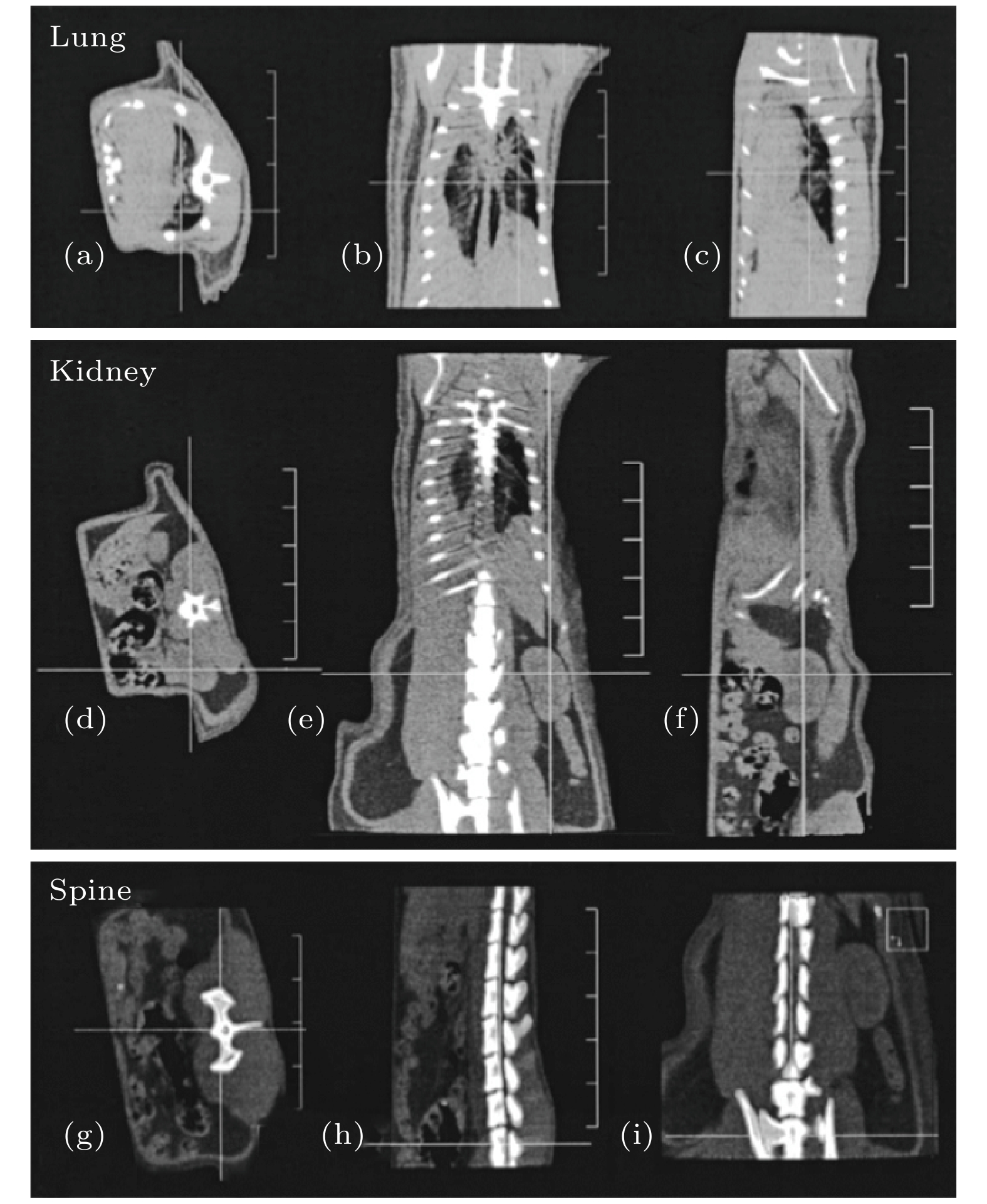
图 12 麻醉小鼠肺、肾和下脊柱区域的基于PFXRL的Micro-CBCT图像. 每个图像的窗宽窗位设置不同以呈现出感兴趣的结构. 横断面((a), (d), (g))、冠状面((b), (e), (h))和矢状面((c), (f), (i))切片展示了各向同性的空间分辨率和好的软组织对比度. 肺内支气管结构、肾脏与周围的肌肉和脂肪、椎骨和椎间隙都清晰可见. 图像中的垂直比例尺显示1 cm的间距
Fig. 12. PFXRL-based Micro-CBCT images of the lung, kidney and lower spine of the anesthetized mice. The window and level settings are varied in each image to allow visualization of the structures of interest. Axial ((a), (d), (g)) , coronal ((b), (e), (h)) , and sagittal ((c), (f), (i)) slices qualitatively demonstrate isotropic spatial resolution, with excellent soft-tissue contrast in each case. Bronchial structure within the lungs is clearly identifiable, the kidney is well delineated from surrounding muscle and fat, and fine detail in the vertebrae and intervertebral spaces is demonstrated. The vertical scale in the images shows 1 cm spacing.
表 1 PFXRL的基本参数
Table 1. Parameters of the PFXRL.
PFXRL Length /mm 69.4 Input focal distance /mm 76.3 Output focal distance /mm 20.1 Diameter of IFS at 17.4 keV /${\mu }\mathrm{m}$ 169.2 Diameter of OFS at 17.4 keV /${\mu }\mathrm{m}$ 46.7 Channel inner diameter of capillary at input/output /μm 10.4  下载: 导出CSV
-
[1] Gregory S G, Sekhon M, Schein J, et al. 2002 Nature 418 743
Google Scholar
[2] Ntziachristos V, Ripoll J, Wang L V, Weissleder R 2005 Nat. Biotechnol. 23 313
Google Scholar
[3] Guerra A D, Belcari N 2007 Nucl. Instrum. Meth. Phys. Res. A 583 119
Google Scholar
[4] Badea C T, Drangova M, Holdsworth D W, Johnson G A 2008 Phys. Med. Biol. 53 319
Google Scholar
[5] Jan M L, Ni Y C, Chen K W, Ching H 2006 Nucl. Instrum. Meth. Phys. Res. A 569 314
Google Scholar
[6] Biederer J, Mirsadraee S, Beer M, Molinari F, Puderbach M 2012 Insights Into Imaging 3 373
Google Scholar
[7] Hoyer C, Gass N, Fahr W W, Sartorius A 2014 Neuropsychobiology 69 187
Google Scholar
[8] Kunjachan S, Ehling J, Storm G, Kiessling F, Lammers T 2015 Chem. Rev. 115 10907
Google Scholar
[9] Eghtedari M, Oraevsky A, Copland J A, Kotov N A, Conjusteau A, Motamedi M 2007 Nano Lett. 7 1914
Google Scholar
[10] Taruttis A, Ntziachristos V 2015 Nat. Photonics 9 219
Google Scholar
[11] Paulus M J, Gleason S S, Kennel S J, Hunsicker P R, Johnson D K 2000 Neoplasia 2 62
Google Scholar
[12] 罗召洋, 杨孝全, 孟远征, 邓勇 2010 物理学报 58 8237
Google Scholar
Luo Z Y, Yang X Q, Meng Y Z, Deng Y 2010 Acta Phys. Sin. 58 8237
Google Scholar
[13] 魏星, 闫镔, 张峰, 李永丽, 席晓琦, 李磊 2014 物理学报 63 058702
Google Scholar
Wei X, Yan B, Zhang F, Li Y L, Xi X Q, Li L 2014 Acta Phys. Sin. 63 058702
Google Scholar
[14] Mazel V, Reiche I, Busignies V, Walter P, Tchoreloff P 2011 Talanta 85 556
Google Scholar
[15] Sun T, Liu Z, Li Y, Lin X, Wang G, Zhu G, Xu Q, Luo P, Pan Q, Liu H 2010 Nucl. Instrum. Meth. Phys. Res. A 622 295
Google Scholar
[16] Macdonald C A, Gibson W M 2003 X-Ray Spectrom. 32 258
Google Scholar
[17] Albertini V R, Paci B, Generosi A, Dabagov S B, Kumakhov M A 2007 Spectrochim. Acta B 62 1203
Google Scholar
[18] Huang R, Bilderback D H 2006 J. Synchrotron Radiat. 13 74
Google Scholar
[19] Balaic D X, Barnea Z, Nugent K A, Garrett R F, Wilkins S W 1996 J. Synchrotron Radiat. 3 289
Google Scholar
[20] MacDonald C A, Owens S M, Gibson W M 1999 J. Appl. Crystallogr. 32 160
Google Scholar
[21] Bjeoumikhov A, Bjeoumikhova S, Langhoff N, Wedell R 2005 Appl. Phys. Lett. 86 144102
Google Scholar
[22] Sun T, Liu Z, Ding X 2007 Nucl. Instrum. Meth. Phys. Res. B 262 153
Google Scholar
[23] Sun T, Peng S, Liu Z, Sun W, Ma Y, Ding X 2013 J. Appl. Crystallogr. 46 1880
Google Scholar
[24] Sun T, Macdonald C A 2013 J. Appl. Phys. 113 053104
Google Scholar
[25] Lamb J S, Bilderback D H, Pollack L, Kwok L, Smilgies D M 2007 J. Appl. Crystallogr. 40 193
Google Scholar
[26] Barrea R A, Huang R, Cornaby S, Bilderback D H, Irving T C 2009 J. Synchrotron Radiat. 16 76
Google Scholar
[27] Zeng X, Duewer F, Feser M, Huang C, Lyon A, Tkachuk A, Yun W 2008 Appl. Opt. 47 2376
Google Scholar
[28] Li F, Liu Z, Sun T, Jiang B, Zhu Y 2016 J. Chem. Phys. 144 104201
Google Scholar
[29] Li F, Liu Z, Sun T 2016 J. Appl. Crystallogr. 49 627
Google Scholar
[30] Li F, Liu Z, Sun T 2016 Rev. Sci. Instrum. 87 093106
Google Scholar
[31] Li F, Liu Z, Sun T 2016 Food Chem. 210 435
Google Scholar
[32] Li F, Liu Z, Sun T, Ma Y, Ding X 2015 Food Control 54 120
Google Scholar
[33] Abreu C C, Kruger D G, MacDonald C A, Mistretta C A, Peppler W W, Xiao Q F 1995 Med. Phys. 22 1793
Google Scholar
[34] Goertzen A L, Nagarkar V, Street R A, Paulus M J, Boone J M, Cherry S R 2004 Phys. Med. Biol. 49 5251
Google Scholar
[35] Kim H K, Min K C, Achterkirchen T, Lee W 2009 IEEE Trans. Nucl. Sci. 56 1179
Google Scholar
[36] Feldkamp L A, Davis L C, Kress J W 1984 J. Opt. Soc. Am. A 1 612
Google Scholar
[37] Flannery B P, Deckman H W, Roberge W G, D'Amico K L 1987 Science 237 1439
Google Scholar
[38] Sun T, Ding X 2005 J. Appl. Phys. 97 124904
Google Scholar
[39] Kai Y, Kwan A L C, Miller D W F, Boone J M 2006 Med. Phys. 33 1695
Google Scholar
[40] Kwan A L C, Boone J M, Yang K, Huang S Y 2007 Med. Phys. 34 275
[41] 余晓锷, 占杰, 李萍, 李婵娟 2006 第四军医大学学报 27 978
Google Scholar
Yu X E, Zhan J, Li P, Li C J 2006 J. Fourth Mil. Med. Univ. 27 978
Google Scholar

 下载:
下载:
计量
- 文章访问数: 10399
- PDF下载量: 127
- 被引次数: 0
