


-
太赫兹波位于氢键和范德瓦耳斯力作用能级范围内, 可以直接与蛋白质耦合激发蛋白质的非线性共振效应, 从而影响蛋白质的构象、神经元的结构和功能. 基于此机制, 体外培养SD大鼠原代皮层神经元, 利用宽带微量太赫兹(0.3—3.0 THz, 最大辐射功率100 μW)短时间累计辐射(3 min/d, 共3 d)皮层神经元; 记录皮层神经元的动态发育参数(胞体面积和突起总长度); 并分析辐射结束后神经元受体相关蛋白(GluA1和GluN1)、突触素(SY-38)和突触后致密蛋白-95(PSD-95)的表达变化. 太赫兹辐射1 d后, 神经元胞体面积增长值提高了144.9% (P< 0.05); 太赫兹辐射的2 d和3 d后, 神经元突起总长度增长值分别提高了65.1% (P <0.05)和109.4% (P < 0.05); 太赫兹辐射3 d后, GluA1和SY-38蛋白表达分别提高了38.1% (P < 0.05)和19.2% (P < 0.05). 结果表明, 宽带微量太赫兹短时累计辐射可以促进皮层神经元胞体和突起的生长, 并且对神经元突起的促进作用存在累计效应; 太赫兹辐射对神经元生长发育的促进作用可能与GluA1和SY-38蛋白表达相关. 这些结果预示着特定频率和能量的太赫兹波可以发展为一种治疗或干预神经发育障碍等疾病的新型神经调控技术.Terahertz waves are located in the energy level range of hydrogen bonding and van der Waals forces, and can directly couple with proteins to excite the nonlinear resonance effect of proteins. Therefore, terahertz wave can affect the conformation of proteins, the structure and function of neurons. Primary cerebral cortex neurons of SD rats are cultured in vitro. Neurons are radiated 3 days by THz wave with 0.3–3.0 THz in frequency and 100 μW in power; the growth and development indicators of neurons (Cell body area, total length of process) are recorded. At the end of a radiation programme, Western blotting is used to detect the protein expressions of GluA1, GluN1, postsynaptic density protein-95 (PSD-95) and synaptophysin 38 (SYP-38). After the first day of terahertz wave radiation, the cell area is increased by 144.9% (P < 0.05); on the second day and third day of terahertz wave radiation, the growth value of the total length of neuronal neurites are increased by 65.1% (P < 0.05) and 109.4% (P < 0.05), respectively. After the three-day terahertz wave irradiation, the protein expressions of GluA1 and SY-38 are increased by 38.1% (P < 0.05) and 19.2% (P < 0.05), respectively. In addition,some results show below. 1) The use of low-intensity broadband terahertz wave in this study will not cause the cortical neurons to die, and will not affect their regular growth. 2) Low-intensity broadband terahertz radiation can promote the growth of cortical neuron cell bodies and processes, but the effects on cortical neuron cell bodies and processes are different. This may be related to the developmental cycle of cultured cortical neurons in vitro, and there is a cumulative effect on the promotion of neuronal processes by low-intensity broadband terahertz. 3) The promotion of neuronal growth and development by low-intensity broadband terahertz wave radiation may be related to the proportion of AMPA receptor subtypes and the expression of presynaptic specific protein SY-38. These results herald a specific-frequency and specific-energy terahertz radiation can be developed into a novel neuromodulation technology for the treatment or intervention of diseases such as neurodevelopmental disorders.
-
Keywords:
- terahertz /
- neuron /
- growth and development /
- cumulative effect
[1] Giovanni A, Capone F, di Biase L, Ferreri F, Florio L, Guerra A, Marano M, Paolucci M, Ranieri F, Salomone G, Tombini M, Thut G, Di Lazzaro V 2017 Front. Aging Neurosci. 9 189
 Google Scholar
[2] Lui JH, Hansen DV, Kriegstein AR 2011 Cell 146 18
Google Scholar
[3] Jones S R, Kerr C E, Wan Q, Pritchett D L, Hämäläinen Moore C I 2010 J. Neurosci. 30 13760
Google Scholar
[4] Kanaan N M, Pigino G F, Brady S T, Lazarov O, Binder L I, Morfini G A 2013 Exp. Neurol 246 44
Google Scholar
[5] Van Battum E Y, Brignani S, Pasterkamp R J 2015 Lancet Neurol. 14 532
Google Scholar
[6] MacLeod D, Dowman J, Hammond R, Leete T, Inoue K, Abeliovich A 2006 Neuron 52 587
Google Scholar
[7] Tang F R, Yu P P, Wang L, Guo S, Yang Q 2017 Acta. Anat. Sin. 48 1
Google Scholar
[8] 郁盛雪, 屈文慧, 隋海娟, 金迎新, 金向楠, 金英 2013 中国药理学通报 29 126
Google Scholar
Yu S X, Qu W H, Sui H J, Jin Y X, Jin X N, Jin Y 2013 Chin. Pharmacol. Bull. 29 126
Google Scholar
[9] Zorkina Y, Abramova O, Ushakova V, Morozova A, Zubkov E, Valikhov M, Melnikov P, Majouga A, Chekhonin V 2020 Molecules 25 5294
Google Scholar
[10] Cullen CL, Young KM 2016 Front. Neural. Circuits 10 26
Google Scholar
[11] Zhang N, Xing M, Wang Y, Tao H, Cheng Y 2015 Neuroscience 311 284
Google Scholar
[12] Bashir S, Uzair M, Abualait T, Arshad M, Khallaf RA, Niaz A, Thani Z, Yoo WK, Túnez I, Demirtas-Tatlidede A, Meo SA 2022 Mol. Med. Rep. 25 109
Google Scholar
[13] Hu Y, Zhong W, Wan J M, Yu A C 2013 Ultrasound. Med. Biol. 39 915
Google Scholar
[14] Peng X Y, Zhou H 2021 Acta Phys. Sin. 70 240701
Google Scholar
[15] Tan S Z, Tan P C, Luo L Q, Chi Y L, Yang Z L, Zhao X L, Zhao L, Dong J, Zhang J, Yao B W, Xu X P, Tian G, Chen J K, Wang H, Peng R Y 2019 Biomed. Environ. Sci. 32 739
Google Scholar
[16] Andrey S, Sergey V P 2013 Bioelectromagnetics 34 133
Google Scholar
[17] Olshevskaya J S, Kozlov A S, Petrov A K, Zapara T A, Ratushnyak A S 2010 Phys. Sci. 5 177
[18] Olshevskaya J S, Kozlov A S, Petrov A K, Zapara T A, Ratushnyak A S 2009 J. Higher Nervous Activ. 59 353
[19] Guo Z Y, Li C Z, Li X J, Wang Y L, Mattson M P, Lu C B 2013 NeuroReport 24 492
Google Scholar
[20] Wang J G, Wang Y L, Xu F, Zhao J X, Zhou S Y, Yu Y, Chazot P L, Wang X F, Lu C B 2016 Acta Pharmacol. Sin. 37 303
Google Scholar
[21] Tiziana M, Rosanna M, Augusto M, Massimo P, Stefano L, Annalisa D 2022 Radiation 2 100
Google Scholar
[22] He Y F, Chen J Y, Knab, Zheng W J, Markelz A G 2010 IEEE Trans. Terahertz. Sci. Technol. 3 149
Google Scholar
[23] Sun L, Zhao L, Peng R Y 2021 Mil. Med. Res. 8 28
[24] Cherkasova O P, Fedorov V I, Nemova E F, Pogodin A S 2009 Opt. Spectrosc. 107 534
Google Scholar
[25] Kummer E, Ban N 2021 Nat. Rev. Mol. Cell. Biol. 22 307
Google Scholar
[26] Masayoshi T 2007 Nat. Photonics 1 97
Google Scholar
[27] Zhao X, Zhang M, Liu Y, Liu H, Ren K, Xue Q, Zhang H, Zhi N, Wang W, Wu S 2021 iScience 24 103485
Google Scholar
[28] Stuart C C, Leah K, Mark F 2006 Curr. Opin. Neurobiol. 16 288
Google Scholar
[29] Jonas P, Racca C, Sakmann B, Seeburg P H, Monyer H 1994 Neuron 12 1281
Google Scholar
[30] Kater S B, Mills L R 1991 J. Neurosci. 14 891
Google Scholar
[31] Greger I H, Watson J F, Cull-Candy S G 2017 Neuron 94 713
Google Scholar
[32] Sun S, Igor T, Jeffrey V, Michael C 2012 J. Radiat. Res. 53 159
Google Scholar
[33] Titushkin I A, Rao V S, Pickard W F, Moros G, Shafirstein G, Cho M R 2009 Radiat. Res. 172 725
Google Scholar
[34] 张欣欣, 何明霞, 赵晋武, 陈勰宇, 刘立媛, 卢晓云, 田甜, 陈孟秋, 王璞 2020 中国激光 47 0207023
Google Scholar
Zhang X X, He M X, Zhao J W, Chen X Y, Liu L Y, Lu X Y, Tian T, Chen M Q, Wang P 2020 Chin. J. Lasers 47 0207023
Google Scholar
[35] Kao H T, Ryoo K, Lin A, Janoschka S R, Augustine G J, Porton B 2017 Eur. J. Neurosci. 45 1085
Google Scholar
[36] Fornasiero E F, Bonanomi D, Benfenati F, Valtorta F 2010 Cell. Mol. Life. Sci. 67 1383
Google Scholar
[37] Bottauscio O, Chiampi M, Zilberti L 2015 IEEE Trans. Magn. 51 7400504
Google Scholar
[38] Anush D, Armenuhi H, Anna N, Erna D, Sinerik A 2012 Electromagn. Biol. Med. 31 132
Google Scholar
[39] Sulatsky M I, Duka M V, Smolyanskaya O A 2014 Phys. Wave Phenom. 22 197
Google Scholar
[40] 查彩慧 2016 博士学位论文(广州: 暨南大学)
Cha C H 2016 Ph. D. Dissertation (Guangzhou: Jinan University) (in Chinese)
[41] Melinda K K, Christopher G L, Vincent L, Hersh L, Bonnie L F 2010 J. Vis. Exp. 45 e2354
Google Scholar
-

图 1 实验平台、实验协议和太赫兹波衰减测试 (a)太赫兹辐射神经元实验平台; (b)太赫兹辐射神经元实验协议; (c)太赫兹波穿透培养液后的时域图; (d)太赫兹波穿透培养液后的频域图
Fig. 1. Experimental platform, protocol and terahertz wave attenuation test: (a) Terahertz radiation neuron experimental platform; (b) experimental protocol for terahertz radiation neurons; (c) time domain diagram of terahertz wave after penetrating culture medium; (d) frequency domain map of terahertz wave after penetrating culture medium.

图 2 太赫兹辐射前后神经元的胞体面积和突起总长度变化 (a)太赫兹辐射1 d后对照组和太赫兹组神经元生长发育状态; (b)太赫兹辐射2 d后对照组和太赫兹组神经元生长发育状态; (c)太赫兹辐射3 d后对照组和太赫兹组神经元生长发育状态(t检验显著性差异:
$ p < 0.05 $ , *;$ p < 0.01 $ , **)Fig. 2. Changes in cell body area and total neurite length of neurons before and after terahertz radiation: (a) The growth and development status of neurons in the control group and the terahertz group after 1 d of terahertz radiation; (b) the growth and development status of neurons in the control group and the terahertz group after 2 d of terahertz radiation; (c) the growth and development status of neurons in the control group and the terahertz group after 3 d of terahertz radiation(significance of t-test:
$ p < 0.05 $ , *;$ p < 0.01 $ , **).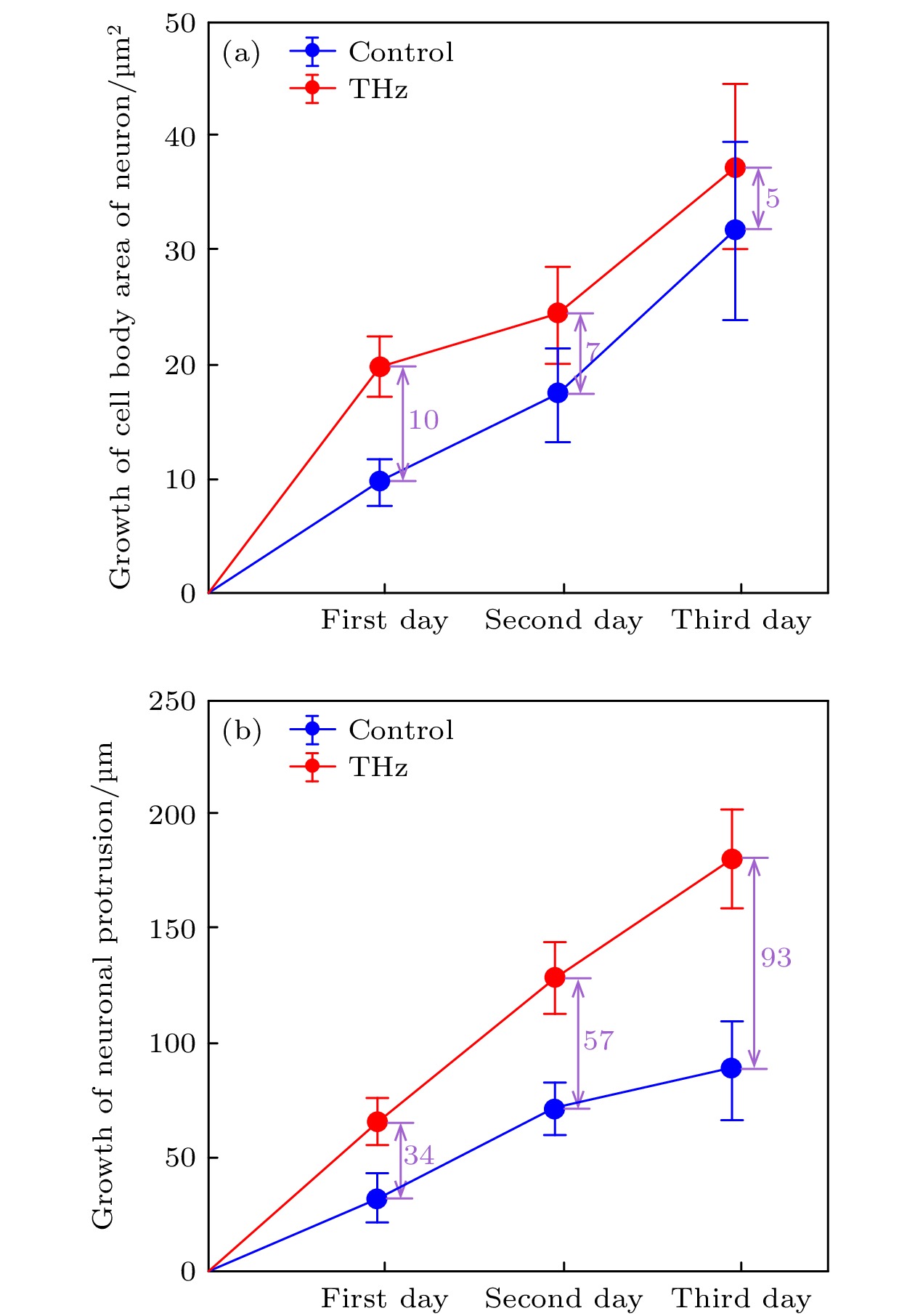
图 3 太赫兹辐射时间与神经元胞体面积和突起总长度的相关性 (a)对照组和太赫兹组神经元胞体面积增长均值误差曲线; (b)对照组和太赫兹组神经元突起总长度增长均值误差曲线
Fig. 3. Correlation between terahertz radiation time and neuronal cell body area and total neurite length: (a) The mean error curve of neuronal cell body area growth in the control group and the terahertz group; (b) the mean error curve of the total length of neurites in the control group and the terahertz group.
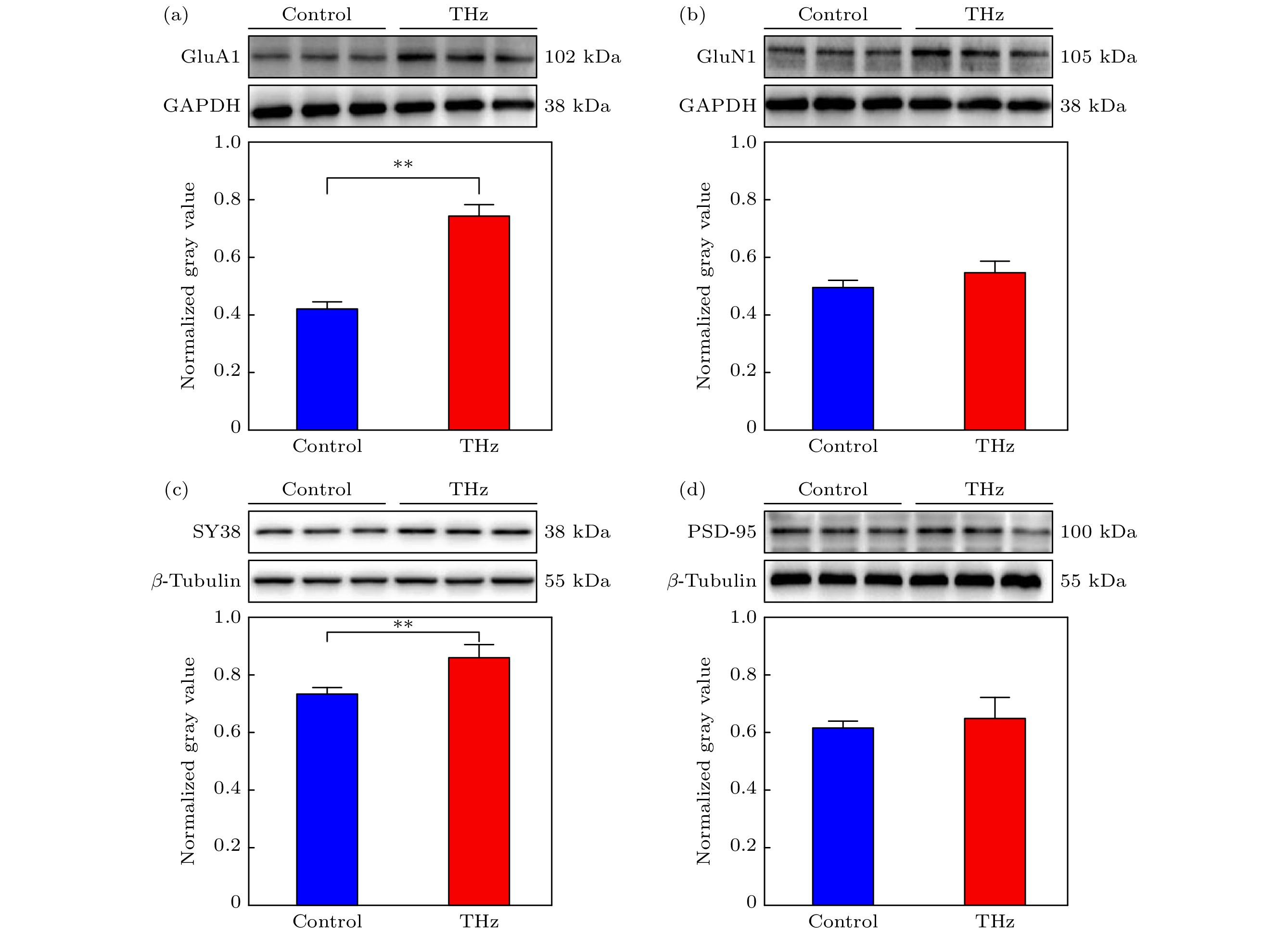
图 4 太赫兹辐射前后神经元相关蛋白表达变化 (a) GluA1蛋白表达变化; (b) GluN1蛋白表达变化; (c) SY38蛋白表达变化; (d) PSD-95蛋白表达变化 (t检验显著性差异:
$ p < 0.05 $ , *;$ p < 0.01 $ , **)Fig. 4. Changes of neuron-related protein expression before and after terahertz radiation: (a) Changes of GluA1 protein expression; (b) changes of GluN1 protein expression; (c) expression changes of SY38 protein; (d) changes of PSD-95 protein expression (significance of t-test:
$ p < 0.05 $ , *;$ p < 0.01 $ , **). -
[1] Giovanni A, Capone F, di Biase L, Ferreri F, Florio L, Guerra A, Marano M, Paolucci M, Ranieri F, Salomone G, Tombini M, Thut G, Di Lazzaro V 2017 Front. Aging Neurosci. 9 189
Google Scholar
[2] Lui JH, Hansen DV, Kriegstein AR 2011 Cell 146 18
Google Scholar
[3] Jones S R, Kerr C E, Wan Q, Pritchett D L, Hämäläinen Moore C I 2010 J. Neurosci. 30 13760
Google Scholar
[4] Kanaan N M, Pigino G F, Brady S T, Lazarov O, Binder L I, Morfini G A 2013 Exp. Neurol 246 44
Google Scholar
[5] Van Battum E Y, Brignani S, Pasterkamp R J 2015 Lancet Neurol. 14 532
Google Scholar
[6] MacLeod D, Dowman J, Hammond R, Leete T, Inoue K, Abeliovich A 2006 Neuron 52 587
Google Scholar
[7] Tang F R, Yu P P, Wang L, Guo S, Yang Q 2017 Acta. Anat. Sin. 48 1
Google Scholar
[8] 郁盛雪, 屈文慧, 隋海娟, 金迎新, 金向楠, 金英 2013 中国药理学通报 29 126
Google Scholar
Yu S X, Qu W H, Sui H J, Jin Y X, Jin X N, Jin Y 2013 Chin. Pharmacol. Bull. 29 126
Google Scholar
[9] Zorkina Y, Abramova O, Ushakova V, Morozova A, Zubkov E, Valikhov M, Melnikov P, Majouga A, Chekhonin V 2020 Molecules 25 5294
Google Scholar
[10] Cullen CL, Young KM 2016 Front. Neural. Circuits 10 26
Google Scholar
[11] Zhang N, Xing M, Wang Y, Tao H, Cheng Y 2015 Neuroscience 311 284
Google Scholar
[12] Bashir S, Uzair M, Abualait T, Arshad M, Khallaf RA, Niaz A, Thani Z, Yoo WK, Túnez I, Demirtas-Tatlidede A, Meo SA 2022 Mol. Med. Rep. 25 109
Google Scholar
[13] Hu Y, Zhong W, Wan J M, Yu A C 2013 Ultrasound. Med. Biol. 39 915
Google Scholar
[14] Peng X Y, Zhou H 2021 Acta Phys. Sin. 70 240701
Google Scholar
[15] Tan S Z, Tan P C, Luo L Q, Chi Y L, Yang Z L, Zhao X L, Zhao L, Dong J, Zhang J, Yao B W, Xu X P, Tian G, Chen J K, Wang H, Peng R Y 2019 Biomed. Environ. Sci. 32 739
Google Scholar
[16] Andrey S, Sergey V P 2013 Bioelectromagnetics 34 133
Google Scholar
[17] Olshevskaya J S, Kozlov A S, Petrov A K, Zapara T A, Ratushnyak A S 2010 Phys. Sci. 5 177
[18] Olshevskaya J S, Kozlov A S, Petrov A K, Zapara T A, Ratushnyak A S 2009 J. Higher Nervous Activ. 59 353
[19] Guo Z Y, Li C Z, Li X J, Wang Y L, Mattson M P, Lu C B 2013 NeuroReport 24 492
Google Scholar
[20] Wang J G, Wang Y L, Xu F, Zhao J X, Zhou S Y, Yu Y, Chazot P L, Wang X F, Lu C B 2016 Acta Pharmacol. Sin. 37 303
Google Scholar
[21] Tiziana M, Rosanna M, Augusto M, Massimo P, Stefano L, Annalisa D 2022 Radiation 2 100
Google Scholar
[22] He Y F, Chen J Y, Knab, Zheng W J, Markelz A G 2010 IEEE Trans. Terahertz. Sci. Technol. 3 149
Google Scholar
[23] Sun L, Zhao L, Peng R Y 2021 Mil. Med. Res. 8 28
[24] Cherkasova O P, Fedorov V I, Nemova E F, Pogodin A S 2009 Opt. Spectrosc. 107 534
Google Scholar
[25] Kummer E, Ban N 2021 Nat. Rev. Mol. Cell. Biol. 22 307
Google Scholar
[26] Masayoshi T 2007 Nat. Photonics 1 97
Google Scholar
[27] Zhao X, Zhang M, Liu Y, Liu H, Ren K, Xue Q, Zhang H, Zhi N, Wang W, Wu S 2021 iScience 24 103485
Google Scholar
[28] Stuart C C, Leah K, Mark F 2006 Curr. Opin. Neurobiol. 16 288
Google Scholar
[29] Jonas P, Racca C, Sakmann B, Seeburg P H, Monyer H 1994 Neuron 12 1281
Google Scholar
[30] Kater S B, Mills L R 1991 J. Neurosci. 14 891
Google Scholar
[31] Greger I H, Watson J F, Cull-Candy S G 2017 Neuron 94 713
Google Scholar
[32] Sun S, Igor T, Jeffrey V, Michael C 2012 J. Radiat. Res. 53 159
Google Scholar
[33] Titushkin I A, Rao V S, Pickard W F, Moros G, Shafirstein G, Cho M R 2009 Radiat. Res. 172 725
Google Scholar
[34] 张欣欣, 何明霞, 赵晋武, 陈勰宇, 刘立媛, 卢晓云, 田甜, 陈孟秋, 王璞 2020 中国激光 47 0207023
Google Scholar
Zhang X X, He M X, Zhao J W, Chen X Y, Liu L Y, Lu X Y, Tian T, Chen M Q, Wang P 2020 Chin. J. Lasers 47 0207023
Google Scholar
[35] Kao H T, Ryoo K, Lin A, Janoschka S R, Augustine G J, Porton B 2017 Eur. J. Neurosci. 45 1085
Google Scholar
[36] Fornasiero E F, Bonanomi D, Benfenati F, Valtorta F 2010 Cell. Mol. Life. Sci. 67 1383
Google Scholar
[37] Bottauscio O, Chiampi M, Zilberti L 2015 IEEE Trans. Magn. 51 7400504
Google Scholar
[38] Anush D, Armenuhi H, Anna N, Erna D, Sinerik A 2012 Electromagn. Biol. Med. 31 132
Google Scholar
[39] Sulatsky M I, Duka M V, Smolyanskaya O A 2014 Phys. Wave Phenom. 22 197
Google Scholar
[40] 查彩慧 2016 博士学位论文(广州: 暨南大学)
Cha C H 2016 Ph. D. Dissertation (Guangzhou: Jinan University) (in Chinese)
[41] Melinda K K, Christopher G L, Vincent L, Hersh L, Bonnie L F 2010 J. Vis. Exp. 45 e2354
Google Scholar

 下载:
下载:


计量
- 文章访问数: 10656
- PDF下载量: 128
- 被引次数: 0
